Anterior Collection (#4)

Exploring the intricate beauty of the anterior: From the delicate heart to neck vascular anatomy, historical artwork unveils its secrets

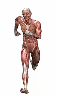
Background imageAnterior Collection: Muscular man standing

Muscular man standing, isolated on white background

Background imageAnterior Collection: Osteichthyans - Aspidorhynchus: Anterior trunk

Osteichthyans - Aspidorhynchus: Anterior trunk

Background imageAnterior Collection: Chondrichthyans - Petalodus: Anterior tooth

Chondrichthyans - Petalodus: Anterior tooth

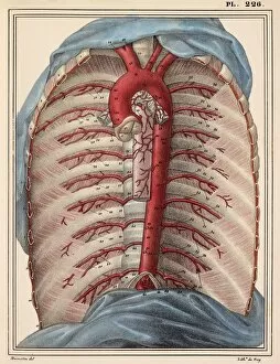
Background imageAnterior Collection: Thoracic aorta, 1825 artwork

Thoracic aorta, 1825 artwork
Thoracic aorta. Dissection of the chest cavity with the heart and lungs and other organs removed, showing the thoracic portion of the aorta (red), the main body artery

Background imageAnterior Collection: Intestinal veins, 1825 artwork

Intestinal veins, 1825 artwork
Intestinal veins. Dissection of the abdominal cavity showing the inferior mesenteric vein (blue) that drains blood from the large intestine

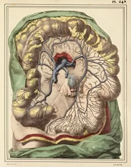
Background imageAnterior Collection: Intestinal arteries, 1825 artwork

Intestinal arteries, 1825 artwork
Intestinal arteries. Dissection of the abdomen, showing the inferior mesenteric artery (red) and its branches that supply oxygenated blood to the large intestine

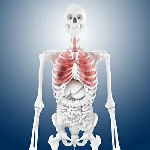
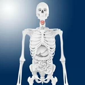
Background imageAnterior Collection: Stomach anatomy, artwork

Stomach anatomy, artwork
Stomach anatomy. Artwork showing the position of the stomach (red) in relation to other abdominal organs and the human skeleton, as seen in an anterior (frontal) view

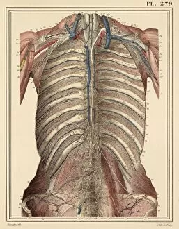
Background imageAnterior Collection: Chest lymph vessels, 1825 artwork

Chest lymph vessels, 1825 artwork
Chest lymph vessels. Dissection of the thoracic cavity showing lymphatic vessels (white) and nodes. The central chest lymph vessel is the thoracic canal, running parallel to the backbone (centre)

Background imageAnterior Collection: Abdominal aorta, 1825 artwork

Abdominal aorta, 1825 artwork
Abdominal aorta. Dissection, with some organs removed, showing the abdominal portion of the aorta (red), the main body artery

Background imageAnterior Collection: Psychic brain, conceptual image

Psychic brain, conceptual image. Artwork showing the human brain and head, surrounded in flames, with the pineal gland highlighted (bright dot in centre of forehead)

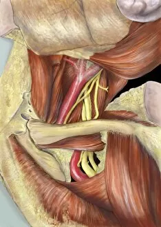
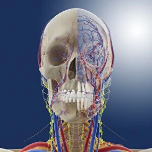
Background imageAnterior Collection: Head and neck anatomy, artwork C014 / 0511

Head and neck anatomy, artwork C014 / 0511
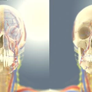
Head and neck anatomy, computer artwork. Arteries are red, veins are blue, nerves are deep yellow and lymph vessels are bright yellow

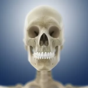
Background imageAnterior Collection: Human skull, artwork C014 / 0422

Human skull, artwork C014 / 0422
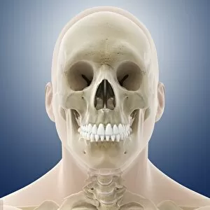
Human skull, computer artwork

Background imageAnterior Collection: Anatomical orientation planes, artwork

Anatomical orientation planes, artwork
Anatomical orientation planes. Computer artwork of a human body, showing the three different orientation planes (green) used as a reference in anatomy

Background imageAnterior Collection: Left hip and nerve plexus, artwork C016 / 6808

Left hip and nerve plexus, artwork C016 / 6808
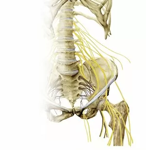
Left hip and nerve plexus. Artwork of the nerves (yellow) and bones of the left hip, seen from the front. This is the lumbar plexus, itself forming part of the lumbosacral plexus

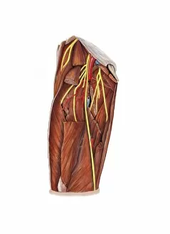
Background imageAnterior Collection: Thigh nerve plexus, artwork C016 / 6804

Thigh nerve plexus, artwork C016 / 6804
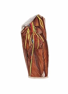
Thigh nerve plexus. Artwork of the nerves (yellow), arteries (red), veins (blue), and muscles (brown) of the upper leg (thigh)

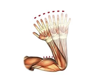
Background imageAnterior Collection: Elbow flexion, artwork C016 / 6790

Elbow flexion, artwork C016 / 6790
Elbow flexion. Artwork of the muscles of the arm from the front, with red arrows showing the direction of movement when flexing (bending) the elbow

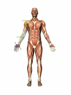
Background imageAnterior Collection: Arm nerve regions, artwork C016 / 6840

Arm nerve regions, artwork C016 / 6840
Arm nerve regions. Artwork of the muscles of the human body from the front, with the right arm (left) labelled with the names of the spinal nerves (cervical, neck, C; and thoracic, chest)

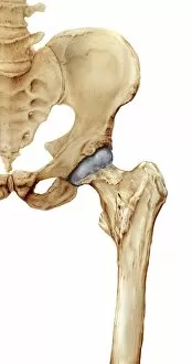
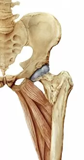
Background imageAnterior Collection: Hip bones, artwork C016 / 7015

Hip bones, artwork C016 / 7015
Hip bones. Artwork of a frontal (anterior) view of the left hip, showing the bones that articulate to form this joint. At lower right is the femur (thigh bone)

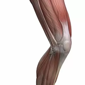
Background imageAnterior Collection: Knee muscles and bones, artwork C016 / 7013

Knee muscles and bones, artwork C016 / 7013
Knee muscles and bones. Artwork of an oblique lateral frontal view of the muscles and bones of a human knee. This view is of the outside of the left leg

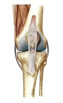
Background imageAnterior Collection: Knee bones and ligaments, artwork C016 / 7012

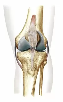
Knee bones and ligaments, artwork C016 / 7012
Knee bones and ligaments. Artwork of a frontal view of the knee joint, showing bones, ligaments, tendons, and cartilage. The bones are the femur (thigh bone, top), patella (knee-cap, upper centre)

Background imageAnterior Collection: Elbow flexion, artwork C016 / 6789

Elbow flexion, artwork C016 / 6789
Elbow flexion. Artwork of the muscles of the arm from the front, with red arrows showing the direction of movement when flexing (bending) the elbow

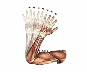
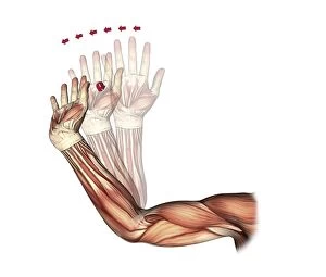
Background imageAnterior Collection: Elbow and wrist extension, artwork C016 / 6791

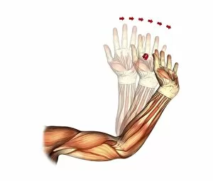
Elbow and wrist extension, artwork C016 / 6791
Elbow and wrist extension. Artwork of the muscles of the arm from the front, with red arrows showing the direction of movement when extending the elbow and wrist joints

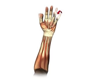
Background imageAnterior Collection: Little and ring finger flexion, artwork C016 / 6794

Little and ring finger flexion, artwork C016 / 6794
Little and ring finger flexion. Artwork of the muscles of the forearm and hand from the front, with red arrows showing the direction of movement of the little and ring (fourth and third)

Background imageAnterior Collection: Right hip and nerve plexus, artwork C016 / 6809

Right hip and nerve plexus, artwork C016 / 6809
Right hip and nerve plexus. Artwork of the nerves (yellow) and bones of the right hip, seen from the front. This is the lumbar plexus, itself forming part of the lumbosacral plexus

Background imageAnterior Collection: Runners anatomy, artwork

Runners anatomy, artwork
Runners anatomy. Artwork showing the musculature of a running human figure. The main muscles used when running are the quadriceps femoris (rectus femoris, vastus medialis, vastus lateralis)

Background imageAnterior Collection: Dental maxillary nerve regions, artwork C016 / 6835

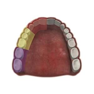
Dental maxillary nerve regions, artwork C016 / 6835
Dental maxillary nerve regions. Artwork of the teeth of the upper jaw (maxilla), seen from below, colour-coded by the branches of the superior alveolar nerve that supply the maxillary teeth

Background imageAnterior Collection: Knee bones and ligaments, artwork C016 / 7011

Knee bones and ligaments, artwork C016 / 7011
Knee bones and ligaments. Artwork of a frontal view of the knee joint, showing bones, ligaments, tendons and cartilage. The bones are the femur (thigh bone, top), patella (knee-cap, upper centre)

Background imageAnterior Collection: Elbow and wrist extension, artwork C016 / 6792

Elbow and wrist extension, artwork C016 / 6792
Elbow and wrist extension. Artwork of the muscles of the arm from the front, with red arrows showing the direction of movement when extending the elbow and wrist joints

Background imageAnterior Collection: Right shoulder nerve plexus, artwork C016 / 6815

Right shoulder nerve plexus, artwork C016 / 6815
Right shoulder nerve plexus. Artwork of the nerves (yellow), arteries (red), muscles (red-brown) and bones (light brown) of the right shoulder, seen from the front

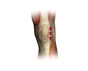
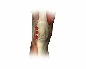
Background imageAnterior Collection: Patella twitch, artwork C016 / 6816

Patella twitch, artwork C016 / 6816
Patella twitch. Artwork of the muscles of the knee from the front, with red arrows showing the direction of movement of the knee cap (patella) when the quadriceps muscles of the thigh contracts

Background imageAnterior Collection: Thigh nerve plexus, artwork C016 / 6805

Thigh nerve plexus, artwork C016 / 6805
Thigh nerve plexus. Artwork of the nerves (yellow), arteries (red), veins (blue), and muscles (brown) of the upper leg (thigh)

Background imageAnterior Collection: Hip bones and thigh muscles, artwork C016 / 7016

Hip bones and thigh muscles, artwork C016 / 7016
Hip bones and thigh muscles, artwork

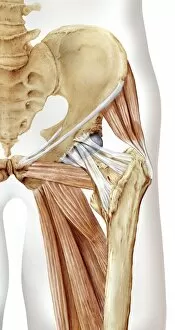
Background imageAnterior Collection: Hip bones, muscles and ligaments, artwork C016 / 7018

Hip bones, muscles and ligaments, artwork C016 / 7018
Hip bones and thigh muscles. Artwork of a frontal (anterior) view of the left hip, showing bones and some thigh muscles (red). At lower right is the femur (thigh bone)

Background imageAnterior Collection: Left armpit nerve plexus, artwork C016 / 6812

Left armpit nerve plexus, artwork C016 / 6812
Left armpit nerve plexus. Artwork of the nerves (yellow), arteries (red), muscles (red-brown) and bones (light brown) of the left armpit, seen from the front

Background imageAnterior Collection: Patella twitch, artwork C016 / 6803

Patella twitch, artwork C016 / 6803
Patella twitch. Artwork of the muscles of the knee from the front, with red arrows showing the direction of movement of the knee cap (patella) when the quadriceps muscles of the thigh contracts

Background imageAnterior Collection: Left shoulder and nerve plexus, artwork C016 / 6810

Left shoulder and nerve plexus, artwork C016 / 6810
Left shoulder and nerve plexus. Artwork of the nerves (yellow), arteries (red), and bones of the left shoulder, seen from the front

Background imageAnterior Collection: Left shoulder nerve plexus, artwork C016 / 6814

Left shoulder nerve plexus, artwork C016 / 6814
Left shoulder nerve plexus. Artwork of the nerves (yellow), arteries (red), muscles (red-brown) and bones (light brown) of the left shoulder, seen from the front

Background imageAnterior Collection: Right armpit nerve plexus, artwork C016 / 6813

Right armpit nerve plexus, artwork C016 / 6813
Right armpit nerve plexus. Artwork of the nerves (yellow), arteries (red), muscles (red-brown) and bones (light brown) of the right armpit, seen from the front

Background imageAnterior Collection: Human skull, artwork C014 / 0427

Human skull, artwork C014 / 0427
Human skull, computer artwork

Background imageAnterior Collection: Human skull, artwork C014 / 0411

Human skull, artwork C014 / 0411
Human skull, computer artwork

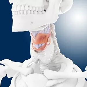
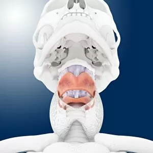
Background imageAnterior Collection: Healthy larynx and thyroid, artwork C014 / 0402

Healthy larynx and thyroid, artwork C014 / 0402
Healthy larynx and thyroid, computer artwork. At centre is the larynx (purple/grey), also known as the voice box, surrounded by the thyroid cartilage (pink).Above the larynx is the hyoid bone (white)

Background imageAnterior Collection: Healthy larynx and thyroid, artwork

Healthy larynx and thyroid, artwork
Healthy larynx and thyroid, computer artwork. At centre is the larynx (purple/grey), also known as the voice box, surrounded by the thyroid cartilage (pink)

Background imageAnterior Collection: Head and neck anatomy, artwork C014 / 0512

Head and neck anatomy, artwork C014 / 0512
Head and neck anatomy, computer artwork. Arteries are red, veins are blue, nerves are deep yellow and lymph vessels are bright yellow

Background imageAnterior Collection: Healthy larynx and thyroid, artwork C014 / 0404

Healthy larynx and thyroid, artwork C014 / 0404
Healthy larynx and thyroid, computer artwork. The larynx, also known as the voice box, is purple/grey. It is surrounded by the thyroid cartilage (pink)

Background imageAnterior Collection: Head and neck anatomy, artwork C014 / 0509

Head and neck anatomy, artwork C014 / 0509
Head and neck anatomy, computer artwork. The skin is transparent allowing arteries (red), veins (blue), nerves (deep yellow) and lymph vessels (bright yellow) to be seen

Background imageAnterior Collection: Healthy larynx and thyroid, artwork C014 / 0400

Healthy larynx and thyroid, artwork C014 / 0400
Healthy larynx and thyroid, computer artwork. At centre is the larynx (purple/grey), also known as the voice box, surrounded by the thyroid cartilage (pink)

Background imageAnterior Collection: Healthy larynx, artwork C014 / 0403

Healthy larynx, artwork C014 / 0403
Healthy larynx, computer artwork. At centre is the larynx (purple/grey), also known as the voice box, surrounded by the thyroid cartilage (pink)

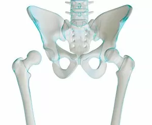
Background imageAnterior Collection: Hip joint bones and anatomy, artwork C014 / 2031

Hip joint bones and anatomy, artwork C014 / 2031
Hip joint bones and anatomy. Artwork of the bones that form the two hip joints (right and left). The hip is a ball-and-socket joint that provides the mobility needed for walking and running


For sale as Licensed Images

Choose your image, Select your licence and Download the media

Exploring the intricate beauty of the anterior: From the delicate heart to neck vascular anatomy, historical artwork unveils its secrets. X-ray reveals the complex network of arteries in the neck and shoulder, while a detailed human foot anatomy showcases skin, veins, arteries, muscles, and bones. The interplay between heart and lungs is captured in stunning visuals. Journey back to 1825 with head and chest artery artwork. Marvel at four views of the magnificent heart itself. Dive into the molecular world with a glimpse of human growth hormone molecule. Discover female heart anatomy through captivating illustrations. Witness the miracle of life as a front view of a nine-month pregnant woman prepares for delivery with her baby phantomed within. Uncover male respiratory system and internal organs' intricacies through anatomical exploration. Finally, explore female body anatomy alongside its internal wonders - an awe-inspiring journey into our anterior realm.