Bacteriology Collection (#3)

"Bacteriology: Exploring the Microscopic World of Skin Disorders and Artwork" Delve into the fascinating realm of bacteriology

Background imageBacteriology Collection: Bacteria research

Bacteria research
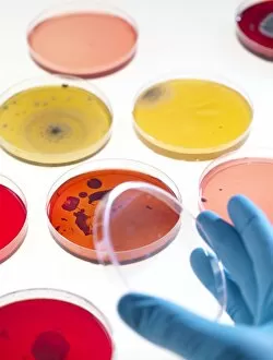
MODEL RELEASED. Bacteria research. Researcher holding an empty petri dish in front of other dishes containing cultured bacteria (dots)

Background imageBacteriology Collection: Bacterial cell structure, artwork

Bacterial cell structure, artwork
Bacterial cell structure. Computer artwork showing the cell structure and components (organelles) of a typical rod-shaped bacteria (bacillus). Not all bacteria have a flagellum (long, tail-like)

Background imageBacteriology Collection: Bacterial lung infection

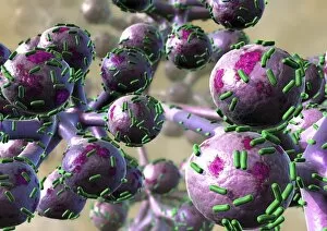
Bacterial lung infection, computer artwork. Rod-shaped bacteria (bacilli, green) on the surface of the alveoli of the lungs (purple)

Background imageBacteriology Collection: Bacillus subtilis bacteria, SEM

Bacillus subtilis bacteria, SEM
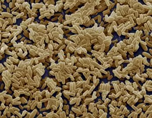
Bacillus subtilis. Coloured scanning electron micrograph (SEM) of Bacillus subtilis bacteria. B. subtilis, or hay bacillus, is an aerobic, gram-positive bacillus(rod-shaped bacterium)

Background imageBacteriology Collection: Lactobacillus bacteria, SEM

Lactobacillus bacteria, SEM
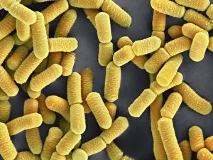
Lactobacillus sp. bacteria, coloured scanning electron micrograph (SEM). Lactobacillus bacteria are probiotic bacteria, which are useful for human and animal health as they produce bacteriocins

Background imageBacteriology Collection: Clostridium perfringens bacterium with spore

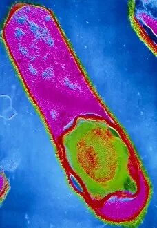
Clostridium perfringens bacterium with spore
False-colour transmission electron micrograph (TEM) of a Clostridium perfringens bacterium with endospore. This species of Gram-positive

Background imageBacteriology Collection: Bacteria on lung hairs

Bacteria on lung hairs
Bacteria in a lung. Computer artwork of bacteria (yellow) on cilia (hair-like structures) in a lung. Bacterial infection of the lungs can cause potentially fatal pneumonia

Background imageBacteriology Collection: Proteus mirabilis bacterium

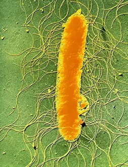
Proteus mirabilis bacterium
False-colour transmission electron micrograph of the bacterium Proteus mirabilis. It is an enterobacterium which is present normally in the human intestine

Background imageBacteriology Collection: Spirochete bacteria, TEM

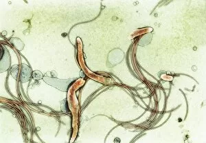
Spirochete bacteria, TEM
Spirochete bacteria, coloured transmission electron micrograph (TEM). Spirochetes are bacteria with helically coiled cells

Background imageBacteriology Collection: Coloured SEM of Clostridium botulinum

Coloured SEM of Clostridium botulinum
Food poisoning bacterium. Coloured scanning electron micrograph of the Gram-positive anaerobic bacteria Clostridium botulinum, responsible for serious food poisoning in humans. C

Background imageBacteriology Collection: Rod-shaped bacteria

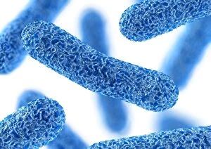
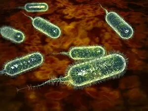
Rod-shaped bacteria. Computer artwork of rod- shaped bacteria (bacilli). Typical bacilli are E. coli and Salmonella bacteria, though there are many others as well

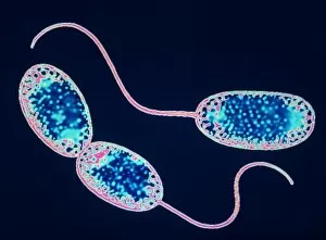
Background imageBacteriology Collection: Nitrobacter sp bacteria, TEM

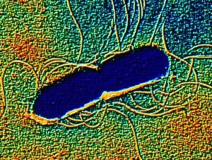
Nitrobacter sp bacteria, TEM

Background imageBacteriology Collection: Inauguration of Louis Pasteurs statue, Paris, 1904

Inauguration of Louis Pasteurs statue, Paris, 1904. An illustration from Le Petit Journal, 24th July 1904

Background imageBacteriology Collection: Emil von Behring, German immunologist and bacteriologist, 1902

Emil von Behring, German immunologist and bacteriologist, 1902. In 1901 von Behring (1854-1917) was awarded the first Nobel Prize for Physiology or Medicine for his development of a serum for

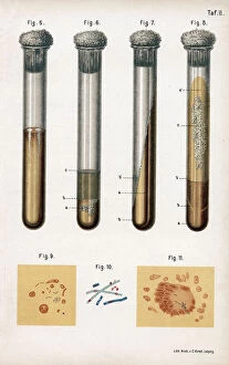
Background imageBacteriology Collection: Various types of bacteria, 1889

Various types of bacteria, 1889. 5: pneumonia culture; 6: albumen from a rotten egg; 7& 8: tuberculosis as prepared by Koch; 9: sputum from a TB patient; 10: anthrax bacillus; 11

Background imageBacteriology Collection: Oswald Theodore Avery, Canadian-born American bacteriologist and molecular biologist

Oswald Theodore Avery, Canadian-born American bacteriologist and molecular biologist. Pictured at work in a laboratory examining a Petri dish of culture

Background imageBacteriology Collection: Bacteriology Laboratory, Nairobi, Kenya, East Africa

Bacteriology Laboratory, Nairobi, Kenya, East Africa. 1931

Background imageBacteriology Collection: Berlin, Ovation of German and foreign doctors to dr. Koch (1

Berlin, Ovation of German and foreign doctors to dr. Koch (1
Robert Koch (1843-1910). German physician. Nobel Prize in Medicine, 1905. Berlin, Ovation of German and foreign doctors to dr. Koch, leaving the hospital. The Spanish and American Illustration, 1890

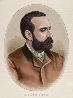
Background imageBacteriology Collection: FERRAN, Jaime (1852-1929). Spanish bacteriologist

FERRAN, Jaime (1852-1929). Spanish bacteriologist. Colored engraving. By Carretero

Background imageBacteriology Collection: PASTEUR, Louis (1822-1895) French chemist and bacteriologist

PASTEUR, Louis (1822-1895) French chemist and bacteriologist
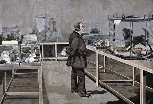
PASTEUR, Louis (Do?le, 1822-Villeneuve-l Etang, 1895) French chemist and bacteriologist. Discovery of rabies vaccine by Dr. Pasteur. Outside the Paris laboratory with the sick together

Background imageBacteriology Collection: PASTEUR, Louis (1822-1895). Pasteur observes the effects of

PASTEUR, Louis (1822-1895). Pasteur observes the effects of
PASTEUR, Louis (1822-1895) French chemist and bacteriologist. Pasteur observes the effects of inoculation of rabies virus, Paris. Engraving by Rico

Background imageBacteriology Collection: Robert Koch - German bacteriologist

Robert Koch - German bacteriologist
Robert Koch (1840-1910) - German bacteriologist. Date: circa 1890

Background imageBacteriology Collection: KOCH, Robert (1843-1910). German physician, discoverer

KOCH, Robert (1843-1910). German physician, discoverer of the tuberculin. Nobel Prize in 1905. Engraving

Background imageBacteriology Collection: PASTEUR, Louis (Dle, 1822-Villeneuve-l Etang, 1895) French chemist and bacteriologist

PASTEUR, Louis (Dle, 1822-Villeneuve-l Etang, 1895) French chemist and bacteriologist
Louis Pasteur (Dole, 1822-Villeneuve-l Etang, 1895) French chemist and bacteriologist. Discoverer of rabies vaccine. Outside the Paris laboratory with the sick together

Background imageBacteriology Collection: PASTEUR, Louis (1822-1895) French chemist and bacteriologist. Vaccination of sheep against anthrax

PASTEUR, Louis (1822-1895) French chemist and bacteriologist. Vaccination of sheep against anthrax
Louis Pasteur (1822-1895). French chemist and bacteriologist. Vaccination of sheep against anthrax. Agerville, France, 1884. Engraving

Background imageBacteriology Collection: HOWARD UNIVERSITY, c1900. A class in the bacteriology laboratory at Howard University

HOWARD UNIVERSITY, c1900. A class in the bacteriology laboratory at Howard University in Washington, D.C. Photograph, c1900

Background imageBacteriology Collection: Conceptual image of antibody attaching and killing bacteria

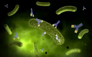
Conceptual image of antibody attaching and killing bacteria

Background imageBacteriology Collection: Microscopic view of histoplasmosis

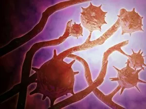
Microscopic view of histoplasmosis. Histoplasmosis is an infection in the lungs caused by inhaling the spores of a fungus. This fungus, called Histoplasma capsulatum

Background imageBacteriology Collection: Microscopic view of diplococcus bacterium

Microscopic view of diplococcus bacterium

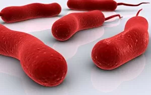
Background imageBacteriology Collection: Conceptual image of cholerae bacteria

Conceptual image of cholerae bacteria

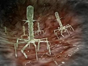
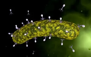
Background imageBacteriology Collection: Microscopic view of bacteriophages on the surface of a bacteria

Microscopic view of bacteriophages on the surface of a bacteria

Background imageBacteriology Collection: Electron micrograph of negative-stained Prochlorococcus

Electron micrograph of negative-stained Prochlorococcus. A cyanophage is a virus that infects cyanobacteria

Background imageBacteriology Collection: Development of fistula

Development of fistula

Background imageBacteriology Collection: Microscopic view of Methicillin-resistant Staphylococcus aureus

Microscopic view of Methicillin-resistant Staphylococcus aureus (MRSA). MRSA is a bacterium responsible for several difficult-to-treat infections in humans

Background imageBacteriology Collection: Conceptual image of RNA virus replication

Conceptual image of RNA virus replication

Background imageBacteriology Collection: Microscopic view of E. coli bacteria cells

Microscopic view of E. coli bacteria cells
Microscopic view of Escherichia coli bacteria cells, commonly known as E. Coli. E. coli is a common type of bacteria that can get into food, such as beef and vegetables

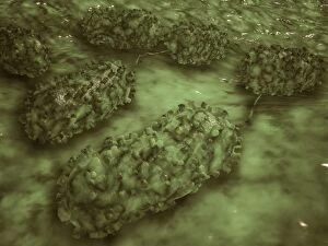
Background imageBacteriology Collection: Microscopic view of Bacilli bacteria

Microscopic view of Bacilli bacteria

Background imageBacteriology Collection: Microscopic view of Anthrax

Microscopic view of Anthrax. Anthrax is an acute disease caused by the bacterium Bacillus anthracis. Most forms of the disease are lethal, and it affects both humans and animals

Background imageBacteriology Collection: Microscopic view of streptococcus

Microscopic view of streptococcus

Background imageBacteriology Collection: A colorized scanning electron micrograph of a white blood cell eating MRSA

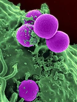
A colorized scanning electron micrograph of a white blood cell eating MRSA
A colorized scanning electron micrograph of a white blood cell eating an antibiotic resistant strain of Staphylococcus aureus bacteria, commonly known as MRSA

Background imageBacteriology Collection: Microscopic view of bacteriophage attacking bacteria

Microscopic view of bacteriophage attacking bacteria

Background imageBacteriology Collection: Conceptual image of Lactobacillus acidophilus

Conceptual image of Lactobacillus acidophilus. Lactobacillus is a gram-positive bacteria that occurs naturally in the human and animal gastrointestinal tract and mouth

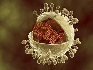
Background imageBacteriology Collection: Conceptual image of human cytomegalovirus

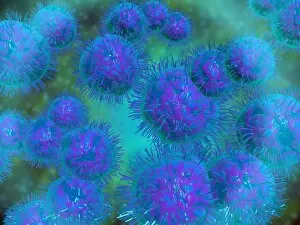
Conceptual image of human cytomegalovirus. Cytomegalovirus is a genus of the viral family Herpesviridae

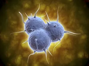
Background imageBacteriology Collection: Microscopic view of cocci bacterium

Microscopic view of cocci bacterium

Background imageBacteriology Collection: Conceptual image of salmonella typhi causing typhoid

Conceptual image of salmonella typhi causing typhoid

Background imageBacteriology Collection: Group of vibrio cholerae bacteria which causes cholera

Group of vibrio cholerae bacteria which causes cholera. Cholera is an infection in the small intestine caused by the bacterium Vibrio cholerae. The main symptoms are diarrhea and vomiting

Background imageBacteriology Collection: Scanning electron micrograph of a human neutrophil ingesting MRSA

Scanning electron micrograph of a human neutrophil ingesting MRSA

Background imageBacteriology Collection: Artistic representation of the immune systems reaction to bacteria invading the tissues

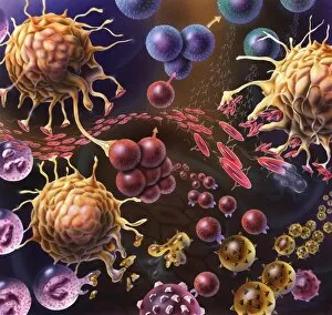
Artistic representation of the immune systems reaction to bacteria invading the tissues


For sale as Licensed Images

Choose your image, Select your licence and Download the media

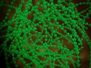
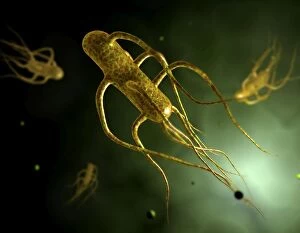
"Bacteriology: Exploring the Microscopic World of Skin Disorders and Artwork" Delve into the fascinating realm of bacteriology, where intricate artwork meets skin disorders. Through scanning electron microscopy (SEM), we witness the captivating beauty of E. Coli bacteria, their distinctive rod-like shapes forming a mesmerizing pattern. Salmonella bacteria also reveal their unique structure under SEM, showcasing their spherical forms with intriguing surface details. In a colored transmission electron micrograph (TEM), Yersinia pestis bacteria come to life in vibrant hues, highlighting the diversity within cell types. This artistic representation allows us to appreciate the complexity and intricacy of these microscopic organisms that can cause severe diseases like plague. Travel back in time as historical diagrams depict Anthrax cultures, revealing how scientists once studied this deadly disease. The tuberculosis bacteria are captured in all their glory; their slender rods painting a somber picture of one of humanity's oldest foes. Witness nature's creativity through the spiral spore chain formation of Streptomyces bacteria – an enchanting display resembling delicate strands woven together with precision. Flagellate bacteria showcase their remarkable motility through whip-like appendages called flagella, propelling themselves forward with grace and agility. Elevating our understanding further is an up-close encounter with Staphylococcus aureus bacteria – notorious for causing various infections ranging from minor skin conditions to life-threatening illnesses. Their distinct clusters become apparent as they thrive amidst human hosts. Amongst this diverse microbial world stands E. coli bacterium - its presence ubiquitous yet often misunderstood due to its association with foodborne illnesses. However, it plays crucial roles in digestion and research breakthroughs alike. Bacteriology unravels the hidden wonders within our microbiome while shedding light on pathogens that challenge human health daily. As we explore these captivating images and delve deeper into this field, we gain insights into both artistry at a microscopic level and the complex interplay between bacteria and human biology.