Bacteriology Collection (#8)

"Bacteriology: Exploring the Microscopic World of Skin Disorders and Artwork" Delve into the fascinating realm of bacteriology

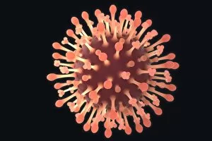
Background imageBacteriology Collection: Flu virus particles, artwork F008 / 3241

Flu virus particles, artwork F008 / 3241
Flu virus particles, computer artwork. In the virus envelope are two types of protein spike, haemagglutinin (H, peg-like) and neuraminidase (N, squares), which determine the strain of virus

Background imageBacteriology Collection: Flu virus particles, artwork F008 / 3254

Flu virus particles, artwork F008 / 3254
Flu virus particles, computer artwork

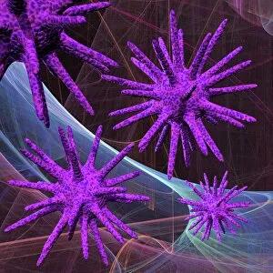
Background imageBacteriology Collection: Virus particles, artwork F008 / 3375

Virus particles, artwork F008 / 3375
Virus particles, computer artwork

Background imageBacteriology Collection: Bacteria research F005 / 0847

Bacteria research F005 / 0847
MODEL RELEASED. Bacteria research. Researcher holding a petri dish containing cultured bacteria

Background imageBacteriology Collection: Bacteria research F005 / 0846

Bacteria research F005 / 0846
MODEL RELEASED. Bacteria research. Researcher holding a petri dish containing cultured bacteria

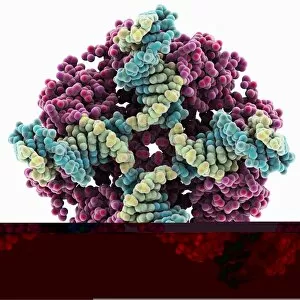
Background imageBacteriology Collection: E. coli Holliday junction complex C014 / 0878

E. coli Holliday junction complex C014 / 0878
E. coli Holliday junction complex. Molecular model of a RuvA protein (dark pink) in complex with a Holliday junction between homologous strands of DNA (deoxyribonucleic acid)

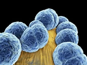
Background imageBacteriology Collection: Staphylococcus aureus MRSA bacteria C014 / 2577

Staphylococcus aureus MRSA bacteria C014 / 2577
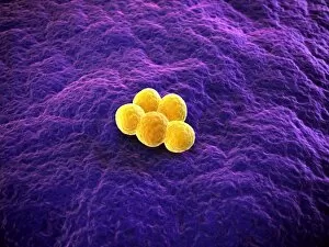
Staphylococcus aureus MRSA bacteria, computer artwork. Methicillin-resistant Staphylococcus aureus (MRSA, orange) is a Gram-positive, round (coccus) bacterium, shown here on a filamentous surface

Background imageBacteriology Collection: Staphylococcus aureus MRSA bacteria C014 / 2578

Staphylococcus aureus MRSA bacteria C014 / 2578
Staphylococcus aureus MRSA bacteria, computer artwork. Methicillin-resistant Staphylococcus aureus (MRSA, orange) is a Gram-positive, round (coccus) bacterium, shown here on a filamentous surface

Background imageBacteriology Collection: Endonuclease IV molecule

Endonuclease IV molecule. Molecular model of the endonuclease IV restriction enzyme EcoRV (grey) bound to a cleaved section of DNA (deoxyribonucleic acid, blue, orange and pink)

Background imageBacteriology Collection: MRSA bacteria, artwork F006 / 3107

MRSA bacteria, artwork F006 / 3107
MRSA bacteria. Computer artwork of methicillin-resistant Staphylococcus aureus (MRSA) bacteria

Background imageBacteriology Collection: MRSA bacteria, artwork F006 / 3106

MRSA bacteria, artwork F006 / 3106
MRSA bacteria. Computer artwork of methicillin-resistant Staphylococcus aureus (MRSA) bacteria

Background imageBacteriology Collection: MRSA bacteria, artwork F006 / 3105

MRSA bacteria, artwork F006 / 3105
MRSA bacteria. Computer artwork of methicillin-resistant Staphylococcus aureus (MRSA) bacteria

Background imageBacteriology Collection: MRSA bacteria, artwork F006 / 3101

MRSA bacteria, artwork F006 / 3101
MRSA bacteria. Computer artwork of methicillin-resistant Staphylococcus aureus (MRSA) bacteria

Background imageBacteriology Collection: MRSA bacteria, artwork F006 / 3103

MRSA bacteria, artwork F006 / 3103
MRSA bacteria. Computer artwork of methicillin-resistant Staphylococcus aureus (MRSA) bacteria

Background imageBacteriology Collection: MRSA bacteria, artwork F006 / 3104

MRSA bacteria, artwork F006 / 3104
MRSA bacteria. Computer artwork of methicillin-resistant Staphylococcus aureus (MRSA) bacteria

Background imageBacteriology Collection: Anthrax protective antigen molecule C014 / 0886

Anthrax protective antigen molecule C014 / 0886
Anthrax protective antigen molecule. Computer model showing the structure of a molecule of protective antigen (PA) produced by anthrax (Bacillus anthracis) bacteria

Background imageBacteriology Collection: Bacteria, artwork F006 / 2321

Bacteria, artwork F006 / 2321
Bacteria. Computer artwork of rod-shaped bacteria

Background imageBacteriology Collection: Bacterial infection, artwork F006 / 2313

Bacterial infection, artwork F006 / 2313
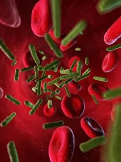
Bacterial infection. Computer artwork of bacteria (green) in the blood stream

Background imageBacteriology Collection: EcoRV restriction enzyme molecule C014 / 2117

EcoRV restriction enzyme molecule C014 / 2117
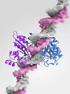
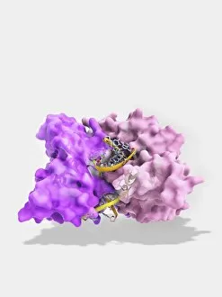
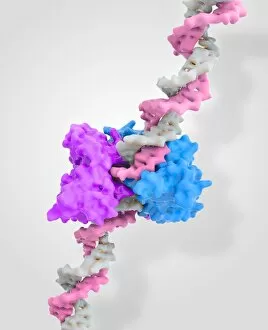
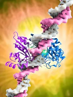
EcoRV restriction enzyme. Molecular model of the type II restriction enzyme EcoRV (purple and blue) bound to a DNA molecule (deoxyribonucleic acid, pink and white)

Background imageBacteriology Collection: Anthrax protective antigen molecule C014 / 0865

Anthrax protective antigen molecule C014 / 0865
Anthrax protective antigen molecule. Computer model showing the structure of a molecule of protective antigen (PA) produced by anthrax (Bacillus anthracis) bacteria

Background imageBacteriology Collection: EcoRV restriction enzyme molecule C014 / 2112

EcoRV restriction enzyme molecule C014 / 2112
EcoRV restriction enzyme. Molecular model of the type II restriction enzyme EcoRV (pink) bound to a cleaved section of DNA (deoxyribonucleic acid, yellow)

Background imageBacteriology Collection: Pseudomonas aeruginosa bacteria, SEM C017 / 7140

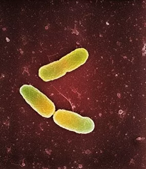
Pseudomonas aeruginosa bacteria, SEM C017 / 7140
Pseudomonas aeruginosa bacteria, coloured scanning electron micrograph (SEM). These Gram-negative rod-shaped bacteria are found in soil, water and as normal flora in the human intestine

Background imageBacteriology Collection: Microbiology research F007 / 0383

Microbiology research F007 / 0383
Microbiology research

Background imageBacteriology Collection: Microbiology research F007 / 0382

Microbiology research F007 / 0382
Microbiology research

Background imageBacteriology Collection: Microbiology research F007 / 0369

Microbiology research F007 / 0369
Microbiology research

Background imageBacteriology Collection: EcoRV restriction enzyme molecule C014 / 2114

EcoRV restriction enzyme molecule C014 / 2114
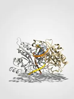
EcoRV restriction enzyme. Molecular model of the type II restriction enzyme EcoRV (white and gold) bound to a cleaved section of DNA (deoxyribonucleic acid, orange and yellow)

Background imageBacteriology Collection: Staphylococcus aureus bacteria, SEM C017 / 7138

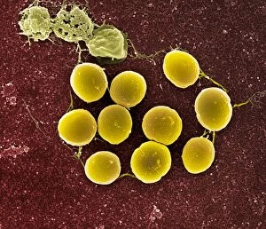
Staphylococcus aureus bacteria, SEM C017 / 7138
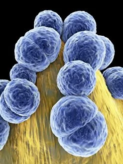
Staphylococcus aureus bacteria, coloured scanning electron micrograph (SEM). These Gram-positive coccus (round) bacteria are found on the skin and mucous membranes of humans and many animals

Background imageBacteriology Collection: EcoRV restriction enzyme molecule C014 / 2116

EcoRV restriction enzyme molecule C014 / 2116
EcoRV restriction enzyme. Molecular model of the type II restriction enzyme EcoRV (purple and blue) bound to a DNA molecule (deoxyribonucleic acid, pink and white)

Background imageBacteriology Collection: EcoRV restriction enzyme molecule C014 / 2115

EcoRV restriction enzyme molecule C014 / 2115
EcoRV restriction enzyme. Molecular model of the type II restriction enzyme EcoRV (purple and blue) bound to a DNA molecule (deoxyribonucleic acid, pink and white)

Background imageBacteriology Collection: Staphylococcus aureus bacteria, SEM C017 / 7136

Staphylococcus aureus bacteria, SEM C017 / 7136
Staphylococcus aureus bacteria, coloured scanning electron micrograph (SEM). These Gram-positive coccus (round) bacteria are found on the skin and mucous membranes of humans and many animals

Background imageBacteriology Collection: Bacteriophage infecting bacterium F006 / 8665

Bacteriophage infecting bacterium F006 / 8665
Bacteriophage infecting bacterium, artwork

Background imageBacteriology Collection: Bacteriophage infecting bacterium F006 / 8664

Bacteriophage infecting bacterium F006 / 8664
Bacteriophage infecting bacterium, artwork

Background imageBacteriology Collection: Bacteria, artwork F006 / 8663

Bacteria, artwork F006 / 8663
Bacteria, computer artwork

Background imageBacteriology Collection: Bacteria, artwork F006 / 8661

Bacteria, artwork F006 / 8661
Bacteria, computer artwork

Background imageBacteriology Collection: Bacteria, artwork F006 / 8662

Bacteria, artwork F006 / 8662
Bacteria, computer artwork

Background imageBacteriology Collection: LAC repressor molecule F006 / 9520

LAC repressor molecule F006 / 9520
LAC repressor. Molecular model of a LAC (lactose) repressor molecule. The LAC repressor inhibits the expression of genes that code for an enzyme which metabolizes lactose in bacteria

Background imageBacteriology Collection: Bacterial infection, artwork F006 / 7933

Bacterial infection, artwork F006 / 7933
Bacterial infection, computer artwork

Background imageBacteriology Collection: Bacterial infection, artwork F006 / 7934

Bacterial infection, artwork F006 / 7934
Bacterial infection, computer artwork

Background imageBacteriology Collection: Restriction enzyme and DNA F006 / 9315

Restriction enzyme and DNA F006 / 9315
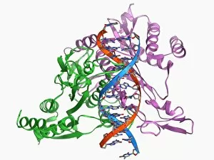
Restriction enzyme and DNA. Molecular model showing an EcoRI endonuclease enzyme (purple and green) bound to a DNA (deoxyribonucleic acid) molecule (red and blue)

Background imageBacteriology Collection: LAC repressor bound to DNA F006 / 9309

LAC repressor bound to DNA F006 / 9309
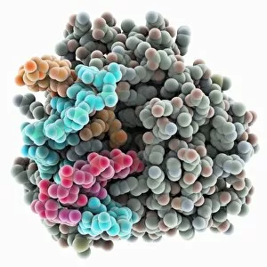
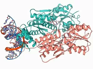
LAC repressor bound to DNA. Molecular model of a LAC (lactose) repressor molecule (pink and turquoise) interacting with bacterial DNA (deoxyribonucleic acid, red and blue)

Background imageBacteriology Collection: Lumazine synthase molecule F006 / 9291

Lumazine synthase molecule F006 / 9291
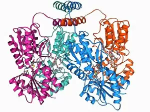
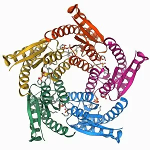
Lumazine synthase molecule. Molecular model showing the structure of a lumazine synthase enzyme molecule from a Brucella abortus bacterium

Background imageBacteriology Collection: E coli Holliday junction complex F006 / 9261

E coli Holliday junction complex F006 / 9261
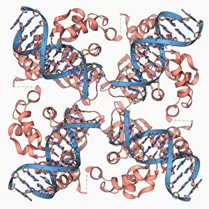
E. coli Holliday junction complex. Molecular model of a RuvA protein (red) in complex with a Holliday junction between homologous strands of DNA (deoxyribonucleic acid, blue) from an E

Background imageBacteriology Collection: Anthrax protective antigen molecule F006 / 9229

Anthrax protective antigen molecule F006 / 9229
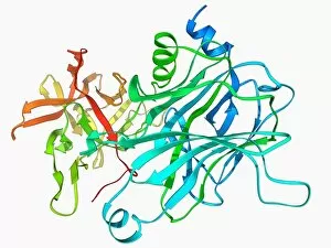
Anthrax protective antigen molecule. Computer model showing the structure of a molecule of protective antigen (PA) produced by anthrax (Bacillus anthracis) bacteria

Background imageBacteriology Collection: Anthrax protective antigen molecule F006 / 9225

Anthrax protective antigen molecule F006 / 9225
Anthrax protective antigen molecule. Computer model showing the structure of a molecule of protective antigen (PA) produced by anthrax (Bacillus anthracis) bacteria

Background imageBacteriology Collection: Tetanus toxin C-fragment molecule F006 / 9223

Tetanus toxin C-fragment molecule F006 / 9223
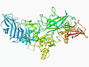
Tetanus toxin C-fragment. Molecular model of a fragment of the neurotoxin protein produced by the bacterium Clostridium tetani that causes tetanus

Background imageBacteriology Collection: T-cell receptor bound to enterotoxin

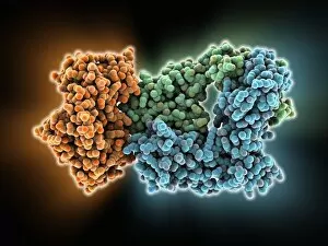
T-cell receptor bound to enterotoxin, molecular model. The T cell receptor (TCR) is a protein complex found on the surface of a type of white blood cell called T lymphocytes (or T cells)

Background imageBacteriology Collection: Antibodies and bacteria, artwork

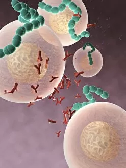
Antibodies and bacteria, artwork
Antibodies and bacteria. Computer artwork showing white blood cells (large, round) and antibody, or immunoglobulin, molecules (red) surrounding bacteria (green)

Background imageBacteriology Collection: Gramicidin antibiotic molecule C015 / 5072

Gramicidin antibiotic molecule C015 / 5072
Gramicidin. Molecular model of the antibiotic gramicidin D, from the bacterium Bacillus brevis. This antibiotic is effective against Gram positive and Gram negative bacteria


For sale as Licensed Images

Choose your image, Select your licence and Download the media

"Bacteriology: Exploring the Microscopic World of Skin Disorders and Artwork" Delve into the fascinating realm of bacteriology, where intricate artwork meets skin disorders. Through scanning electron microscopy (SEM), we witness the captivating beauty of E. Coli bacteria, their distinctive rod-like shapes forming a mesmerizing pattern. Salmonella bacteria also reveal their unique structure under SEM, showcasing their spherical forms with intriguing surface details. In a colored transmission electron micrograph (TEM), Yersinia pestis bacteria come to life in vibrant hues, highlighting the diversity within cell types. This artistic representation allows us to appreciate the complexity and intricacy of these microscopic organisms that can cause severe diseases like plague. Travel back in time as historical diagrams depict Anthrax cultures, revealing how scientists once studied this deadly disease. The tuberculosis bacteria are captured in all their glory; their slender rods painting a somber picture of one of humanity's oldest foes. Witness nature's creativity through the spiral spore chain formation of Streptomyces bacteria – an enchanting display resembling delicate strands woven together with precision. Flagellate bacteria showcase their remarkable motility through whip-like appendages called flagella, propelling themselves forward with grace and agility. Elevating our understanding further is an up-close encounter with Staphylococcus aureus bacteria – notorious for causing various infections ranging from minor skin conditions to life-threatening illnesses. Their distinct clusters become apparent as they thrive amidst human hosts. Amongst this diverse microbial world stands E. coli bacterium - its presence ubiquitous yet often misunderstood due to its association with foodborne illnesses. However, it plays crucial roles in digestion and research breakthroughs alike. Bacteriology unravels the hidden wonders within our microbiome while shedding light on pathogens that challenge human health daily. As we explore these captivating images and delve deeper into this field, we gain insights into both artistry at a microscopic level and the complex interplay between bacteria and human biology.