Bacteriology Collection (#5)

"Bacteriology: Exploring the Microscopic World of Skin Disorders and Artwork" Delve into the fascinating realm of bacteriology

Background imageBacteriology Collection: Microscopic view of staphylococcus

Microscopic view of staphylococcus

Background imageBacteriology Collection: Scanning electron micrograph of Staphylococcus and a dead human neutrophil

Scanning electron micrograph of Staphylococcus and a dead human neutrophil
Scanning electron micrograph of methicillin-resistant Staphylococcus aureus and a dead human neutrophil

Background imageBacteriology Collection: Microscopic view of coronavirus

Microscopic view of coronavirus

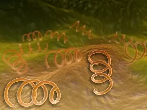
Background imageBacteriology Collection: Microscopic view of Syphillis

Microscopic view of Syphillis. Syphilis is a sexually transmitted infection caused by the spirochete bacterium, Treponema pallidum

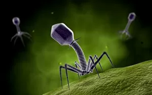
Background imageBacteriology Collection: Microscopic view of bacteriophage

Microscopic view of bacteriophage

Background imageBacteriology Collection: Microscopic view of Rickettsia

Microscopic view of Rickettsia. Rickettsia is a gram-negative bacteria carried by parasites such as ticks, fleas and lice

Background imageBacteriology Collection: Microscopic view of corncob formation in dental plaque

Microscopic view of corncob formation in dental plaque, believed to be limited to strains of Corynebacterium matruchotii and Streptococcus sanguis

Background imageBacteriology Collection: Conceptual image of the coronavirus

Conceptual image of the coronavirus. Coronaviruses primarily infect the upper respiratory and gastrointestinal tract

Background imageBacteriology Collection: Microscopic view of herpes virus

Microscopic view of herpes virus

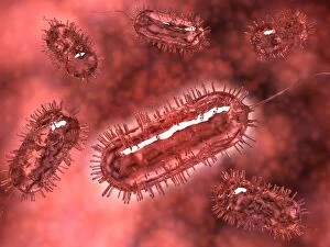
Background imageBacteriology Collection: Group of Escherichia coli bacteria cells, known as E. Coli

Group of Escherichia coli bacteria cells, known as E. Coli
Group of Escherichia coli bacteria cells, commonly known as E. Coli. E. coli is a common type of bacteria that can get into food, like beef and vegetables

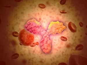
Background imageBacteriology Collection: Microscopic view of human anitbodies with red blood cells

Microscopic view of human anitbodies with red blood cells. Human antibodies are the Y-shaped proteins used by the immune system to defend against foreign objects like bacteria and viruses

Background imageBacteriology Collection: Microscopic view of a group of Borrelia burgdorferi

Microscopic view of a group of Borrelia burgdorferi, the bacterial agent of Lyme disease transmitted by ticks

Background imageBacteriology Collection: Microscopic view of Rubella virus

Microscopic view of Rubella virus
Microscopic view of Rubella. Rubella is an acute, contagious viral infection. While the illness is generally mild in children, it has serious consequences in pregnant women causing fetal death

Background imageBacteriology Collection: Microscopic view of Legionella pneumophila

Microscopic view of Legionella pneumophila. Legionella pneumophila is a thin, aerobic, pleomorphic, flagellated, non-spore forming, gram-negative bacterium of the genus Legionella

Background imageBacteriology Collection: Conceptual image of bacteria

Conceptual image of bacteria

Background imageBacteriology Collection: Microscopic view of Yersinia

Microscopic view of Yersinia. Yersinia is a gram-negative, rod-shaped bacteria in the family Enterobacteriaceae

Background imageBacteriology Collection: Conceptual image of salmonella

Conceptual image of salmonella. Salmonella is a genus of rod-shaped, gram-negative bacteria. There are two main species of Salmonella, Salmonella bongori and Salmonella enterica

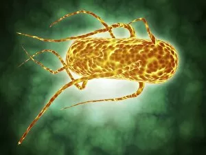
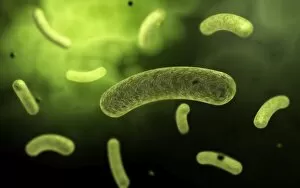
Background imageBacteriology Collection: Conceptual image of flagellate bacterium

Conceptual image of flagellate bacterium

Background imageBacteriology Collection: Microscopic view of bacteria

Microscopic view of bacteria

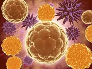
Background imageBacteriology Collection: Conceptual image of a ubiquitous virus

Conceptual image of a ubiquitous virus. A ubiquitous virus is contagious in early childhood through the respiratory tract

Background imageBacteriology Collection: Microscopic view of Henipavirus

Microscopic view of Henipavirus. Henipavirus is a established group of paramyxoviruses comprising the Hendra virus, Nipah virus and Cedar virus

Background imageBacteriology Collection: Conceptual image of vibrio cholerae causing cholera

Conceptual image of vibrio cholerae causing cholera

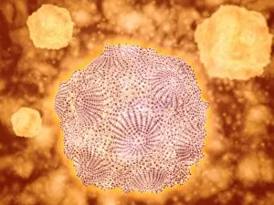
Background imageBacteriology Collection: Microscopic view of Canine Parvovirus

Microscopic view of Canine Parvovirus. Canine Parvovirus is a highly contagious viral disease that is one of the most common causes of diarrhea in dogs under 6 months of age

Background imageBacteriology Collection: Conceptual image of common bacteria

Conceptual image of common bacteria

Background imageBacteriology Collection: Professor Robert Wurz (1858-1919) 1901 (oil on card)

Professor Robert Wurz (1858-1919) 1901 (oil on card)
XIR223829 Professor Robert Wurz (1858-1919) 1901 (oil on card) by Toulouse-Lautrec, Henri de (1864-1901); Musee Toulouse-Lautrec, Albi

Background imageBacteriology Collection: ILYA ILICH MECHNIKOV (1845-1916). Russian bacteriologist and zoologist

ILYA ILICH MECHNIKOV (1845-1916). Russian bacteriologist and zoologist

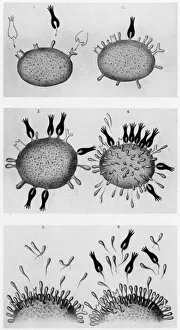
Background imageBacteriology Collection: Drawings used to illustrate German bacteriologist Paul Ehrlichs theory of how blood cells produce

Drawings used to illustrate German bacteriologist Paul Ehrlichs theory of how blood cells produce antibodies to
EHRLICHs THEORY, 1900. Drawings used to illustrate German bacteriologist Paul Ehrlichs theory of how blood cells produce antibodies to neutralize invading bacteria

Background imageBacteriology Collection: Test tube containing a culture of tuberculosis bacilli grown by Robert Koch. Line engraving, 1890

Test tube containing a culture of tuberculosis bacilli grown by Robert Koch. Line engraving, 1890
ROBERT KOCH: BACILLI, 1890. Test tube containing a culture of tuberculosis bacilli grown by Robert Koch. Line engraving, 1890

Background imageBacteriology Collection: Test tube containing a culture of cholera bacilli grown by Robert Koch. Line engraving, 1890

Test tube containing a culture of cholera bacilli grown by Robert Koch. Line engraving, 1890
ROBERT KOCH: BACILLI, 1890. Test tube containing a culture of cholera bacilli grown by Robert Koch. Line engraving, 1890

Background imageBacteriology Collection: Engraving after a drawing by Robert Koch of fresh bacilli from the lungs of a tuberculosis patient

Engraving after a drawing by Robert Koch of fresh bacilli from the lungs of a tuberculosis patient
ROBERT KOCH: BACILLI. Engraving after a drawing by Robert Koch of fresh bacilli from the lungs of a tuberculosis patient, as seen under Kochs microscope

Background imageBacteriology Collection: Engraving after a drawing by Robert Koch of tuberculosis bacilli

Engraving after a drawing by Robert Koch of tuberculosis bacilli, after two weeks growth under a culture
ROBERT KOCH: BACILLI. Engraving after a drawing by Robert Koch of tuberculosis bacilli, after two weeks growth under a culture, as seen under the microscope

Background imageBacteriology Collection: Louis Pasteur (1822-1895) French chemist. Bacteriology. Hydrophobia. Inoculation

Louis Pasteur (1822-1895) French chemist. Bacteriology. Hydrophobia. Inoculation by attenuated culture. Popular tribute from French Le Petit Journal, Paris, at the time of his death
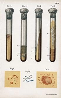
Background imageBacteriology Collection: 5: Pneumonia culture. 6 Albumen from rotten egg. 7& 8: Tuberculosis as prepared by Koch

5: Pneumonia culture. 6 Albumen from rotten egg. 7& 8: Tuberculosis as prepared by Koch
5:Pneumonia culture. 6 Albumen from rotten egg. 7& 8:Tuberculosis as prepared by Koch. 9:Sputum from TB patient. 10: Anthrax bacillus. 11:TB bacillus stained blue

Background imageBacteriology Collection: Louis Pasteur (1822-185) French chemist and founder of modern bacteriology, here

Louis Pasteur (1822-185) French chemist and founder of modern bacteriology, here holding rabbits used in his work on Hydrophobia (Rabies). Cartoon from Vanity Fair, January 1887

Background imageBacteriology Collection: Robert Koch (1843-1910)

Robert Koch (1843-1910) German bacteriologist and physician in his laboratory. In 1890 Koch introduced Tuberculin which he thought was a cure for Tuberculosis
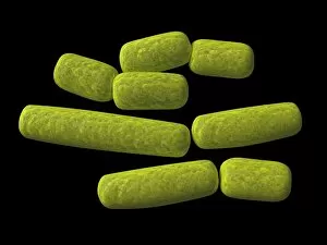
Background imageBacteriology Collection: Biology - medicine - microbiology - bacteriology - bacteria - Hemolytic Streptococci

Biology - medicine - microbiology - bacteriology - bacteria - Hemolytic Streptococci, microscope view
Biology - Medicine - Microbiology - Bacteriology - Microscopy - Bacteria, hemolytic streptococci

Background imageBacteriology Collection: Haemophilus, artwork F007 / 6739

Haemophilus, artwork F007 / 6739
Haemophilus bacteria, computer artwork

Background imageBacteriology Collection: Staphylococcus aureus bacteria, SEM C017 / 7137

Staphylococcus aureus bacteria, SEM C017 / 7137
Staphylococcus aureus bacteria, coloured scanning electron micrograph (SEM). These Gram-positive coccus (round) bacteria are found on the skin and mucous membranes of humans and many animals

Background imageBacteriology Collection: Bacteria, artwork F006 / 3717

Bacteria, artwork F006 / 3717
Bacteria, computer artwork

Background imageBacteriology Collection: Bacteria, artwork F006 / 3716

Bacteria, artwork F006 / 3716
Bacteria, computer artwork

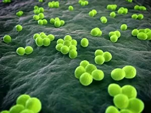
Background imageBacteriology Collection: MRSA bacteria, artwork F007 / 7431

MRSA bacteria, artwork F007 / 7431
MRSA (methicillin-resistant Staphylococcus aureus) bacteria, computer artwork

Background imageBacteriology Collection: Bacillus bacteria, artwork F007 / 6735

Bacillus bacteria, artwork F007 / 6735
Bacillus bacteria, computer artwork

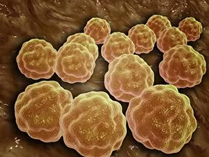
Background imageBacteriology Collection: Coccus bacteria, artwork F007 / 6761

Coccus bacteria, artwork F007 / 6761
Coccus bacteria, Computer artwork

Background imageBacteriology Collection: Propionibacterium acnes, artwork F007 / 6754

Propionibacterium acnes, artwork F007 / 6754
Propionibacterium acnes bacteria, computer artwork

Background imageBacteriology Collection: Chlamydia, artwork F007 / 6763

Chlamydia, artwork F007 / 6763
Chlamydia bacteria, computer artwork

Background imageBacteriology Collection: Chlamydia, artwork F007 / 6746

Chlamydia, artwork F007 / 6746
Chlamydia bacteria, computer artwork

Background imageBacteriology Collection: Streptococcus, artwork F007 / 6760

Streptococcus, artwork F007 / 6760
Streptococcus bacteria, computer artwork

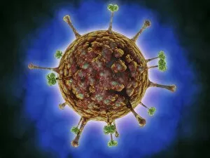
Background imageBacteriology Collection: Infectious salmon anaemia virus particle F007 / 8160

Infectious salmon anaemia virus particle F007 / 8160
Infectious salmon anaemia virus particle, computer artwork


For sale as Licensed Images

Choose your image, Select your licence and Download the media

"Bacteriology: Exploring the Microscopic World of Skin Disorders and Artwork" Delve into the fascinating realm of bacteriology, where intricate artwork meets skin disorders. Through scanning electron microscopy (SEM), we witness the captivating beauty of E. Coli bacteria, their distinctive rod-like shapes forming a mesmerizing pattern. Salmonella bacteria also reveal their unique structure under SEM, showcasing their spherical forms with intriguing surface details. In a colored transmission electron micrograph (TEM), Yersinia pestis bacteria come to life in vibrant hues, highlighting the diversity within cell types. This artistic representation allows us to appreciate the complexity and intricacy of these microscopic organisms that can cause severe diseases like plague. Travel back in time as historical diagrams depict Anthrax cultures, revealing how scientists once studied this deadly disease. The tuberculosis bacteria are captured in all their glory; their slender rods painting a somber picture of one of humanity's oldest foes. Witness nature's creativity through the spiral spore chain formation of Streptomyces bacteria – an enchanting display resembling delicate strands woven together with precision. Flagellate bacteria showcase their remarkable motility through whip-like appendages called flagella, propelling themselves forward with grace and agility. Elevating our understanding further is an up-close encounter with Staphylococcus aureus bacteria – notorious for causing various infections ranging from minor skin conditions to life-threatening illnesses. Their distinct clusters become apparent as they thrive amidst human hosts. Amongst this diverse microbial world stands E. coli bacterium - its presence ubiquitous yet often misunderstood due to its association with foodborne illnesses. However, it plays crucial roles in digestion and research breakthroughs alike. Bacteriology unravels the hidden wonders within our microbiome while shedding light on pathogens that challenge human health daily. As we explore these captivating images and delve deeper into this field, we gain insights into both artistry at a microscopic level and the complex interplay between bacteria and human biology.