Biochemical Collection (#84)

"Unveiling the Intricacies of Biochemical World: From Anaesthetic Inhibiting Ion Channels to DNA Discoveries" Delving into the depths wonders

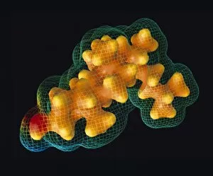
Background imageBiochemical Collection: Cholesterol

Cholesterol. Surface maps (wire-frame and colour-coded) of the electrostatic potential around a cholesterol molecule (C27. H46.O)

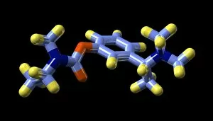
Background imageBiochemical Collection: Rivastigmine Alzheimers drug molecule

Rivastigmine Alzheimers drug molecule
Rivastigmine Alzheimers drug. Molecular model of the Alzheimers drug rivastigmine. Atoms are represented as rods and are colour-coded: carbon (light blue), hydrogen (yellow)

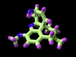
Background imageBiochemical Collection: Galantamine Alzheimers drug molecule

Galantamine Alzheimers drug molecule
Galantamine. Molecular model of the Alzheimers drug galantamine, marketed under the name Reminyl. Atoms are represented as rods and are colour-coded: carbon (light green), hydrogen (purple)

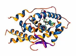
Background imageBiochemical Collection: THG anabolic steroid and receptor

THG anabolic steroid and receptor
THG bound to receptor. Molecular model of a molecule of the anabolic steroid drug tetrahydrogestrinone (THG, ball-and-stick primary structure) bound to a human androgen receptor (secondary structure)

Background imageBiochemical Collection: Donepezil Alzheimers drug, molecular model

Donepezil Alzheimers drug, molecular model
Donepezil Alzheimers drug. Molecular model of the Alzheimers drug donepezil. Atoms are represented as rods and are colour-coded: carbon (blue), hydrogen (yellow)

Background imageBiochemical Collection: Harmine drug molecule

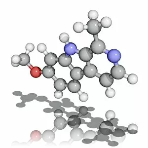
Harmine drug molecule. Molecular model of Harmine, a hallucinogenic alkaloid found in the seed coats of a plant (Peganum harmala) of the Mediterranean region and the Middle East

Background imageBiochemical Collection: Nitrous oxide molecule

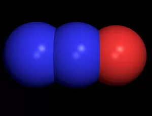
Nitrous oxide molecule
Nitrous oxide. Computer model of a molecule of nitrous oxide. Atoms are represented as spheres and are colour-coded: nitrogen (blue) and oxygen (red)

Background imageBiochemical Collection: Bisphenol A organic pollutant molecule

Bisphenol A organic pollutant molecule
Bisphenol A, molecular model. This chemical is used in the plastics industry, both as an antioxidant and as a component of epoxy resins and polycarbonate plastics

Background imageBiochemical Collection: Glucose sugar molecule

Glucose sugar molecule
Glucose, molecular model. Atoms are represented as spheres (blue). Glucose is a simple sugar (monosaccharide) that is utilised by the body as an energy source

Background imageBiochemical Collection: Hyaluronic acid, molecular model

Hyaluronic acid, molecular model. Hyaluronic acid (or hyaluronon) is a glycosaminoglycan, a type of biological polymer made up of repeating units of a disaccharide (two sugar molecules)

Background imageBiochemical Collection: Sucrose molecule

Sucrose molecule
Sucrose. Computer-generated molecular model of sucrose (C12H22O11). Sucrose is composed of carbon (green), hydrogen (grey-white) and oxygen (red). Sucrose is a disaccharide (sugar)

Background imageBiochemical Collection: Maltose molecule

Maltose molecule
Maltose. Computer-generated molecular model of maltose (C12H22O11). Maltose is composed of carbon (green), hydrogen (grey-white) and oxygen (red)

Background imageBiochemical Collection: Lactose molecule

Lactose molecule
Lactose. Computer-generated molecular model of lactose. Lactose is composed of carbon(green), hydrogen (grey-white) and oxygen (red)

Background imageBiochemical Collection: Glucose molecule

Glucose molecule
Glucose. Computer-generated model of glucose (C6H12O6). Glucose is composed of carbon (green), hydrogen (grey-white) and oxygen (red). Glucose is a monosaccharide (sugar)

Background imageBiochemical Collection: Cellulose molecule

Cellulose molecule
Cellulose. Computer-generated molecular model of cellulose. Cellulose is composed of carbon (green), hydrogen (white-grey) and oxygen (red)

Background imageBiochemical Collection: Amylose molecule

Amylose molecule
Amylose. Computer-generated molecular model of amylose. Amylose is composed of carbon (green), hydrogen (grey-white) and oxygen (red). Amylose is a polysaccharide that is a component of starch

Background imageBiochemical Collection: Rotigotine drug molecule

Rotigotine drug molecule
Rotigotine, computer model. This drug, marketed as Neupro, is used in the treatment of Parkinsons disease. Parkinsons disease is a movement disorder caused by the loss of cells in the brain that

Background imageBiochemical Collection: Perphenazine antipsychotic drug

Perphenazine antipsychotic drug, molecular model. This drug, marketed as Trilafon, Triavil and Etrafon, is a common treatment for patients suffering from psychoses

Background imageBiochemical Collection: Lisinopril high blood pressure drug

Lisinopril high blood pressure drug, molecular model. This drug, marketed as Prinivil, Zestril and Lisodur, is used to treat hypertension (high blood pressure) and heart disease

Background imageBiochemical Collection: Amlodipine high blood pressure drug

Amlodipine high blood pressure drug, molecular model. This drug, marketed as Istin, is used to treat hypertension (high blood pressure) and angina pectoris (heart pain)

Background imageBiochemical Collection: Flumazenil benzodiazepine antidote

Flumazenil benzodiazepine antidote
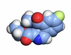
Flumazenil benzodiazepine antagonist drug, molecular model. This drug, marketed as Anexate, is used to treat overdoses of benzodiazepine drugs, a class of sedatives that includes valium

Background imageBiochemical Collection: Buspirone anti-anxiety drug molecule

Buspirone anti-anxiety drug molecule
Buspirone anti-anxiety drug. Computer model of a molecule of buspirone, marketed as Buspar, an anxiolytic (anti-anxiety) drug

Background imageBiochemical Collection: HIV antibody therapy

HIV antibody therapy. Molecular model of the interaction of the HIV surface protein gp120 (green, lower right) as it interacts with a human white blood cell surface protein (CD4, blue)

Background imageBiochemical Collection: Cholesterol enzyme affected by a drug

Cholesterol enzyme affected by a drug
Cholesterol enzyme being affected by a drug. Molecular model of the shape of the human enzyme Hmg-Coa reductase interacting (complexed) with the anti-cholesterol drug Fluvastatin (not seen)

Background imageBiochemical Collection: Amprenavir drug molecule

Amprenavir drug molecule
Amprenavir. Computer model of a molecule of the drug amprenavir. It is also known as VX-478, and is sold under the brand name Agenerase

Background imageBiochemical Collection: Theobromine drug molecule

Theobromine drug molecule
Theobromine. Computer model of a molecule of the alkaloid drug theobromine. This substance is most often consumed in products of the cacao tree, such as chocolate and cocoa

Background imageBiochemical Collection: Quinapril drug molecule

Quinapril drug molecule
Quinapril. Computer model of a molecule of the drug quinapril. It is sold under the brand name Accupril. It is an ACE (angiotensin converting enzyme) inhibitor drug

Background imageBiochemical Collection: HIV enzyme being affected by a drug

HIV enzyme being affected by a drug. Molecular model of HIVs reverse transcriptase enzyme as it interacts with a drug (not seen)

Background imageBiochemical Collection: Nitroglycerin drug molecule

Nitroglycerin drug molecule
Nitroglycerin. Computer model of a molecule of the drug nitroglycerine. It is also known as nitroglycerin, trinitroglycerin and glyceryl trinitrate

Background imageBiochemical Collection: Ampalex drug molecule

Ampalex drug molecule
Ampalex. Computer model of a molecule of the drug made under the brand name Ampalex. It is also known by its trial name of CX-516. It is an ampakine drug, used to treat Alzheimers disease

Background imageBiochemical Collection: Tacrine drug molecule

Tacrine drug molecule
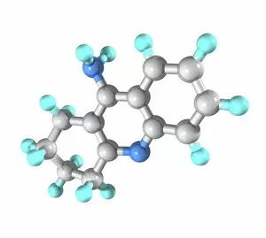
Tacrine. Computer model of a molecule of the drug tacrine. It is sold under the brand name Cognex. It is an acetylcholinesterase inhibitor drug

Background imageBiochemical Collection: Cephalexin drug molecule

Cephalexin drug molecule
Cephalexin. Computer model of a molecule of the drug cephalexin. It is sold under the brand name Keflex. It is a cephalosporin antibiotic drug, used to treat bacterial infections

Background imageBiochemical Collection: Atorvastatin drug molecule

Atorvastatin drug molecule
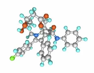
Atorvastatin. Computer model of a molecule of the drug atorvastatin. It is sold under the brand name Lipitor. It is a statin drug

Background imageBiochemical Collection: Azithromycin drug molecule

Azithromycin drug molecule
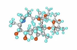
Azithromycin. Computer model of a molecule of the drug azithromycin. It is sold under the brand names Zithromax (Zmax) and Sumamed

Background imageBiochemical Collection: Atenolol drug molecule

Atenolol drug molecule
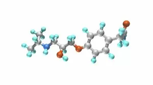
Atenolol. Computer model of a molecule of the drug atenolol. It is sold under the brand name Tenormin. It is a beta blocker drug, used to treat hypertension (high blood pressure)

Background imageBiochemical Collection: Methamphetamine molecule

Methamphetamine molecule. Computer graphic of the drug methamphetamine (formula C10.H15.N), a quicker acting and longer lasting derivative of amphetamine

Background imageBiochemical Collection: Mescaline molecule

Mescaline molecule. Computer graphic of the drug mescaline (formula C11.H17.N.O3). The atoms (tubes) are colour-coded: carbon (yellow), oxygen (red), nitrogen (blue) and hydrogen (white)

Background imageBiochemical Collection: Ephedrine molecule

Ephedrine molecule
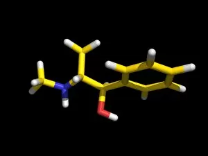
Ephedrine. Computer graphic of a molecule of the drug ephedrine (C10.H15.N.O). The atoms (tubes) of the molecule are colour-coded: hydrogen (white), carbon (yellow), oxygen (red) and nitrogen (blue)

Background imageBiochemical Collection: Morphine molecule

Morphine molecule
Morphine. Computer model of a molecule of morphine (C17.H19.N.O3). The atoms (tubes) of this drug are colour-coded: carbon (purple), oxygen (red), hydrogen (grey) and nitrogen (yellow)

Background imageBiochemical Collection: Hydrocodone drug

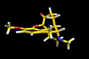
Hydrocodone drug. Molecular model of the analgesic drug hydrocodone (C18H21NO3). Atoms (sticks) are colour-coded: carbon (yellow), hydrogen (white), nitrogen (blue) and oxygen (red)

Background imageBiochemical Collection: Cortisol hormone molecule

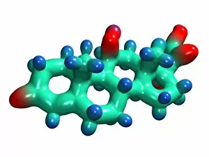
Cortisol hormone molecule. Computer model of a molecule of the steroid hormone cortisol. Atoms are represented as blobs and are colour-coded: carbon (green), hydrogen (blue) and oxygen (red)

Background imageBiochemical Collection: Follicle stimulating hormone molecule

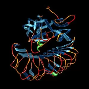
Follicle stimulating hormone molecule. Computer artwork showing the secondary structure of a molecule of follicle stimulating hormone (FSH, bottom) and its receptor (FSHR, top)

Background imageBiochemical Collection: Corticosterone hormone molecule

Corticosterone hormone molecule, computer model. Atoms are represented as spheres and are colour- coded: carbon (pink), hydrogen (white) and oxygen (blue)

Background imageBiochemical Collection: Erythropoietin molecule bound to receptor

Erythropoietin molecule bound to receptors. Computer model of a molecule of erythropoietin (EPO) (orange) bound to two extracellular EPO receptors (pink and purple)

Background imageBiochemical Collection: Prostacyclin, molecular model

Prostacyclin, molecular model. This is a hormone that acts to widen blood vessels (thus lowering blood pressure) and to prevent aggregation of platelets (preventing blood clotting)


For sale as Licensed Images

Choose your image, Select your licence and Download the media

"Unveiling the Intricacies of Biochemical World: From Anaesthetic Inhibiting Ion Channels to DNA Discoveries" Delving into the depths wonders, scientists have uncovered an anaesthetic that inhibits an ion channel (C015 / 6718), shedding light on new possibilities for pain management. The enigmatic double-stranded RNA molecule reveals its secrets, captivating researchers with its role in gene regulation and potential therapeutic applications. Peering into the intricate world of DNA transcription through a molecular model, scientists unravel the mysteries behind genetic information transfer and cellular function. Captured under a microscope's gaze, caffeine crystals dazzle with their vibrant beauty, reminding us of this ubiquitous stimulant's impact on our daily lives. The iconic DNA molecule stands tall as a symbol of life's blueprint, holding within it the key to our genetic heritage and evolutionary history. Immunoglobulin G antibody molecule emerges as a formidable defender against pathogens, showcasing nature's ingenious immune system at work. Through mesmerizing crystal formations seen under intense magnification, EDTA crystals reveal their significance in chelation therapy and metal ion sequestration processes. Oxytocin hormone crystals shimmer like precious gems when observed through polarized light microscopy (PLM C016 / 7196), highlighting its crucial role in social bonding and reproductive functions. Watson and Crick forever etched their names in scientific history by unravelling the structure of DNA; their groundbreaking discovery paved the way for countless advancements in genetics research. Celebrated author Isaac Asimov not only captivated readers with his science fiction tales but also left an indelible mark as a biochemist who popularized complex scientific concepts for all to comprehend and appreciate. Artistic renditions bring metabolic enzymes to life as they orchestrate vital chemical reactions within cells – true catalysts that drive life's intricate processes.