
Biomechanics Collection (#2)
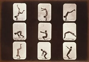
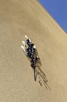
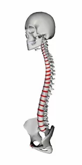
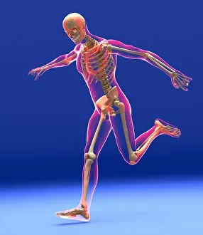
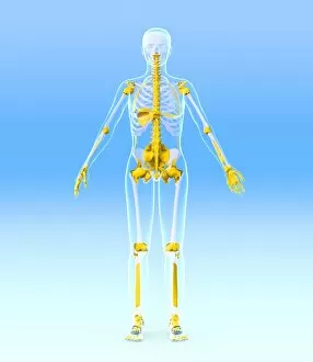
"Unlocking the Secrets of Movement: Exploring the Fascinating World of Biomechanics" Running skeleton in body


For sale as Licensed Images
Choose your image, Select your licence and Download the media
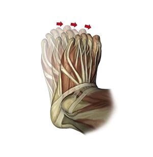
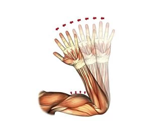
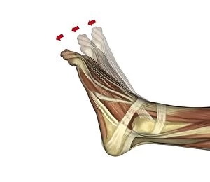
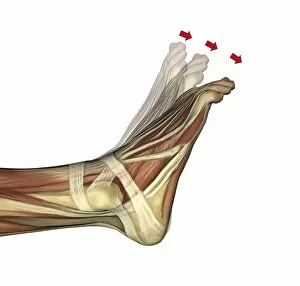
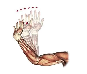
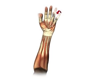
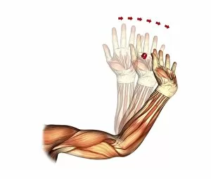
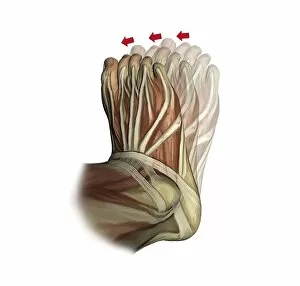
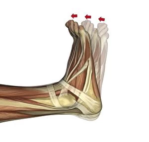
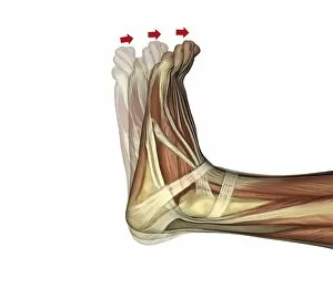
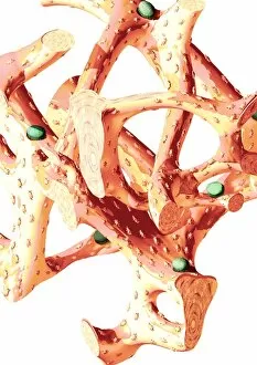
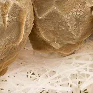
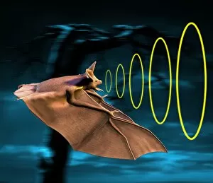
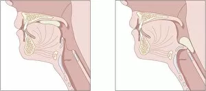
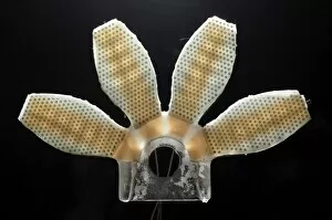
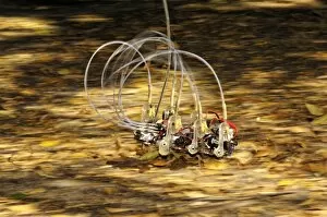
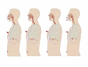
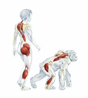
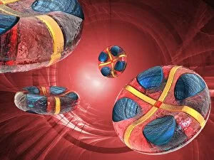
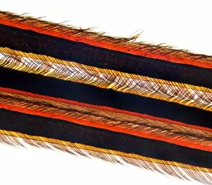
"Unlocking the Secrets of Movement: Exploring the Fascinating World of Biomechanics" Running skeleton in body, artwork: Unveiling the hidden mechanics behind our every step - a captivating glimpse into biomechanics. Mechanics of respiration, diagram: Breathing life into understanding: Delving deep into the intricate workings of our respiratory system through biomechanical analysis. Leg muscles in running, artwork: Muscles in motion: A visual exploration of how our legs power us forward during running, courtesy of biomechanics. Namaqua chameleon catching prey: Nature's precision engineering at play: Witnessing the remarkable hunting techniques of a Namaqua chameleon through a biomechanical lens. Housefly foot, SEM: The tiny wonders beneath our feet: Examining the extraordinary structure and gripping abilities found on a housefly's foot using scanning electron microscopy (SEM). Gecko foot, SEM: Adhesive mastery revealed: Unraveling the secrets behind geckos' incredible climbing abilities with high-resolution SEM imaging and biomechanical insights. Bat sonar: Echolocation demystified: Peering into how bats navigate their surroundings using sound waves – an awe-inspiring example of nature-inspired biomechanics. Fist clenching movement, artwork C016 / 6795: From strength to dexterity - Understanding the complex movements involved in clenching your fist through artistic representation and biophysical analysis. Little and ring finger flexion, artwork C016 / 6793: A tale told by fingers - Illuminating the intricacies behind little and ring finger flexion via stunning artistry combined with detailed biomechanical research. Cicada noise mechanism, diagram C018 / 0296: Unmasking nature's orchestra conductor - Decoding how cicadas produce their unique sounds with an insightful diagram showcasing the biomechanics behind their noise mechanism.