
Bundles Collection (#5)
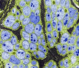
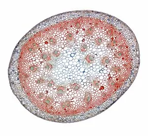
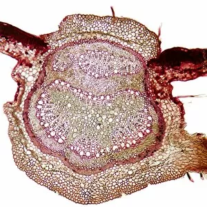
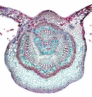
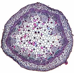
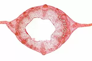
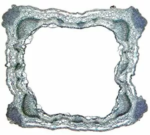
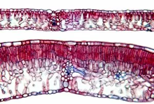
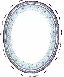
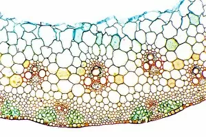
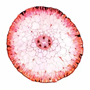
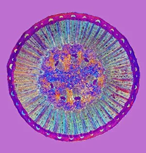
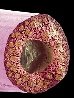
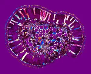
"Bundles: Nature's Intricate Weavings Unveiled" Delicate Collagen Bundles: Revealing the intricate beauty of collagen fibers under a Scanning Electron Micrograph (SEM


For sale as Licensed Images
Choose your image, Select your licence and Download the media
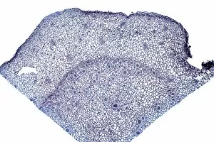
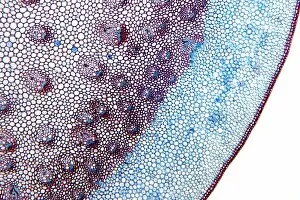
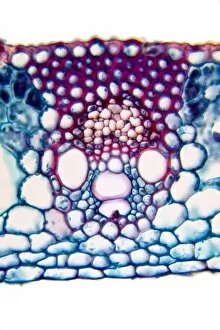
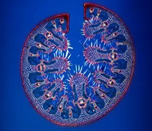
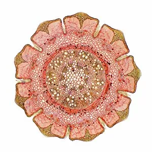
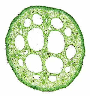
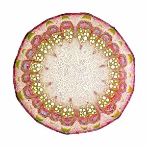
"Bundles: Nature's Intricate Weavings Unveiled" Delicate Collagen Bundles: Revealing the intricate beauty of collagen fibers under a Scanning Electron Micrograph (SEM), nature's own masterpieces. Power in Motion: Witness the strength and resilience of leg muscles in running, tightly bundled to propel us forward with every stride. Artistry Unleashed: Like carefully woven bundles, artwork captures emotions and stories, inviting us into a world where imagination knows no bounds. Serenity on Water: Behold the ethereal elegance of a water lily leaf captured through a light micrograph, showcasing nature's delicate bundle of tranquility. Tendon Tales Untangled: Dive deep into the fascinating realm of tendons as seen under an SEM, unraveling their role in connecting muscle to bone with remarkable precision. Visionary Strengths: Discover the intricate network of eye muscles magnified by an SEM, reminding us how these tiny bundles enable our sight to explore life's wonders. Cultural Treasures Wrapped Up: Explore vibrant prayer flags for sale at Swayambhunath Buddhist temple (Monkey Temple), each bundle carrying wishes for peace and prosperity across the world. Golden Hour Harvest: As evening light bathes hay bales in its warm embrace, witness nature's bounty bundled up before winter arrives - a picturesque scene that whispers tales from rural landscapes. Warmth Shared Anew: Scouts distributing blankets in Zakynthos, Greece remind us that compassion can be found even amidst challenging times when communities come together to support one another - bundling warmth and hope for those in need. The Return Of Traditions Past: Step back into history as "The Return Of The Washhouse" depicts laundresses skillfully bundling clothes while reviving memories of simpler times when cleanliness was an art form itself. The Gathering Hands.