Cell Biology Collection (page 7)

"Exploring the Intricacies of Cell Biology: A Fascinating Journey into Cellular Life" Nerve and glial cells, captured through a mesmerizing light micrograph

Background imageCell Biology Collection: Male reproductive sperm entering the egg

Male reproductive sperm entering the egg

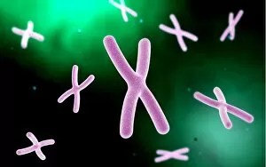
Background imageCell Biology Collection: Microscopic view of chromosome

Microscopic view of chromosome

Background imageCell Biology Collection: Scanning electron micrograph of Staphylococcus and a dead human neutrophil

Scanning electron micrograph of Staphylococcus and a dead human neutrophil
Scanning electron micrograph of methicillin-resistant Staphylococcus aureus and a dead human neutrophil

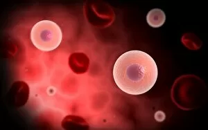
Background imageCell Biology Collection: Microscopic view of plasma cell inside blood vessel

Microscopic view of plasma cell inside blood vessel

Background imageCell Biology Collection: Close-up view of the synapse in the nervous system

Close-up view of the synapse in the nervous system. A synapse is a structure that permits a neuron (or nerve cell) to pass an electrical or chemical signal to another cell

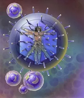
Background imageCell Biology Collection: Vitruvian Man inside virus particle

Vitruvian Man inside virus particle

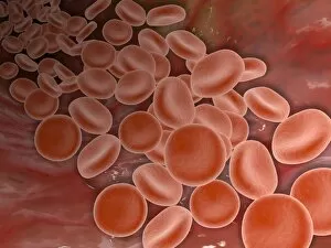
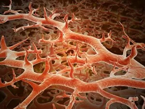
Background imageCell Biology Collection: Inside view of the artery with red blood cells

Inside view of the artery with red blood cells

Background imageCell Biology Collection: Clathrin Mediated Endocytosis

Clathrin Mediated Endocytosis

Background imageCell Biology Collection: Microscopic view of a mast cell

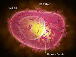
Microscopic view of a mast cell. Mast cells are found resident in tissues throughout the body, particularly in association with structures such as blood vessels and nerves

Background imageCell Biology Collection: Generic vials with chemicals in research lab

Generic vials with chemicals in research lab

Background imageCell Biology Collection: Microscopic view of birch tree pollen

Microscopic view of birch tree pollen. Birch tree pollen is considered to be an important allergenic tree pollen
Background imageCell Biology Collection: Microscopic view of Paramecium bursaria

Microscopic view of Paramecium bursaria, a species of ciliate protozoan that has a mutualistic symbiotic relationship with green algae called Zoochlorella

Background imageCell Biology Collection: Microscopic view of protozoa

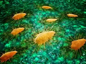
Microscopic view of protozoa. Leishmaniasis and dysentery are examples of diseases involving protozoa, often transmitted to humans by the bite of a parasite-carrying insect

Background imageCell Biology Collection: Microscopic view of stem cell development

Microscopic view of stem cell development. In adult organisms, stem cells and progenitor cells act as a repair system for the body, replenishing adult tissues

Background imageCell Biology Collection: Conceptual image of mitosis

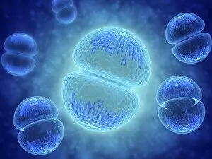
Conceptual image of mitosis. Mitosis is the process in the cell cycle by which a cell duplicates into two genetically identical sets of chromosomes

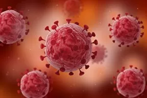
Background imageCell Biology Collection: Microscopic view of coronavirus

Microscopic view of coronavirus

Background imageCell Biology Collection: Conceptual image of cancer virus

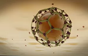
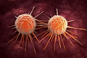
Conceptual image of cancer virus

Background imageCell Biology Collection: Microscopic view of a group of macrophages

Microscopic view of a group of macrophages. Macrophages contribute to tumor growth and progression. Attracted to oxygen-starved (hypoxic) and necrotic tumor cells they promote chronic inflammation

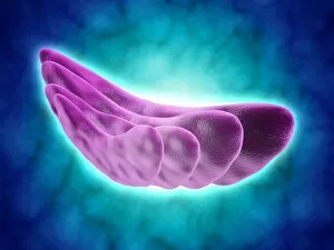
Background imageCell Biology Collection: Conceptual image of the Golgi apparatus

Conceptual image of the Golgi apparatus. The Golgi apparatus is an organelle found in most eukaryotic cells

Background imageCell Biology Collection: Microscopic view of athletes foot fungus

Microscopic view of athletes foot fungus. Athletes foot fungus, also known as ringworm of the foot and tinea pedis, is a fungal infection of the skin

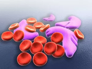
Background imageCell Biology Collection: Conceptual image of sickle cell anemia

Conceptual image of sickle cell anemia. Sickle cell anemia is a disease in which your body produces abnormally shaped red blood cells. The cells are shaped like a crescent or sickle

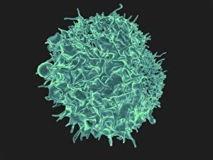
Background imageCell Biology Collection: Colorized scanning electron micrograph of a T lymphocyte

Colorized scanning electron micrograph of a T lymphocyte

Background imageCell Biology Collection: Primary cultures of superior cervical ganglia cells

Primary cultures of superior cervical ganglia cells

Background imageCell Biology Collection: Researcher in lab uses pipette and measures RNA

Researcher in lab uses pipette and measures RNA

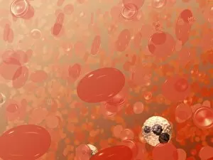
Background imageCell Biology Collection: Field of blood cells illustration

Field of blood cells illustration
This is a field of blood cells. The bi-concave disks are red blood cells or erythrocytes. The white cell with the dark purplish, multi-lobed nucleus is a neutrophil

Background imageCell Biology Collection: Installing a flow cell on an Illumina cluster station

Installing a flow cell on an Illumina cluster station

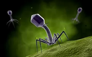
Background imageCell Biology Collection: Microscopic view of bacteriophage

Microscopic view of bacteriophage

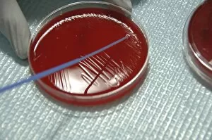
Background imageCell Biology Collection: Staphylococcus Bacteria from human skin grown on agar

Staphylococcus Bacteria from human skin grown on agar
Staphylococcus bacteria from human skin grown on agar in the laboratory

Background imageCell Biology Collection: Patient positioned in computed tomography scanner

Patient positioned in computed tomography scanner
A patient model wearing a short face mask is positioned in a computed tomography (CT) scanner to obtain images of a tumor before radiation therapy

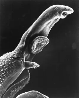
Background imageCell Biology Collection: Scanning electron micrograph of a schistosome parasite

Scanning electron micrograph of a schistosome parasite, which enters the body through the skin of persons coming in contact with infested waters. The adult worm lives in the veins of its host

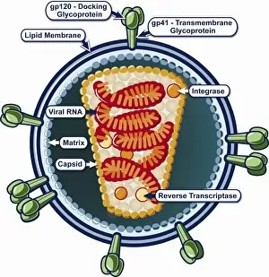
Background imageCell Biology Collection: Structure of HIV

Structure of HIV

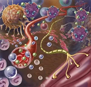
Background imageCell Biology Collection: Nerve with myelin sheath, seen in lower right, connects with muscle

Nerve with myelin sheath, seen in lower right, connects with muscle. Blood vessel and immune cells are seen in the center and upper right of image

Background imageCell Biology Collection: Grouping of virus particles

Grouping of virus particles

Background imageCell Biology Collection: Conceptual image of the coronavirus

Conceptual image of the coronavirus. Coronaviruses primarily infect the upper respiratory and gastrointestinal tract

Background imageCell Biology Collection: Microscopic view of virus

Microscopic view of virus

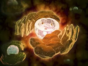
Background imageCell Biology Collection: Conceptual image of endoplasmic reticulum around a cell nucleus

Conceptual image of endoplasmic reticulum around a cell nucleus. Endoplasmic reticulum is an organelle that forms a continuous membrane system of flattened sacs within the cytoplasm of a eukaryotic

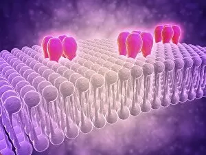
Background imageCell Biology Collection: Conceptual image of GABA receptors

Conceptual image of GABA receptors. The GABA receptors are a class of receptors that respond to the neurotransmitter gamma-aminobutyric acid

Background imageCell Biology Collection: Microscopic view of herpes virus

Microscopic view of herpes virus

Background imageCell Biology Collection: Conceptual image of Trypanosoma

Conceptual image of Trypanosoma

Background imageCell Biology Collection: Conceptual image of influenza causing flu virus

Conceptual image of influenza causing flu virus

Background imageCell Biology Collection: 3D rendering of macrophage phagocytosis

3D rendering of macrophage phagocytosis

Background imageCell Biology Collection: Microscopic view of phagocytic macrophages

Microscopic view of phagocytic macrophages, which are involved in the immune response within the body

Background imageCell Biology Collection: Microscopic view of sperm swimming towards egg

Microscopic view of sperm swimming towards egg

Background imageCell Biology Collection: Close-up view of athletes foot fungus

Close-up view of athletes foot fungus, also known as ringworm of the foot and tinea pedis. Athletes foot fungus is a fungal infection of the skin

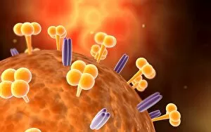
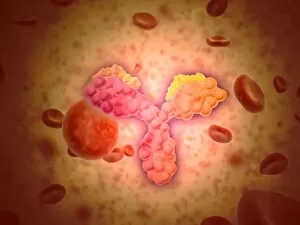
Background imageCell Biology Collection: Microscopic view of human anitbodies with red blood cells

Microscopic view of human anitbodies with red blood cells. Human antibodies are the Y-shaped proteins used by the immune system to defend against foreign objects like bacteria and viruses

Background imageCell Biology Collection: Conceptual image of a neuron ligntning signal passing

Conceptual image of a neuron ligntning signal passing

Background imageCell Biology Collection: Conceptual image of cell nucleus

Conceptual image of cell nucleus. The cell nucleus acts like the brain of the cell. It helps control eating, movement, and reproduction

Background imageCell Biology Collection: Virus landing on cell membrane

Virus landing on cell membrane


For sale as Licensed Images

Choose your image, Select your licence and Download the media

"Exploring the Intricacies of Cell Biology: A Fascinating Journey into Cellular Life" Nerve and glial cells, captured through a mesmerizing light micrograph, reveal the intricate network that supports our nervous system. T lymphocytes battling cancer cells - witness this epic battle in stunning detail with SEM C001 / 1679 imagery. Anaesthetic inhibiting an ion channel - delve into the world of cellular communication as we uncover how anaesthetics affect these vital channels (C015 / 6718). Glial cells take center stage in a captivating confocal light micrograph, showcasing their crucial role in supporting neurons. Witness the beauty of glial stem cell culture under a microscope; it's like exploring an enchanting galaxy within our own bodies (light micrograph). Dendritic cells come to life through exquisite artwork, highlighting their pivotal role in immune response and defense mechanisms. Pine pollen grains unveil their intricate structure under a light microscope – nature's tiny marvels never cease to amaze us. Dive deep into the inner workings of pine stems with a captivating light micrograph – discover the building blocks that support these majestic trees. Explore the hidden wonders of lime tree stems through a mesmerizing light micrograph – witness nature's architectural masterpiece up close. Uncover various cell types brought to life through breathtaking artwork – each one playing its unique part in maintaining life's delicate balance. Immerse yourself in neural stem cell culture and witness firsthand how these remarkable cells hold immense potential for regenerative medicine. The artistic portrayal of cell membranes (C013 / 7467) invites you to ponder upon their crucial role as gatekeepers controlling molecular traffic within living organisms. Embark on this awe-inspiring journey into cell biology and unlock the secrets that shape all living beings from within.