Cell Biology Collection (page 8)

"Exploring the Intricacies of Cell Biology: A Fascinating Journey into Cellular Life" Nerve and glial cells, captured through a mesmerizing light micrograph

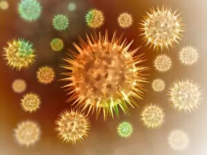
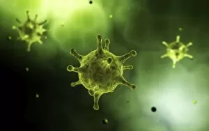
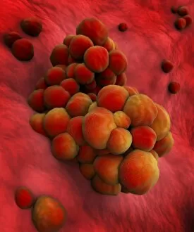
Background imageCell Biology Collection: Grouping of virus particles

Grouping of virus particles

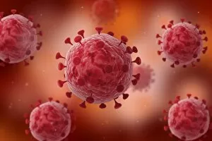
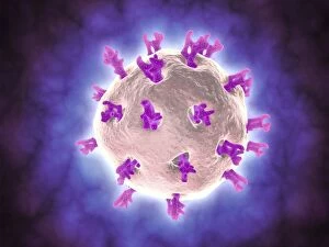
Background imageCell Biology Collection: Conceptual image of the coronavirus

Conceptual image of the coronavirus. Coronaviruses primarily infect the upper respiratory and gastrointestinal tract

Background imageCell Biology Collection: Microscopic view of virus

Microscopic view of virus

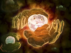
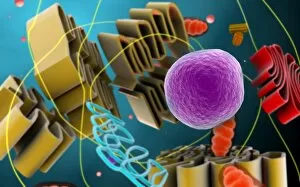
Background imageCell Biology Collection: Conceptual image of endoplasmic reticulum around a cell nucleus

Conceptual image of endoplasmic reticulum around a cell nucleus. Endoplasmic reticulum is an organelle that forms a continuous membrane system of flattened sacs within the cytoplasm of a eukaryotic

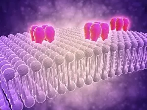
Background imageCell Biology Collection: Conceptual image of GABA receptors

Conceptual image of GABA receptors. The GABA receptors are a class of receptors that respond to the neurotransmitter gamma-aminobutyric acid

Background imageCell Biology Collection: Microscopic view of herpes virus

Microscopic view of herpes virus

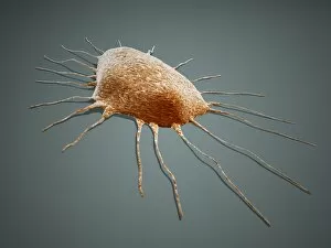
Background imageCell Biology Collection: Conceptual image of Trypanosoma

Conceptual image of Trypanosoma

Background imageCell Biology Collection: Conceptual image of influenza causing flu virus

Conceptual image of influenza causing flu virus

Background imageCell Biology Collection: 3D rendering of macrophage phagocytosis

3D rendering of macrophage phagocytosis

Background imageCell Biology Collection: Microscopic view of phagocytic macrophages

Microscopic view of phagocytic macrophages, which are involved in the immune response within the body

Background imageCell Biology Collection: Microscopic view of sperm swimming towards egg

Microscopic view of sperm swimming towards egg

Background imageCell Biology Collection: Close-up view of athletes foot fungus

Close-up view of athletes foot fungus, also known as ringworm of the foot and tinea pedis. Athletes foot fungus is a fungal infection of the skin

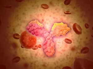
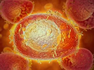
Background imageCell Biology Collection: Microscopic view of human anitbodies with red blood cells

Microscopic view of human anitbodies with red blood cells. Human antibodies are the Y-shaped proteins used by the immune system to defend against foreign objects like bacteria and viruses

Background imageCell Biology Collection: Conceptual image of a neuron ligntning signal passing

Conceptual image of a neuron ligntning signal passing

Background imageCell Biology Collection: Conceptual image of cell nucleus

Conceptual image of cell nucleus. The cell nucleus acts like the brain of the cell. It helps control eating, movement, and reproduction

Background imageCell Biology Collection: Virus landing on cell membrane

Virus landing on cell membrane

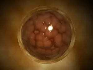
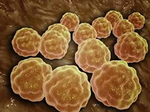
Background imageCell Biology Collection: Microscopic view of a blastula during pregnancy

Microscopic view of a blastula during pregnancy. After the cleavage has produced over 100 cells, the embryo is called a blastula

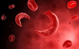
Background imageCell Biology Collection: Microscopic view of sicke cells causing anemia disease

Microscopic view of sicke cells causing anemia disease

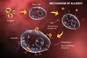
Background imageCell Biology Collection: Mast cell releasing histamine due to allergic reaction

Mast cell releasing histamine due to allergic reaction

Background imageCell Biology Collection: Microscopic view of rotavirus

Microscopic view of rotavirus. Rotavirus is the most common cause of severe diarrhea among infants and young children. It is a genus of double-stranded RNA virus in the family Reoviridae

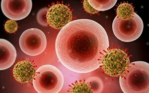
Background imageCell Biology Collection: Microscopic view of cell and virus

Microscopic view of cell and virus

Background imageCell Biology Collection: Microscopic view of blood cells

Microscopic view of blood cells

Background imageCell Biology Collection: Microscopic view of cancer virus

Microscopic view of cancer virus

Background imageCell Biology Collection: Conceptual image of a blood vessel

Conceptual image of a blood vessel

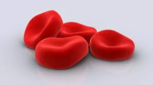
Background imageCell Biology Collection: Conceptual image of red blood cells

Conceptual image of red blood cells

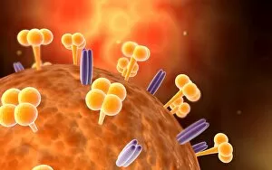
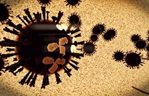
Background imageCell Biology Collection: A black swarm of H5N1 avian flu viruses are attacked by antibodies

A black swarm of H5N1 avian flu viruses are attacked by antibodies (the three legged elements), which mark the virus for destruction

Background imageCell Biology Collection: Microscopic view of Rubella virus

Microscopic view of Rubella virus
Microscopic view of Rubella. Rubella is an acute, contagious viral infection. While the illness is generally mild in children, it has serious consequences in pregnant women causing fetal death

Background imageCell Biology Collection: Microscopic view of centrioles within a human cell

Microscopic view of centrioles within a human cell
Microscopic view of barrel shaped structure of centrioles within a human cell. A centriole is a cylinder shaped cell structure found in most eukaryotic cells

Background imageCell Biology Collection: Isolated cancer cell Macrophage

Isolated cancer cell Macrophage. Macrophages contribute to tumor growth and progression. Attracted to oxygen-starved (hypoxic) and necrotic tumor cells they promote chronic inflammation

Background imageCell Biology Collection: Neurofibromatosis, a genetic disorder of the nervous system

Neurofibromatosis, a genetic disorder of the nervous system
A genetic disorder of the nervous system, neurofibromatosis causes tumors to form on nerves throughout the body, including a type of tumor called an optic nerve glioma that can result in childhood

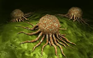
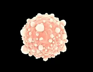
Background imageCell Biology Collection: Microscopic view of cancer cells

Microscopic view of cancer cells. Cancer occurs when a cells gene mutations make the cell unable to correct DNA damage

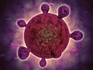
Background imageCell Biology Collection: Conceptual image of common virus

Conceptual image of common virus

Background imageCell Biology Collection: Colorized scanning electron micrograph of filamentous Ebola virus particles

Colorized scanning electron micrograph of filamentous Ebola virus particles (blue) budding from a chronically infected VERO E6 cell (yellow-green)

Background imageCell Biology Collection: Rack of pipettes

Rack of pipettes

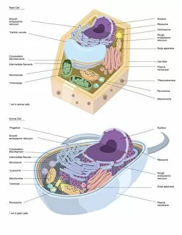
Background imageCell Biology Collection: Comparative illustration of plant and animal cell anatomy (with labels)

Comparative illustration of plant and animal cell anatomy (with labels)

Background imageCell Biology Collection: Conceptual image illustrating the process of bone metastasis

Conceptual image illustrating the process of bone metastasis

Background imageCell Biology Collection: Components of blood vessel interact with intracellular fluid

Components of blood vessel interact with intracellular fluid; white blood cells, red blood cells, oxygen, platelets and hormones

Background imageCell Biology Collection: Conceptual image of the dengue virus

Conceptual image of the dengue virus. The dengue virus is a mosquito-borne RNA virus that causes dengue fever

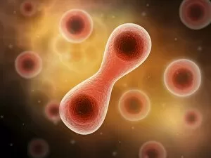
Background imageCell Biology Collection: Microscopic view of cell division

Microscopic view of cell division

Background imageCell Biology Collection: Microscopic view of Yersinia

Microscopic view of Yersinia. Yersinia is a gram-negative, rod-shaped bacteria in the family Enterobacteriaceae

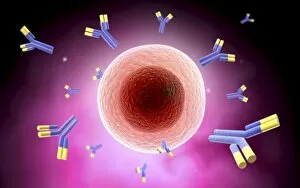
Background imageCell Biology Collection: Microscopic view of cell and antibody

Microscopic view of cell and antibody

Background imageCell Biology Collection: Microscopic view of a microbe

Microscopic view of a microbe. Microbes are single-cell organisms so tiny that millions can fit into the eye of a needle

Background imageCell Biology Collection: Microscopic view of tumor

Microscopic view of tumor

Background imageCell Biology Collection: Microscopic view of animal cell nucleus

Microscopic view of animal cell nucleus

Background imageCell Biology Collection: Conceptual image of flagellate bacterium

Conceptual image of flagellate bacterium

Background imageCell Biology Collection: Microscopic view of pancreatic cancer cell

Microscopic view of pancreatic cancer cell

Background imageCell Biology Collection: Microscopic view of male sperm cells

Microscopic view of male sperm cells

Background imageCell Biology Collection: Microscopic view of a mast cell found in connective tissue

Microscopic view of a mast cell found in connective tissue that contains numerous basophilic granules and releases substances such as heparin and histamine in response to injury


For sale as Licensed Images

Choose your image, Select your licence and Download the media

"Exploring the Intricacies of Cell Biology: A Fascinating Journey into Cellular Life" Nerve and glial cells, captured through a mesmerizing light micrograph, reveal the intricate network that supports our nervous system. T lymphocytes battling cancer cells - witness this epic battle in stunning detail with SEM C001 / 1679 imagery. Anaesthetic inhibiting an ion channel - delve into the world of cellular communication as we uncover how anaesthetics affect these vital channels (C015 / 6718). Glial cells take center stage in a captivating confocal light micrograph, showcasing their crucial role in supporting neurons. Witness the beauty of glial stem cell culture under a microscope; it's like exploring an enchanting galaxy within our own bodies (light micrograph). Dendritic cells come to life through exquisite artwork, highlighting their pivotal role in immune response and defense mechanisms. Pine pollen grains unveil their intricate structure under a light microscope – nature's tiny marvels never cease to amaze us. Dive deep into the inner workings of pine stems with a captivating light micrograph – discover the building blocks that support these majestic trees. Explore the hidden wonders of lime tree stems through a mesmerizing light micrograph – witness nature's architectural masterpiece up close. Uncover various cell types brought to life through breathtaking artwork – each one playing its unique part in maintaining life's delicate balance. Immerse yourself in neural stem cell culture and witness firsthand how these remarkable cells hold immense potential for regenerative medicine. The artistic portrayal of cell membranes (C013 / 7467) invites you to ponder upon their crucial role as gatekeepers controlling molecular traffic within living organisms. Embark on this awe-inspiring journey into cell biology and unlock the secrets that shape all living beings from within.