Cell Biology Collection (#9)

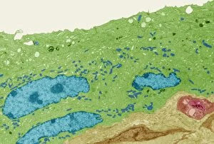
"Exploring the Intricacies of Cell Biology: A Fascinating Journey into Cellular Life" Nerve and glial cells, captured through a mesmerizing light micrograph

Background imageCell Biology Collection: Colorized scanning electron micrograph of filamentous Ebola virus particles

Colorized scanning electron micrograph of filamentous Ebola virus particles (blue) budding from a chronically infected VERO E6 cell (yellow-green)

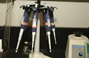
Background imageCell Biology Collection: Rack of pipettes

Rack of pipettes

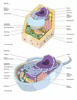
Background imageCell Biology Collection: Comparative illustration of plant and animal cell anatomy (with labels)

Comparative illustration of plant and animal cell anatomy (with labels)

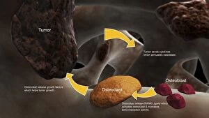
Background imageCell Biology Collection: Conceptual image illustrating the process of bone metastasis

Conceptual image illustrating the process of bone metastasis

Background imageCell Biology Collection: Components of blood vessel interact with intracellular fluid

Components of blood vessel interact with intracellular fluid; white blood cells, red blood cells, oxygen, platelets and hormones

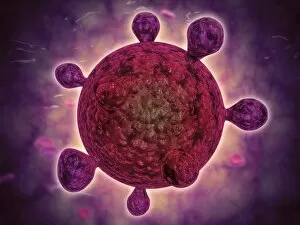
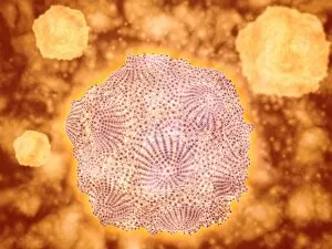
Background imageCell Biology Collection: Conceptual image of the dengue virus

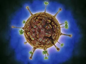
Conceptual image of the dengue virus. The dengue virus is a mosquito-borne RNA virus that causes dengue fever

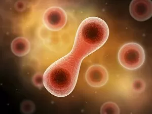
Background imageCell Biology Collection: Microscopic view of cell division

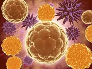
Microscopic view of cell division

Background imageCell Biology Collection: Microscopic view of Yersinia

Microscopic view of Yersinia. Yersinia is a gram-negative, rod-shaped bacteria in the family Enterobacteriaceae

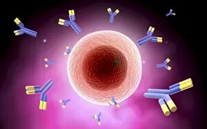
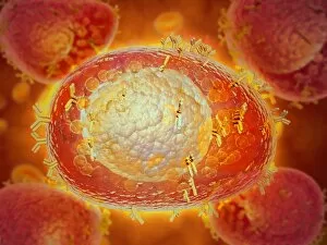
Background imageCell Biology Collection: Microscopic view of cell and antibody

Microscopic view of cell and antibody

Background imageCell Biology Collection: Microscopic view of a microbe

Microscopic view of a microbe. Microbes are single-cell organisms so tiny that millions can fit into the eye of a needle

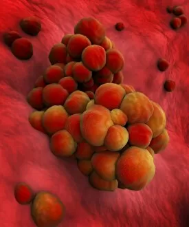
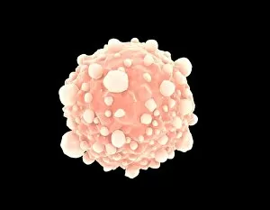
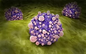
Background imageCell Biology Collection: Microscopic view of tumor

Microscopic view of tumor

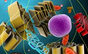
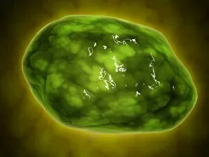
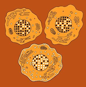
Background imageCell Biology Collection: Microscopic view of animal cell nucleus

Microscopic view of animal cell nucleus

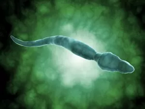
Background imageCell Biology Collection: Conceptual image of flagellate bacterium

Conceptual image of flagellate bacterium

Background imageCell Biology Collection: Microscopic view of pancreatic cancer cell

Microscopic view of pancreatic cancer cell

Background imageCell Biology Collection: Microscopic view of male sperm cells

Microscopic view of male sperm cells

Background imageCell Biology Collection: Microscopic view of a mast cell found in connective tissue

Microscopic view of a mast cell found in connective tissue that contains numerous basophilic granules and releases substances such as heparin and histamine in response to injury

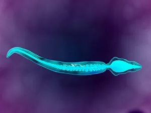
Background imageCell Biology Collection: Microscopic view showing bone structure of sperm

Microscopic view showing bone structure of sperm

Background imageCell Biology Collection: Malria spores in the human blood stream

Malria spores in the human blood stream
The malaria spores move from the mosquito to the human blood stream. the spores divide many times and produce large numbers of spores

Background imageCell Biology Collection: Conceptual image of lyssavirus

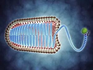
Conceptual image of lyssavirus. Lyssavirus is a genus of viruses belonging to the family Rhabdoviridae. This group of RNA viruses includes the rabies virus traditionally associated with the disease

Background imageCell Biology Collection: Artistic rendering of the surface of a human dendritic cell

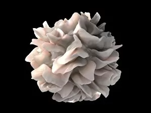
Artistic rendering of the surface of a human dendritic cell illustrating the unexpected discovery of sheet-like processes that fold back onto the membrane surface

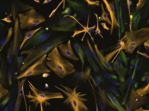
Background imageCell Biology Collection: Microscopic view of a multipolar neuron

Microscopic view of a multipolar neuron. Multipolar neurons possess a single axon and many dendrites
Background imageCell Biology Collection: Conceptual image of a ubiquitous virus

Conceptual image of a ubiquitous virus. A ubiquitous virus is contagious in early childhood through the respiratory tract
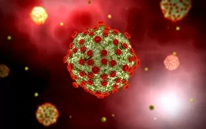
Background imageCell Biology Collection: Microscopic view of Henipavirus

Microscopic view of Henipavirus. Henipavirus is a established group of paramyxoviruses comprising the Hendra virus, Nipah virus and Cedar virus

Background imageCell Biology Collection: Microscipic view of pancreatic cancer cells

Microscipic view of pancreatic cancer cells

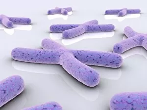
Background imageCell Biology Collection: Conceptual image of chromosome

Conceptual image of chromosome

Background imageCell Biology Collection: Scientist prepares a Western blot

Scientist prepares a Western blot, a technique used to detect a specific protein in a blood or tissue sample

Background imageCell Biology Collection: Illustration of action potential of a nerve cell

Illustration of action potential of a nerve cell

Background imageCell Biology Collection: Conceptual image of vibrio cholerae causing cholera

Conceptual image of vibrio cholerae causing cholera

Background imageCell Biology Collection: Artery cross section with red blood cell flow

Artery cross section with red blood cell flow

Background imageCell Biology Collection: Conceptual image of lysosome

Conceptual image of lysosome. Lysosomes are cellular organelles that contain acid hydrolase enzymes that break down waste materials and cellular debris

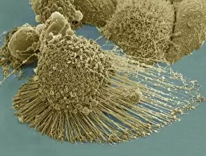
Background imageCell Biology Collection: Scanning electron micrograph of an apoptotic HeLa cell

Scanning electron micrograph of an apoptotic HeLa cell. Zeiss Merlin HR-SEM

Background imageCell Biology Collection: Conceptual image of a neuron

Conceptual image of a neuron

Background imageCell Biology Collection: Microscopic view of Canine Parvovirus

Microscopic view of Canine Parvovirus. Canine Parvovirus is a highly contagious viral disease that is one of the most common causes of diarrhea in dogs under 6 months of age

Background imageCell Biology Collection: Microscopic view of a single male sperm cell

Microscopic view of a single male sperm cell

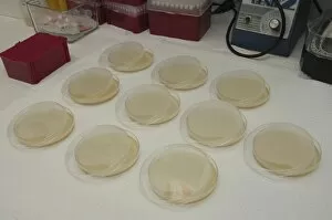
Background imageCell Biology Collection: Cloning dishes in research lab

Cloning dishes in research lab

Background imageCell Biology Collection: Vials in research lab

Vials in research lab

Background imageCell Biology Collection: Directed differentiation of multipotential human neural progenitor cells

Directed differentiation of multipotential human neural progenitor cells
Human neural progenitor cells were isolated under selective culture conditions from the developing human brain and directed through lineage differentiation to GFAP + (glial fibrillary acid protein)

Background imageCell Biology Collection: Microscopic view of HIV virus

Microscopic view of HIV virus

Background imageCell Biology Collection: Surface of HIV infected microphage

Surface of HIV infected microphage
3D representation of the surface and interior of an HIV-infected macrophage obtained using newly developed tools for 3D imaging using ion-abrasion scanning electron microscopy

Background imageCell Biology Collection: Torn muscle fibers with healing stages surrounding

Torn muscle fibers with healing stages surrounding. 1. Macrophage clean-up. 2. New cells migrate to repair. 3. Cells differentiate to become muscle cells

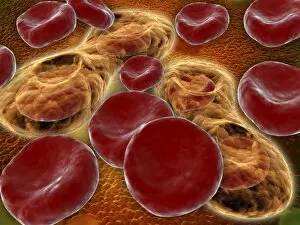
Background imageCell Biology Collection: Red blood cells with white blood cells

Red blood cells with white blood cells

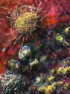
Background imageCell Biology Collection: Influenza virus, glassy look

Influenza virus, glassy look

Background imageCell Biology Collection: Stochastic gene expression, illustration C018 / 0906

Stochastic gene expression, illustration C018 / 0906
Stochastic gene expression, illustration. Every cell in an organism contains every single gene that makes up the organisms genome. However, they are not all active (expressed) in each cell

Background imageCell Biology Collection: Cancer cell, artwork C016 / 9843

Cancer cell, artwork C016 / 9843
Cancer cell. Computer artwork of a cancerous (malignant) cell (top left) in tissue. The uneven surface and cytoplasmic projections are characteristic of cancer cells

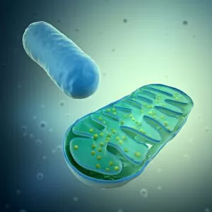
Background imageCell Biology Collection: Mitochondria, artwork

Mitochondria, artwork
Mitochondria, computer artwork

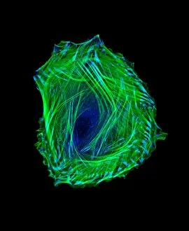
Background imageCell Biology Collection: Embryonic smooth muscle cell C018 / 8595

Embryonic smooth muscle cell C018 / 8595
Embryonic smooth muscle cell, immunofluorescence micrograph. Actin filaments are green, the protein vinculin is light blue. Actin is part of the cytoskeleton

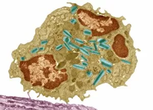
Background imageCell Biology Collection: Eosinophil white blood cell, TEM C014 / 1438

Eosinophil white blood cell, TEM C014 / 1438
Eosinophil white blood cell. Transmission electron micrograph (TEM) of a section through an eosinophil. Eosinophils, like all white blood cells, are part of the bodys immune system

Background imageCell Biology Collection: Bladder lining, TEM C014 / 1473

Bladder lining, TEM C014 / 1473
Bladder lining. Transmission electron micrograph (TEM) of a section through the epithelial cells that line of a human bladder, showing large flat vesicles (large, blue)


For sale as Licensed Images

Choose your image, Select your licence and Download the media

"Exploring the Intricacies of Cell Biology: A Fascinating Journey into Cellular Life" Nerve and glial cells, captured through a mesmerizing light micrograph, reveal the intricate network that supports our nervous system. T lymphocytes battling cancer cells - witness this epic battle in stunning detail with SEM C001 / 1679 imagery. Anaesthetic inhibiting an ion channel - delve into the world of cellular communication as we uncover how anaesthetics affect these vital channels (C015 / 6718). Glial cells take center stage in a captivating confocal light micrograph, showcasing their crucial role in supporting neurons. Witness the beauty of glial stem cell culture under a microscope; it's like exploring an enchanting galaxy within our own bodies (light micrograph). Dendritic cells come to life through exquisite artwork, highlighting their pivotal role in immune response and defense mechanisms. Pine pollen grains unveil their intricate structure under a light microscope – nature's tiny marvels never cease to amaze us. Dive deep into the inner workings of pine stems with a captivating light micrograph – discover the building blocks that support these majestic trees. Explore the hidden wonders of lime tree stems through a mesmerizing light micrograph – witness nature's architectural masterpiece up close. Uncover various cell types brought to life through breathtaking artwork – each one playing its unique part in maintaining life's delicate balance. Immerse yourself in neural stem cell culture and witness firsthand how these remarkable cells hold immense potential for regenerative medicine. The artistic portrayal of cell membranes (C013 / 7467) invites you to ponder upon their crucial role as gatekeepers controlling molecular traffic within living organisms. Embark on this awe-inspiring journey into cell biology and unlock the secrets that shape all living beings from within.