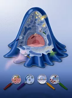
Cell Collection (#97)

"Exploring the Intricacies of Life: Unveiling the Wonders of Cells" Delving into the microscopic world

Background imageCell Collection: Tree-of-heaven stem, light micrograph

Tree-of-heaven stem, light micrograph
Tree-of-heaven stem. Polarised light micrograph of a cross-section through the stem of the tree-of-heaven (Ailanthus glandulosa). Below the outer layers (red) is a ring of vascular bundles

Background imageCell Collection: Stomach section, light micrograph

Stomach section, light micrograph
Stomach section. Light micrograph of a vertical section through a stomach. The upper layer (pink-purple) is the glandular mucosa

Background imageCell Collection: Skin section, light micrograph

Skin section, light micrograph
Skin section. Semi-polarised light micrograph of a vertical section through human skin. The outer epidermis is the thin layer across top, with the dermis layer beneath it

Background imageCell Collection: Aralia leaf stalk, light micrograph

Aralia leaf stalk, light micrograph
Aralia leaf stalk. Light micrograph of a cross-section through a leaf stalk (petiole) from an Araliatree. The outer layer directly below the epidermis is a supporting collenchyma ring (red)

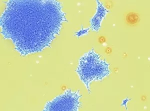
Background imageCell Collection: Human embryonic stem cells, TEM

Human embryonic stem cells, TEM
Stem cells. Coloured transmission electron micrograph (TEM) of a human embryonic stem cells (HESC, blue). HESCs are pluripotent

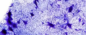
Background imageCell Collection: Motor neurones, light micrograph

Motor neurones, light micrograph
Motor neurones. Light micrograph of motor nerve cells (motor neurones) and their support cells. The cell bodies of the neurones (large deep purple areas) have a central nucleus

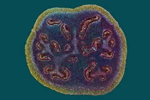
Background imageCell Collection: Bracken fern rhizome, light micrograph

Bracken fern rhizome, light micrograph
Bracken fern rhizome. Polarised light micrograph of a cross-section through a rhizome from a bracken fern (Pteris aquilinum). The outer layer (yellow-green) consists of supporting fibres

Background imageCell Collection: Tongue section, light micrograph

Tongue section, light micrograph
Tongue section. Light micrograph of a vertical section through a mammalian tongue. At top, the surface is covered with circumvallate papillae

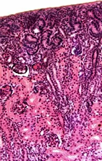
Background imageCell Collection: Kidney cortex, light micrograph

Kidney cortex, light micrograph
Kidney cortex. Light micrograph of a vertical section through the cortex (outer layer) of a kidney. The cortex tissue (across top) is stained a darker red

Background imageCell Collection: Nettle stinging hair, light micrograph

Nettle stinging hair, light micrograph
Nettle stinging hair. Polarised light micrograph of a stinging hair from a Roman nettle (Urtica pilulifera). This tapering needle-shaped cell consists of a round basal part embedded in a

Background imageCell Collection: Strychnine seed tissue, light micrograph

Strychnine seed tissue, light micrograph
Strychnine seed tissue. Light micrograph of a section through a seed of the strychnine plant (Strychnos nux vomica), showing the cellular bridges (plasmodesma)

Background imageCell Collection: Fallopian tube, SEM

Fallopian tube, SEM
Fallopian tube. Coloured scanning electron micrograph (SEM) of the lining of a fallopian tube (oviduct). The fallopian tubes carry the egg from the ovary to the uterus (womb)

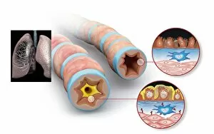
Background imageCell Collection: Asthma pathology, artwork

Asthma pathology, artwork
Asthma pathology. Computer artwork showing cross-sections through a normal airway (right) and an asthmatic, constricted airway (left)

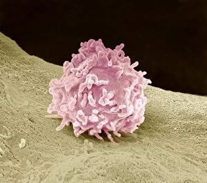
Background imageCell Collection: Granulocyte, SEM

Granulocyte, SEM
Granulocyte. Coloured scanning electron micrograph (SEM) of a granulocyte in bone marrow. Granulocytes are a category of white blood cell characterized by the presence of granules in their cytoplasm

Background imageCell Collection: Hippocampal neuron fluorescent micrograph

Hippocampal neuron fluorescent micrograph
Hippocampal neuron. Fluorescent micrograph of a neuron (nerve cell, centre) from the hippocampus, surrounded by glial cells (support cells)

Background imageCell Collection: Granulocytes, SEM

Granulocytes, SEM
Granulocytes. Coloured scanning electron micrograph (SEM) of granulocytes in bone marrow. Granulocytes are a category of white blood cell characterized by the presence of granules in their cytoplasm

Background imageCell Collection: Nerve bundle, SEM

Nerve bundle, SEM
Nerve bundle. Coloured scanning electron micrograph (SEM) of a freeze-fractured section through a bundle of myelinated nerve fibres. Myelin sheaths (yellow) can be seen surrounding the axons (blue)

Background imageCell Collection: Blood-filled pulmonary vein, SEM

Blood-filled pulmonary vein, SEM
Blood-filled pulmonary vein. Coloured scanning electron micrograph (SEM) of a section through a pulmonary vein (green) in lung tissue (purple), showing the red blood cells (erythrocytes)

Background imageCell Collection: Salmonella bacteria, TEM

Salmonella bacteria, TEM
Salmonella bacteria. Coloured transmission electron micrograph (TEM) of a Salmonella sp. bacteria (green) in the cytoplasm of an infected cell

Background imageCell Collection: Passive transport, artwork

Passive transport, artwork
Passive transport. Artwork showing diffusion of molecules across a cell membrane, a process known as passive transport. Diffusion is the movement of molecules from an area of high concentration to an

Background imageCell Collection: Woundcare gel therapy, artwork

Woundcare gel therapy, artwork
Woundcare gel therapy. Artwork showing the cellular effects of woundcare therapy using a platelet-leucocyte enriched gel to support soft tissue and bone healing

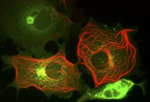
Background imageCell Collection: Cell structure, fluorescent micrograph

Cell structure, fluorescent micrograph
Cell structure. Fluorescent light micrograph of cultured cells from a cell line derived from African green monkey kidney cells. Microtubules, part of the cells cytoskeleton, are red

Background imageCell Collection: Antique battery

Antique battery

Background imageCell Collection: Small intestine structures, artwork

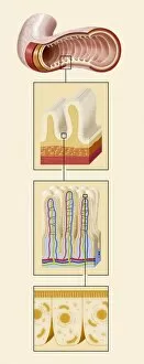
Small intestine structures, artwork
Small intestine structures. Artwork sequence of magnified views of the small intestine, with the magnification increasing from top to bottom

Background imageCell Collection: Bacteria with integrated foreign bacteria

Bacteria with integrated foreign bacteria. Computer artwork showing a section of foreign DNA (deoxyribonucleic acid, blue) integrated into a bacterial chromosome (green)

Background imageCell Collection: Plant cell wall, diagram

Plant cell wall, diagram

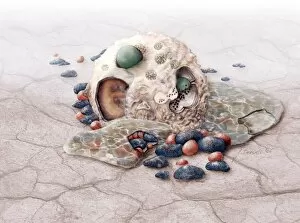
Background imageCell Collection: Parasite and cells

Parasite and cells
Concept image. An aggresive unknown Parasite has entered a cell and consumes it from within

Background imageCell Collection: HIV replication cycle, diagram

HIV replication cycle, diagram
HIV replication cycle. Diagram and magnified views showing the retroviral process by which the human immunodeficiency virus (HIV) replicates

Background imageCell Collection: Bone growth, artwork

Bone growth, artwork
Bone growth. Computer artwork showing the development and growth of a long bone. Behind the epiphysis (head) is an area of spongy bone consisting of trabeculae filled with bone marrow (green)

Background imageCell Collection: Chloroplast structures, artwork

Chloroplast structures, artwork
Chloroplast structures. Cutaway artwork showing the internal structure of a chloroplast, the organelle in plant cells responsible for photosynthesis

Background imageCell Collection: Burdock stem, light micrograph

Burdock stem, light micrograph
Burdock stem. Polarised light micrograph of a cross-section through the stem of a burdock plant (Arctium lappa). The outer layers below the epidermis are the collenchyma (red patches)

Background imageCell Collection: Bacterial DNA, conceptual artwork

Bacterial DNA, conceptual artwork
Bacterial DNA, conceptual computer artwork. Bacterial cells containing two molecules of DNA (deoxyribonucleic acid, represented by letters)

Background imageCell Collection: Aquaporins, artwork

Aquaporins, artwork
Aquaporins. Computer artwork of water molecules (red and white) passing through pores (yellow) in a cell membrane (blue). These pores are proteins known as aquaporins that are embedded in

Background imageCell Collection: Nasal mucosa, artwork

Nasal mucosa, artwork
Nasal mucosa, cross-section. Artwork of a sequence (left to right) showing mucus production in response to infection and inflammation

Background imageCell Collection: Tongue anatomy, artwork

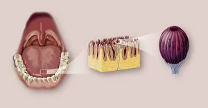
Tongue anatomy, artwork
Tongue anatomy. Artwork sequence of magnified views of the human mouth and tongue, with the magnification increasing from left to right

Background imageCell Collection: DNA replication fork, artwork

DNA replication fork, artwork
DNA replication fork. Diagram showing the cyclic sequence (right) for replication of DNA (deoxyribonucleic acid). Details of the DNA are at left

Background imageCell Collection: Cell manipulation

Cell manipulation. Cells being manipulated with laboratory instruments as part of research into cell biology. The dark areas are the cell nuclei

Background imageCell Collection: Eukaryotic flagellum structure, artwork

Eukaryotic flagellum structure, artwork
Eukaryotic flagellum structure. Cutaway artwork showing the internal structure of the flagellum in eukaryotic cells. Flagella are tail-like projections used for cell locomotion

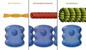
Background imageCell Collection: Cytoskeleton components, diagram

Cytoskeleton components, diagram. The cytoskeleton is the internal support structure of a cell, composed of filaments of various diameters in nanometres (nm)

Background imageCell Collection: Kidney cells, light micrograph

Kidney cells, light micrograph
Kidney cells. Quantum dot fluorescence micrograph of a section through kidney tissue showing its cells

Background imageCell Collection: Mitochondrion, artwork

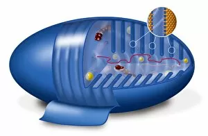
Mitochondrion, artwork
Mitochondrion. Cutaway artwork showing the internal structure of a mitochondrion. This structure, found in eukaryotic cells, is the site of energy production

Background imageCell Collection: Intestinal villi anatomy, artwork

Intestinal villi anatomy, artwork
Intestinal villi anatomy. Artwork showing six types of cell found on the surface of villi in the small intestine. Clockwise from upper right they are: enterocytes (green)

Background imageCell Collection: Viral infection

Viral infection

Background imageCell Collection: Animal cell processes, artwork

Animal cell processes, artwork
Animal cell processes. Cutaway artwork showing the structures inside an animal cell and four different processes that take place inside it or on its membrane (all marked by magnifying glasses)


For sale as Licensed Images

Choose your image, Select your licence and Download the media

"Exploring the Intricacies of Life: Unveiling the Wonders of Cells" Delving into the microscopic world, a histological diagram of a mammalian retina reveals the intricate structure and organization of cells that enable us to perceive light. The cerebellum tissue, captured in a light micrograph, showcases its complex network of cells responsible for coordinating movement and balance. Intriguingly, even philosophers find solace in contemplating cellular existence. "Philosopher in Meditation, " an oil painting from 1632, reminds us that pondering life's mysteries often leads us back to our fundamental building blocks – cells. Zooming further into this realm, nerve and glial cells come alive under the lens. This captivating light micrograph highlights their vital role in transmitting signals throughout our nervous system. Meanwhile, a lavender pollen grain captured through scanning electron microscopy (SEM) displays nature's exquisite design at the cellular level. The battle against diseases takes center stage as T lymphocytes confront cancer cells in another SEM image. Science and evolution intertwine as we witness these tiny warriors fighting for survival within our bodies. Artistic expressions also shed light on cell significance; "The Death of Socrates, " painted in 1787, serves as a poignant reminder that even great minds are ultimately composed of countless individual cells. Santiago Ramon y Cajal's meticulous drawing from 1894 unveils various cell types within the mammalian cerebellum. His work not only represents scientific progress but also exemplifies how art can aid understanding by visually capturing complexity. Stepping away from biology momentarily, we encounter Rolls Royce/Snecma Olympus 593 Mk602 engine undergoing testing within a controlled environment called a test cell. Here too, precision engineering relies on understanding cellular mechanics to achieve optimal performance. Returning to biological marvels - HeLa cells take center stage under yet another microscope lens. These immortalized human cells have revolutionized medical research, paving the way for countless breakthroughs.