Chromosomes Collection (#3)

Chromosomes are the intricate threads that hold the blueprint of life within our cells

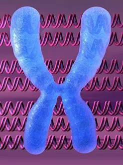
Background imageChromosomes Collection: Chromosome and DNA molecules

Chromosome and DNA molecules. Computer artwork of a condensed chromosome with molecules of DNA (deoxyribonucleic acid) behind it

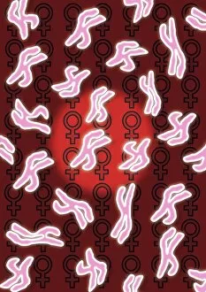
Background imageChromosomes Collection: Female sex chromosomes

Female sex chromosomes. Computer artwork of human X chromosomes (pink). In the background are female gender symbols. The X chromosome is one of two sex chromosomes

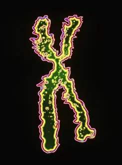
Background imageChromosomes Collection: Computer artwork of the human X chromosome

Computer artwork of the human X chromosome
X chromosome. Computer graphic based on a light micrograph of a human X chromosome, one of the two sex chromosomes. Each human cell has 46 chromosomes of which 22 pairs are identical in females

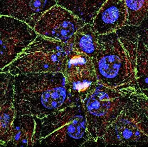
Background imageChromosomes Collection: Mitosis, fluorescence micrograph

Mitosis, fluorescence micrograph
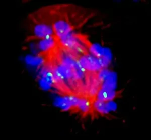
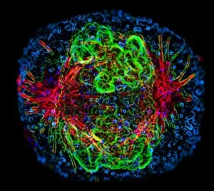
Mitosis. Fluorescence micrograph of a cell (centre) in anaphase during mitosis (nuclear division). During mitosis two daughter nuclei are formed from one parent nucleus

Background imageChromosomes Collection: Meiosis, SEM

Meiosis, SEM
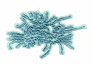
Meiosis. Coloured scanning electron micrograph (SEM) of chromosomes during metaphase (I) of meiosis (gamete formation). Chromosomes consist of deoxyribonucleic acid (DNA) and proteins

Background imageChromosomes Collection: Computer-enhanced LM of human chromosomes

Computer-enhanced LM of human chromosomes
Chromosomes. Computer-enhanced light micrograph of human chromosomes. Chromosomes are thread-like structures in the cell nucleus that carry genetic information in the form of genes

Background imageChromosomes Collection: Human chromosome 3

Human chromosome 3, computer artwork. Humans have 23 pairs of chromosomes in total. Chromosomes are made up of DNA (Deoxyribonucleic acid), which carries the bodys genetic information

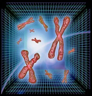
Background imageChromosomes Collection: Computer artwork of chromosomes over grids

Computer artwork of chromosomes over grids

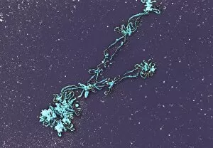
Background imageChromosomes Collection: Lampbrush chromosomes, TEM

Lampbrush chromosomes, TEM
Lampbrush chromosomes. Coloured transmission electron micrograph (TEM) of lampbrush chromosomes (LBCs). A chromosome consists of proteins and DNA (deoxyribonucleic acid)

Background imageChromosomes Collection: Abnormal mitosis

Abnormal mitosis
Mitosis. Fluorescence micrograph of a cell during abnormal anaphase of mitosis (nuclear division). During mitosis two daughter nuclei are formed from one parent nucleus

Background imageChromosomes Collection: Dividing cell

Dividing cell. Differential interference contrast (DIC) light micrograph of a cell (lower left) in the metaphase stage of mitosis (cell division). The cells nuclei are stained with fluorescent dye

Background imageChromosomes Collection: Male chromosome set

Male chromosome set
Male human chromosome set. Computer artwork of the complete set (karyotype) of healthy male human chromosomes. Humans have 46 chromosomes in most cells

Background imageChromosomes Collection: Gender determination

Gender determination, computer artwork. Gender is determined by the sex chromosomes, X and Y. Females have 2 X chromosomes and men an X and a Y chromosome

Background imageChromosomes Collection: Male sex chromosomes

Male sex chromosomes. Computer artwork of an X and Y symbolising the male sex chromosomes. An individuals sex is determined by the presence or absence of the Y chromosome

Background imageChromosomes Collection: Mitosis, TEM

Mitosis, TEM
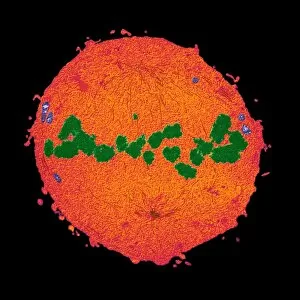
Mitosis, coloured transmission electron micrograph (TEM). Longitudinal section through a human T cell in metaphase. During mitosis two daughter nuclei are formed from one parent nucleus

Background imageChromosomes Collection: Cell mitosis

Cell mitosis. Confocal fluorescence light micrograph composite showing 6 stages of mitotic cell division. At far left, the cell has completed the first stage of cell division known as interphase in

Background imageChromosomes Collection: Mitosis

Mitosis. Fluorescence micrograph of a cell during prometaphase of mitosis (nuclear division). During mitosis two daughter nuclei are formed from one parent nucleus

Background imageChromosomes Collection: Normal female chromosomes

Normal female chromosomes
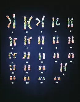
False-colour light micrograph of a normal trypsin/leishman banded female karyotype, the full complement of female chromosomes arranged in numbered homologous pairs

Background imageChromosomes Collection: Human chromosomes

Human chromosomes
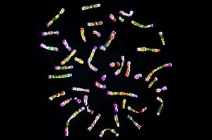
False-colour light micrograph of a normal set of human chromosomes, the strands of DNA which lie within the nucleus of each human cell and carry the genetic code

Background imageChromosomes Collection: Sperm fertilising an egg, artwork

Sperm fertilising an egg, artwork
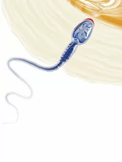
Sperm fertilising an egg. Cutaway artwork of a human sperm cell (spermatozoon) penetrating an egg cells (ovum) thick outer layer (zona pellucida). The egg cells membrane is at top right

Background imageChromosomes Collection: Genetic modification

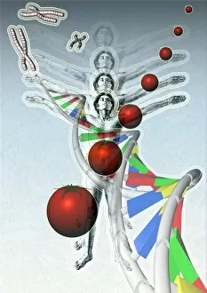
Genetic modification, conceptual artwork. Human figures and DNA (deoxyribonucleic acid, upper left to bottom right corner), which carries the genetic code. Chromosomes are seen at top left corner

Background imageChromosomes Collection: Mitosis, artwork

Mitosis, artwork


For sale as Licensed Images

Choose your image, Select your licence and Download the media

Chromosomes are the intricate threads that hold the blueprint of life within our cells. Among them, the X and Y chromosomes determine our biological sex, with males having one X and one Y chromosome. Through processes like mitosis, these chromosomes replicate themselves to ensure proper cell division. Under a light micrograph, we can witness this incredible phenomenon in action. In fluorescent micrographs capturing cell division, vibrant colors illuminate the intricate dance as they separate into two new cells. This mesmerizing display showcases the beauty hidden within our bodies. Examining a full set of male chromosomes under a scanning electron microscope (SEM) reveals their unique structure and arrangement. These microscopic images provide us with invaluable insights into our genetic makeup. Dividing cells captured in fluorescent micrographs remind us of the constant renewal happening inside us. Each split brings forth new possibilities for growth and development. Biomedical illustrations vividly depict DNA replication—a crucial process where bases attach to strands forming two identical double DNA strands. The twisting motion symbolizes how life's building blocks come together to create something extraordinary. A cross-sectional biomedical illustration unveils protein synthesis and ribosome activity—essential components for cellular function. This complex mechanism ensures that proteins are produced accurately according to instructions encoded in our chromosomes. The image of a single chromosome evokes wonder at its intricacy—an elegant structure carrying vital information that shapes who we are at a fundamental level. Astrocyte nerve cells intertwine with our understanding of chromosomes' role beyond reproduction—they play critical roles in brain function and communication between neurons. Under SEM imaging techniques, human chromosomes reveal their awe-inspiring complexity up close—each strand holding countless secrets waiting to be unraveled by scientists seeking answers about life itself.