
Conjunctiva Collection
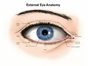
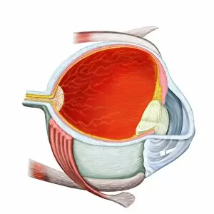
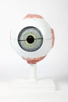
The conjunctiva, an essential part of the anatomy of the human eye, is depicted in a captivating lithograph published in 1874


For sale as Licensed Images
Choose your image, Select your licence and Download the media
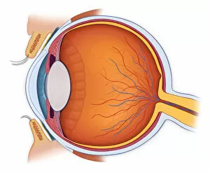
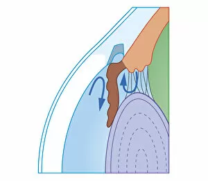
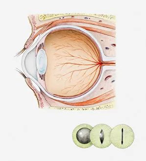
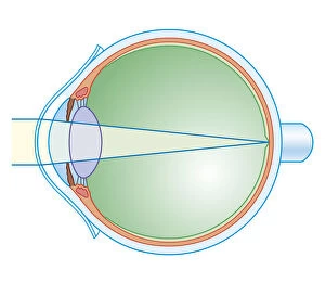
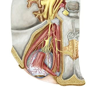
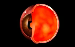
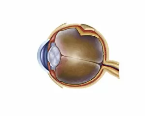
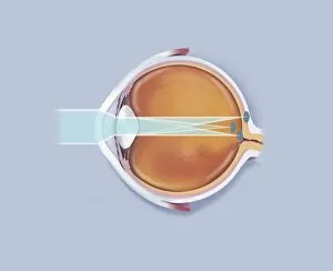
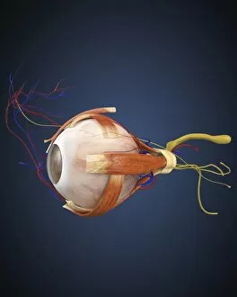
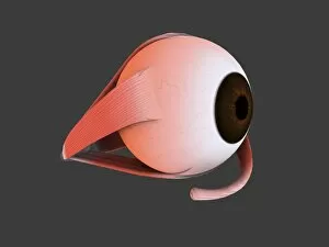
The conjunctiva, an essential part of the anatomy of the human eye, is depicted in a captivating lithograph published in 1874. This external anatomy illustration showcases the intricate details of this delicate tissue layer that covers the front surface of the eyeball and lines the inner surface of the eyelids. In another cross-section biomedical illustration, we witness a normal anatomy view of the eye. The complexity and beauty of this structure are highlighted as we explore its various components, including the cornea, iris, lens, retina, and optic nerve. Moving on to more specific conditions affecting vision health, a detailed cross-section biomedical illustration demonstrates fluid flow in chronic glaucoma with blocked trabecular meshwork. This visual representation sheds light on how this condition can lead to increased intraocular pressure and potential damage to the optic nerve if left untreated. For cat lovers out there, an intriguing cross-section illustration showcases not only feline ocular structures but also highlights their mesmerizing green irises and black pupils. It serves as a reminder that animals too possess unique anatomical features worthy of exploration. To address glaucoma-related concerns further, additional illustrations depict both surgical techniques used for treatment. A laser iridectomy technique is showcased alongside a trabeculectomy procedure aimed at relieving intraocular pressure caused by chronic glaucoma. These illustrations provide insight into cutting-edge medical interventions designed to preserve vision health. Lastly, key anatomical features are emphasized in yet another cross-sectional biomedical illustration focused solely on eye anatomy. By highlighting critical structures such as blood vessels and nerves within this complex organ system, viewers gain a deeper understanding of its functionality.