Home > Stocktrek Images > Medical
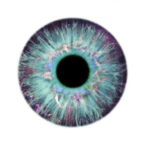
Anatomy of human eye

![]()
Wall Art and Photo Gifts from Stocktrek
Anatomy of human eye
Stocktrek Images specializes in Astronomy, Dinosaurs, Medical, Military Forces, Ocean Life, & Sci-Fi
Media ID 13009827
© Stocktrek Images
Anatomy Anterior Chamber Aqueous Humour Biology Biomedical Illustrations Bulbar Sheath Canal Of Schlemm Cartilage Choroid Ciliary Body Ciliary Processes Conjunctiva Connective Tissue Cornea Cross Section Cutaway View Cutout Detail Dissection Dura Mater Eyeball Eyes Eyesight Fascia Bulbi Fovea Centralis Healthcare Human Anatomy Human Body Parts Human Eyes Human Organs Internal Organs Iris Lens Macula Medical Medicine Nucleus Ophthalmology Optic Disc Optic Nerves Ora Serrata Physiology Pupil Retina Retinal Artery Retinal Veins Schlemms Canal Sclera Sensory System Sight Structure Superior Rectus Muscle Vision Vitreous Body Vorticose Veins
EDITORS COMMENTS
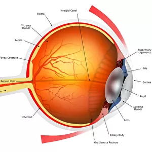
This print showcases the intricate and fascinating anatomy of the human eye. Against a pristine white background, this horizontally oriented image captures the essence of healthcare and medical science. Digitally generated with impeccable detail, it presents an artistic illustration of the eyeball, iris, and various components that contribute to our eyesight. The cross-section view reveals the complexity of the retina, cornea, and other internal organs within this sensory system. With a close-up cutout perspective, we are able to appreciate every minute structure responsible for our vision. The lens dissection unveils its role in focusing light onto the retina while highlighting connective tissues such as ciliary processes. From Schlemm's canal to optic nerves and vitreous body, each element is meticulously portrayed in this artwork. The anterior chamber provides insight into fluid dynamics through which aqueous humor circulates. This composition also highlights important features like ora serrata, conjunctiva, optic disc, dura mater along with retinal arteries and veins. Moreover, attention is drawn towards bulbar sheath surrounding vorticose veins as well as macula and fovea centralis - crucial areas for sharp visual acuity. The sclera encloses these delicate structures alongside choroid while superior rectus muscle aids in eye movement. Overall, this stunning portrayal serves as a testament to human anatomy's complexity while emphasizing its significance in ophthalmology and medicine at large. A true masterpiece capturing both scientific precision and aesthetic appeal.
MADE IN THE USA
Safe Shipping with 30 Day Money Back Guarantee
FREE PERSONALISATION*
We are proud to offer a range of customisation features including Personalised Captions, Color Filters and Picture Zoom Tools
SECURE PAYMENTS
We happily accept a wide range of payment options so you can pay for the things you need in the way that is most convenient for you
* Options may vary by product and licensing agreement. Zoomed Pictures can be adjusted in the Cart.