Home > Stocktrek Images > Medical
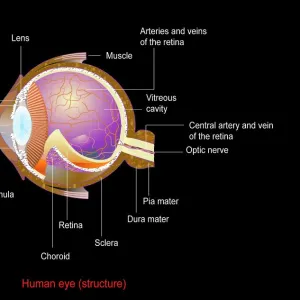
Anatomy of human eye showing focal points

![]()
Wall Art and Photo Gifts from Stocktrek
Anatomy of human eye showing focal points
Stocktrek Images specializes in Astronomy, Dinosaurs, Medical, Military Forces, Ocean Life, & Sci-Fi
Media ID 13009097
© TriFocal Communications/Stocktrek Images
Anatomy Anterior Chamber Aqueous Humour Biology Biomedical Illustrations Bulbar Sheath Canal Of Schlemm Cartilage Choroid Ciliary Body Ciliary Processes Colored Background Conjunctiva Connective Tissue Cornea Cross Section Cutaway View Cutout Detail Digitally Enhanced Dissection Dura Mater Eyeball Eyes Eyesight Fascia Bulbi Focus Fovea Centralis Healthcare Human Anatomy Human Body Parts Human Eyes Human Organs Illustration Technique Internal Organs Iris Lens Macula Medical Medicine Nucleus Ophthalmology Optic Disc Optic Nerves Ora Serrata Physiology Pupil Retina Retinal Artery Retinal Veins Schlemms Canal Sclera Sensory System Sight Structure Superior Rectus Muscle Vision Vitreous Body Vorticose Veins Watercolor Painting
EDITORS COMMENTS
This watercolor painting, digitally enhanced to perfection, showcases the intricate anatomy of the human eye with a focus on its focal points. The artist's biomedical illustrations skillfully bring this artwork to life, presenting a colorful and detailed image that is both visually stunning and educational. The vibrant colors of the illustration pop against the colored background, drawing attention to every element of the eye's structure. From the cornea to the retina, from the sclera to the macula, each component is meticulously depicted with precision and accuracy. This mesmerizing print offers a close-up view of various parts such as the lens, iris, and pupil while also providing a cutaway view for an in-depth understanding. The cross-section reveals internal organs like cartilage and nucleus alongside important structures like optic nerves and connective tissue. With its emphasis on detail and clarity, this artwork serves as an invaluable resource for medical professionals in ophthalmology or anyone interested in exploring human anatomy. Its informative nature makes it ideal for educational purposes or enhancing healthcare settings. TriFocal Communications has masterfully captured not only the physical aspects but also delves into physiology by highlighting sensory systems responsible for sight. This composition beautifully combines artistry with scientific knowledge to create an engaging visual experience that sparks curiosity about our remarkable vision system.
MADE IN THE USA
Safe Shipping with 30 Day Money Back Guarantee
FREE PERSONALISATION*
We are proud to offer a range of customisation features including Personalised Captions, Color Filters and Picture Zoom Tools
SECURE PAYMENTS
We happily accept a wide range of payment options so you can pay for the things you need in the way that is most convenient for you
* Options may vary by product and licensing agreement. Zoomed Pictures can be adjusted in the Cart.