
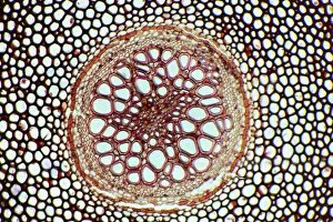
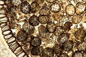
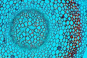
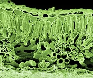
Cross Section Collection (#78)
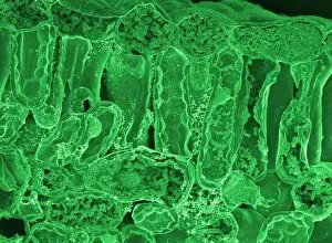
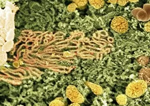
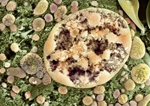
"Unveiling the Hidden World: Exploring Cross Sections in Nature and Architecture" Delving into the intricate world of honey bees


For sale as Licensed Images
Choose your image, Select your licence and Download the media
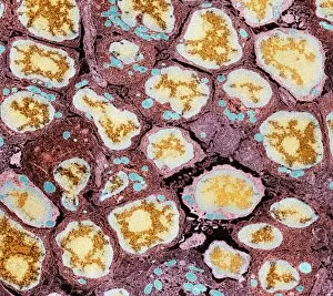
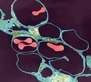
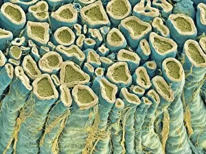
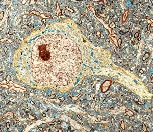
"Unveiling the Hidden World: Exploring Cross Sections in Nature and Architecture" Delving into the intricate world of honey bees, witness the mesmerizing cross section of a honeycomb revealing their remarkable life cycle. 🐝🍯 Journey deep within a Honey Bee (Apis mellifera) to discover its internal anatomy through a fascinating cross-section exploration. Marvel at the complexity of human hair follicles and skin as we unveil their hidden secrets through an enlightening cross-sectional view. Embark on a captivating expedition through the human brain, where a cross section exposes the enigmatic limbic system and primitive forebrain. As twilight descends upon Kathmandu's Boudhanath Stupa, immerse yourself in its beauty with an awe-inspiring cross-sectional perspective. Witness nature's underground marvels as we reveal a mole devouring worms beneath our feet—a captivating glimpse into their subterranean world. Step back in time to admire the architectural genius behind Florence Cathedral's dome, revealed through an intricate plan and cutaway drawing from the 16th century. Prayer flags flutter alongside Nepal's iconic Boudhanath Stupa—experience this spiritual haven from an extraordinary cross-sectional viewpoint. Encounter Australia's vibrant wildlife like never before as you explore the inner workings of a Green Tree Python through its stunning cross section. Take flight aboard Pan-American Clipper Boeing 314 with an exclusive cutaway view that unveils every detail of this magnificent aircraft from c1940. Explore Tab X’s mysterious contents by peering into its intriguing cross-section—an invitation to unravel its enigmatic nature.