Cross Section Collection (page 85)

"Unveiling the Hidden World: Exploring Cross Sections in Nature and Architecture" Delving into the intricate world of honey bees

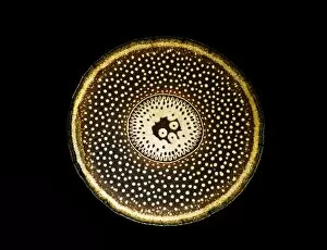
Background imageCross Section Collection: Light Micrograph (LM): A transverse section of a root of Conifer (Pandanus sp)

Light Micrograph (LM): A transverse section of a root of Conifer (Pandanus sp. ); Magnification x30 (on 10)
LRDS-210 Light Micrograph (LM): A transverse section of a root of Conifer Magnification x30 (on 10.5 cm width print) Pandanus sp

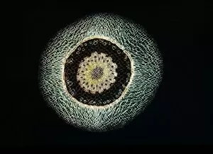
Background imageCross Section Collection: Light Micrograph (LM): A transverse section of an aerial root of Orchid (Dendrobium sp)

Light Micrograph (LM): A transverse section of an aerial root of Orchid (Dendrobium sp. ); Magnification x30 (on 10)
LRDS-209 Light Micrograph (LM): A transverse section of an aerial root of Orchid Magnification x30 (on 10.5 cm width print) Dendrobium sp

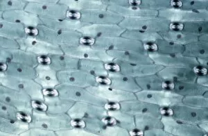
Background imageCross Section Collection: Light Micrograph (LM): A transverse section of a leaf of a Tulip (Tulipa sp)

Light Micrograph (LM): A transverse section of a leaf of a Tulip (Tulipa sp)
LRDS-202 Light Micrograph (LM): A transverse section of a leaf of a Tulip (Tulipa sp.) showing Stomata Magnification x600 (on 10.5 cm width print) Tulipa sp

Background imageCross Section Collection: Light Micrograph (LM): Longitudinal section of old sunflower stem(Helianthus sp)

Light Micrograph (LM): Longitudinal section of old sunflower stem(Helianthus sp)
LRDS-199 Light Micrograph (LM): Longitudinal section of old sunflower stem(Helianthus sp.) showing spiral chloroplasts Magnification x1200 (on 10.5 cm width print)

Background imageCross Section Collection: Light Micrograph (LM): A concentration of cells on the epidermis of a plant showing stomata

Light Micrograph (LM): A concentration of cells on the epidermis of a plant showing stomata
LRDS-197 Light Micrograph (LM): A concentration of cells on the epidermis of a plant showing stomata David Spears (Last Refuge)

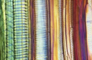
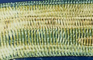
Background imageCross Section Collection: Light Micrograph (LM): Longitudinal section shows Scalariform vessels of Fern; Magnification x1200

Light Micrograph (LM): Longitudinal section shows Scalariform vessels of Fern; Magnification x1200 (on 10)
LRDS-196 Light Micrograph (LM): Longitudinal section shows Scalariform vessels of Fern Magnification x1200 (on 10.5 cm width print) David Spears (Last Refuge)

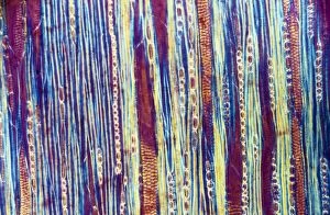
Background imageCross Section Collection: Light Micrograph (LM): A longitudinal section f a Ribes sp. stem; Magnification x600 (on 10)

Light Micrograph (LM): A longitudinal section f a Ribes sp. stem; Magnification x600 (on 10. 5 cm width print)
LRDS-194 Light Micrograph (LM): A longitudinal section of a Ribes sp. stem Magnification x600 (on 10.5 cm width print) Ribes sp

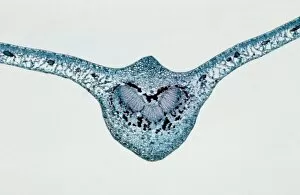
Background imageCross Section Collection: Light Micrograph (LM): transverse section of Prunus Leaf; Magnification x18 (on 10)

Light Micrograph (LM): transverse section of Prunus Leaf; Magnification x18 (on 10. 5 cm width print)
LRDS-181 Light Micrograph (LM): transverse section of Prunus Leaf Magnification x18 (on 10.5 cm width print) Prunus sp David Spears (Last Refuge)

Background imageCross Section Collection: Light Micrograph (LM): A transverse section shows Vascular Bundle in Helianthus stem;

Light Micrograph (LM): A transverse section shows Vascular Bundle in Helianthus stem; Magnification x600 (on 10)
LRDS-177 Light Micrograph (LM): A transverse section shows Vascular Bundle in Helianthus stem Magnification x600 (on 10.5 cm width print) Helianthus sp David Spears (Last Refuge)

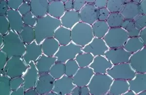
Background imageCross Section Collection: Light Micrograph (LM): transvers section shows Parenchyma Ground Tissue in Buttercup stem;

Light Micrograph (LM): transvers section shows Parenchyma Ground Tissue in Buttercup stem; Magnification x600 (on 10)
LRDS-176 Light Micrograph (LM): transvers section shows Parenchyma Ground Tissue in Buttercup stem Magnification x600 (on 10.5 cm width print) David Spears (Last Refuge)

Background imageCross Section Collection: Light Micrograph (LM): tranverse section shows Parenchyma or packing tissue from Cucurbita Stem;

Light Micrograph (LM): tranverse section shows Parenchyma or packing tissue from Cucurbita Stem; Magnification x600
LRDS-174 Light Micrograph (LM): tranverse section shows Parenchyma or packing tissue from Cucurbita Stem Magnification x600 (on 10.5 cm width print) David Spears (Last Refuge)

Background imageCross Section Collection: Seymchan Meteorite Cross-section - St. Petersburg Paleontological Lab - Russia

Seymchan Meteorite Cross-section - St. Petersburg Paleontological Lab - Russia
CAN-3456 Seymchan Meteorite Cross-section St. Petersburg Paleontological Lab - Russia John Cancalosi Please note that prints are for personal display purposes only

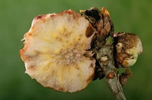
Background imageCross Section Collection: Gall Wasp - cross section

Gall Wasp - cross section
PPG-285 Gall Wasp - cross section Biorrhiza pallida Pascal Goetgheluck Please note that prints are for personal display purposes only and may not be reproduced in any way

Background imageCross Section Collection: Termite - nest cut in half

Termite - nest cut in half
PPG-1661 Termite - nest cut in half. Africa Apicotermes Pascal Goetgheluck Please note that prints are for personal display purposes only and may not be reproduced in any way

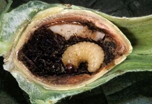
Background imageCross Section Collection: Hazelnut Weevil - larva in an hazelnut. Europe

Hazelnut Weevil - larva in an hazelnut. Europe
PPG-1614 Hazelnut Weevil - larva in an hazelnut Europe Curculio nucum Pascal Goetgheluck Please note that prints are for personal display purposes only and may not be reproduced in any way

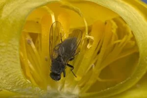
Background imageCross Section Collection: Globe flower - The Chiastocheta fly is inside the flower and will fertilize it. Europe

Globe flower - The Chiastocheta fly is inside the flower and will fertilize it. Europe
PPG-1501 Globe flower - with Chiastocheta fly inside, cross-section Europe. Trollius europaeus The fly pollinates the host plant and then lays eggs on the developing fruit

Background imageCross Section Collection: Bacterial cell

Bacterial cell, computer artwork. This is ageneralised bacterium, illustrating features ofdifferent bacterial types that would not becombined in a single cell

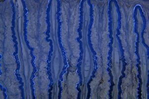
Background imageCross Section Collection: Fossil - Mammoth Tooth - cross-section. Pleistocene South Carolina, USA E50T4155

Fossil - Mammoth Tooth - cross-section. Pleistocene South Carolina, USA E50T4155
CAN-2181 Fossil - Mammoth Tooth - cross-section. Pleistocene South Carolina, USA Mammuthus meridionalis John Cancalosi Please note that prints are for personal display purposes only


For sale as Licensed Images

Choose your image, Select your licence and Download the media

"Unveiling the Hidden World: Exploring Cross Sections in Nature and Architecture" Delving into the intricate world of honey bees, witness the mesmerizing cross section of a honeycomb revealing their remarkable life cycle. 🐝🍯 Journey deep within a Honey Bee (Apis mellifera) to discover its internal anatomy through a fascinating cross-section exploration. Marvel at the complexity of human hair follicles and skin as we unveil their hidden secrets through an enlightening cross-sectional view. Embark on a captivating expedition through the human brain, where a cross section exposes the enigmatic limbic system and primitive forebrain. As twilight descends upon Kathmandu's Boudhanath Stupa, immerse yourself in its beauty with an awe-inspiring cross-sectional perspective. Witness nature's underground marvels as we reveal a mole devouring worms beneath our feet—a captivating glimpse into their subterranean world. Step back in time to admire the architectural genius behind Florence Cathedral's dome, revealed through an intricate plan and cutaway drawing from the 16th century. Prayer flags flutter alongside Nepal's iconic Boudhanath Stupa—experience this spiritual haven from an extraordinary cross-sectional viewpoint. Encounter Australia's vibrant wildlife like never before as you explore the inner workings of a Green Tree Python through its stunning cross section. Take flight aboard Pan-American Clipper Boeing 314 with an exclusive cutaway view that unveils every detail of this magnificent aircraft from c1940. Explore Tab X’s mysterious contents by peering into its intriguing cross-section—an invitation to unravel its enigmatic nature.