Cross Section Collection (#80)

"Unveiling the Hidden World: Exploring Cross Sections in Nature and Architecture" Delving into the intricate world of honey bees

Background imageCross Section Collection: Lily bud, light micrograph

Lily bud, light micrograph
Lily bud. Light micrograph of a cross-section through an immature lily bud (Lilium sp.), showing the male and female reproductive organs of the flower

Background imageCross Section Collection: Butchers broom stem, light micrograph

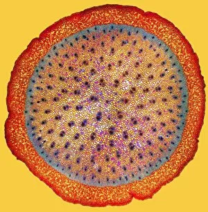
Butchers broom stem, light micrograph
Butchers broom stem. Polarised light micrograph of a section through a butchers broom stem (Ruscus aculeatis) showing many vascular bundles (dark clusters) within the plant cortex (orange)

Background imageCross Section Collection: Flax plant stem, light micrograph

Flax plant stem, light micrograph
Flax plant stem. Light micrograph of a transverse section through a stem of the flax plant (Linum usitatissimum). The layers from outer to inner (some very thin) are the epidermis (bottom)

Background imageCross Section Collection: Clover plant stem, light micrograph

Clover plant stem, light micrograph
Clover plant stem. Light micrograph of a transverse section through a stem of the clover plant (Trifolium pratense). Magnification: x100 when printed at 10 centimetres wide

Background imageCross Section Collection: Wheat stem, light micrograph

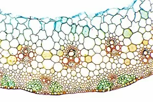
Wheat stem, light micrograph
Wheat stem. Light micrograph of a section through the stem of a wheat grass (Triticum aestivium). The circular structures (orange and green) are vascular bundles

Background imageCross Section Collection: Lily anther, polar light micrograph

Lily anther, polar light micrograph
Lily anther. Polar light micrograph of a cross-section through a lily flower anther (Lilium sp.), showing four anther sacs (round structures)

Background imageCross Section Collection: Cotton plant root, light micrograph

Cotton plant root, light micrograph
Cotton plant root. Light micrograph of a transverse section through a root of the cotton plant (Gossypium hirsutum). The layers from outer to centre (some very thin)

Background imageCross Section Collection: Peanut plant stem, light micrograph

Peanut plant stem, light micrograph
Peanut plant stem. Light micrograph of a transverse section through a stem of the peanut plant (Arachis hypogaea). Below the stems outer layer (epidermis) is a cortex of parenchyma cells (blue)

Background imageCross Section Collection: Lily anther, light micrograph

Lily anther, light micrograph
Lily anther. Light micrograph of a cross-section through a lily flower anther (Lilium sp.), showing four anther sacs (curled structures)

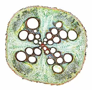
Background imageCross Section Collection: Cucurbita plant stem, light micrograph

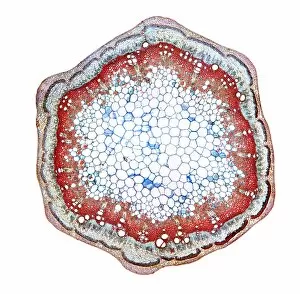
Cucurbita plant stem, light micrograph
Cucurbita plant stem. Light micrograph of a transverse section through a stem of a Cucurbita plant. Magnification: x14 when printed at 10 centimetres wide

Background imageCross Section Collection: Lily carpel, LM

Lily carpel, LM
Lily carpel. Light micrograph of a cross-section through a lily flower carpel (Lilium sp.), showing the three fused leaves that make up the pistil

Background imageCross Section Collection: Lily bud pistil and anthers, LM

Lily bud pistil and anthers, LM
Lily bud. Light micrograph of a cross-section through an immature lily bud (Lilium sp.), showing the male and female reproductive organs of the flower

Background imageCross Section Collection: Stinging nettle stem, light micrograph

Stinging nettle stem, light micrograph
Stinging nettle stem. Polarised light micrograph of a transverse section through a stem of the stinging nettle plant (Urtica dioica)

Background imageCross Section Collection: Dandelion plant stem, light micrograph

Dandelion plant stem, light micrograph
Dandelion plant stem. Light micrograph of a transverse section through a stem of a dandelion (Taraxacum officinalis). The outer layer is the epidermis and cuticle (red-blue)

Background imageCross Section Collection: Tea leaf, light micrograph

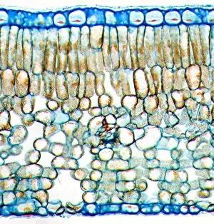
Tea leaf, light micrograph
Tea leaf. Light micrograph of a cross-section through a tea (Camellia sinensis) leaf. The upper and lower epidermis on the surfaces of the leaf are blue

Background imageCross Section Collection: Kidney bean stem, light micrograph

Kidney bean stem, light micrograph
Kidney bean stem. Light micrograph of a section through the stem of a kidney bean (Phaseolus vulgaris) plant. The outer layer is the cuticle (brown), with a cortex of parenchyma (yellow) beneath it

Background imageCross Section Collection: Plasmodesma plant cells, light micrograph

Plasmodesma plant cells, light micrograph
Plasmodesma plant cells. Light micrograph of a transverse section through intracellular bridges (plasmodema cell) in the fruit of a strychnine plant (Strychnos nux-vomica)

Background imageCross Section Collection: Broad bean root, light micrograph

Broad bean root, light micrograph
Broad bean root. Light micrograph of a transverse section through the young root of a broad bean (Vicia faba) plant. The outer epidermis is covered in root hairs

Background imageCross Section Collection: Wallflower bud, light micrograph

Wallflower bud, light micrograph
Wallflower bud. Light micrograph of a cross-section through an immature wallflower bud (Cheiranthus cheiri), showing the male and female reproductive organs of the flower

Background imageCross Section Collection: Kidney bean root, light micrograph

Kidney bean root, light micrograph
Kidney bean root. Light micrograph of a section through the root of a kidney bean (Phaseolus vulgaris) plant. At centre is the pith (blue)

Background imageCross Section Collection: Plant breathing pore, light micrograph

Plant breathing pore, light micrograph
Plant breathing pore. Light micrograph of a transverse section through a young woody stem from a lime tree (Tilia europaeus), showing a young breathing pore (lenticel) at centre left

Background imageCross Section Collection: Collenchyma plant cells, light micrograph

Collenchyma plant cells, light micrograph
Collenchyma plant cells. Light micrograph of a transverse section through collenchyma tissue in the outer stem of a common nettle (Urtica dioica)

Background imageCross Section Collection: Oleander stem, light micrograph

Oleander stem, light micrograph
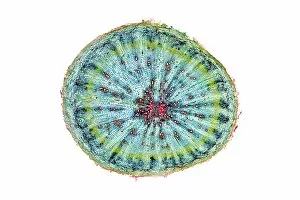
Oleander stem. Light micrograph of a cross-section through the stem of an oleander (Nerium oleander) tree. The outer layer is a thick cuticle (yellow) and then cork cambium (blue-green)
Background imageCross Section Collection: Black burdock stem, light micrograph

Black burdock stem, light micrograph
Black burdock stem. Polarised light micrograph of a transverse section through the stem of a black burdock (Arctium lappa) plant
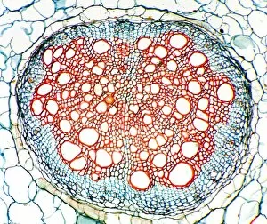
Background imageCross Section Collection: Goosefoot root, light micrograph

Goosefoot root, light micrograph
Goosefoot root. Light micrograph of a section through the root of a goosefoot (Cheopodium sp.) plant. Xylem cells, which transport water around the plant are red

Background imageCross Section Collection: Kidney bean, light micrograph

Kidney bean, light micrograph
Kidney bean. Light micrograph of a transverse section through the fruit pod of the kidney bean (Phaseolus vulgaris), showing a seed (red)

Background imageCross Section Collection: Protea plant stem, light micrograph

Protea plant stem, light micrograph
Protea plant stem. Light micrograph of a transverse section through a stem from the protea plant (Roupala polhii). The outer layer is the epidermis and cuticle (red)

Background imageCross Section Collection: Begonia stem, light mictograph

Begonia stem, light mictograph
Begoinia stem. Light micrograph of a transverse section through the stem of a begonia (Begonia sp.) plant. At centre is a large area of pith, which contains starch grains (orange) a store of energy

Background imageCross Section Collection: Marsh marigold flower bud

Marsh marigold flower bud. Light micrograph of a cross-section through the anther of an immature marsh marigold (Caltha palustris) flower bud

Background imageCross Section Collection: Dicotyledon leaf, light micrograph

Dicotyledon leaf, light micrograph
Dicotyledon leaf. Light micrograph of a transverse section through a typical dicotyledon leaf. The section passes through the midrib

Background imageCross Section Collection: Blackcurrant plant stem, light micrograph

Blackcurrant plant stem, light micrograph
Blackcurrant plant stem. Light micrograph of a cross-section through the stem of a blackcurrant (Ribes nigrum) plant. The outer cortex (blue and pale brown) is made of cork cambium tissue

Background imageCross Section Collection: Sunflower root, light micrograph

Sunflower root, light micrograph
Sunflower root. Polarised light micrograph of a transverse section through the root of a sunflower (Helianthus annuus) plant

Background imageCross Section Collection: Spikenard stem, light micrograph

Spikenard stem, light micrograph
Spikenard stem. Light micrograph of a cross-section through the stem of a Spikenard (Aralia sp.) tree. The dark red cells just beneath the surface bark are collenchyma cells

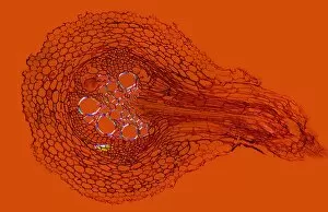
Background imageCross Section Collection: Buttercup root, light micrograph

Buttercup root, light micrograph
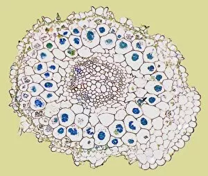
Buttercup root. Light micrograph of a transverse section through the root of a buttercup (Ranunculus sp.) plant. At centre is the vascular bundle, comprising xylem (red) and phloem (blue) tissues

Background imageCross Section Collection: Potato starch grains, light micrograph

Potato starch grains, light micrograph
Potato starch grains. Polarised light micrograph of a transverse section through a potato tuber (Solanum tuberosum), showing the starch grains

Background imageCross Section Collection: Orchid starch grains, light micrograph

Orchid starch grains, light micrograph
Orchid starch grains. Polarised light micrograph of a transverse section through an orchid tuber (Phaius grandifolius), showing the starch grains

Background imageCross Section Collection: Rose stem, light micrograph

Rose stem, light micrograph
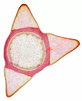
Rose stem. Light micrograph of a cross-section through the stem of a rose (Rosa sp.). The three triangular protrusions are thorns, which protect the plant from being eaten by animals

Background imageCross Section Collection: Tomato root, light micrograph

Tomato root, light micrograph
Tomato root. Light micrograph of a transverse section through the centre root of a tomato (Lycopersicum esculentum) plant. Xylem cells, which transport water around the plant, are red

Background imageCross Section Collection: Buttercup flower bud, light micrograph

Buttercup flower bud, light micrograph
Buttercup flower bud. Light micrograph of a cross-section through an immature wallflower bud (Ranunculus sp.), showing the male and female reproductive organs of the flower

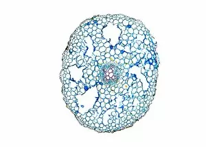
Background imageCross Section Collection: Squash root, light micrograph

Squash root, light micrograph
Squash root. Light micrograph of a transverse section through a squash (Cucubita sp.) root. The large vessels (red) are xylem cells, which transport water. Parenchyma tissue is blue

Background imageCross Section Collection: Xylem and phloem plant tissue, SEM

Xylem and phloem plant tissue, SEM
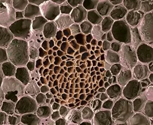
Xylem and phloem plant tissue. Coloured scanning electron micrograph (SEM) of a transverse section through the stem of a woody plant

Background imageCross Section Collection: Sunflower stem, light micrograph

Sunflower stem, light micrograph
Sunflower stem. Light micrograph of a transverse section through the stem of a sunflower (Helianthus annuus) plant, showing a vascular bundle

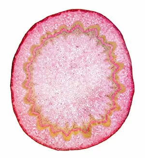
Background imageCross Section Collection: Radish root, light micrograph

Radish root, light micrograph
Radish root. Light micrograph of a transverse section through the root of a radish (Raphanus sativa) plant. The outer layer is cork cambium (red)

Background imageCross Section Collection: Root fungus, light micrograph

Root fungus, light micrograph
Root fungus. Light micrograph of a cross-section through a root containing fungal hyphae called endotrophic mycorrhiza. The hyphae (green and blue)

Background imageCross Section Collection: Human ear anatomy, artwork

Human ear anatomy, artwork
Human ear anatomy. Computer artwork of the structure of the human ear, showing the outer ear, middle ear and inner ear. The inner ear contains the cochlea (coiled, upper centre)

Background imageCross Section Collection: Dog rose stem, light micrograph

Dog rose stem, light micrograph
Dog rose stem. Polarised light micrograph of a cross-section through the stem of a dog rose (Rosa canina) plant. The outer cortex (black and red) is made of cork cambium tissue

Background imageCross Section Collection: Fools parsley fruit, light micrograph

Fools parsley fruit, light micrograph
Fools parsley fruit. Light micrograph of a transverse section through the fruit (cremocarp) of a fools parsley (Aethusa cynapium)

Background imageCross Section Collection: Alexander flowers, light micrograph

Alexander flowers, light micrograph
Alexander flowers. Light micrograph of a section through a cluster of alexander flowers (Smyrnium olusatrum). Seen here are cross-sections of the central umbels


For sale as Licensed Images

Choose your image, Select your licence and Download the media

"Unveiling the Hidden World: Exploring Cross Sections in Nature and Architecture" Delving into the intricate world of honey bees, witness the mesmerizing cross section of a honeycomb revealing their remarkable life cycle. 🐝🍯 Journey deep within a Honey Bee (Apis mellifera) to discover its internal anatomy through a fascinating cross-section exploration. Marvel at the complexity of human hair follicles and skin as we unveil their hidden secrets through an enlightening cross-sectional view. Embark on a captivating expedition through the human brain, where a cross section exposes the enigmatic limbic system and primitive forebrain. As twilight descends upon Kathmandu's Boudhanath Stupa, immerse yourself in its beauty with an awe-inspiring cross-sectional perspective. Witness nature's underground marvels as we reveal a mole devouring worms beneath our feet—a captivating glimpse into their subterranean world. Step back in time to admire the architectural genius behind Florence Cathedral's dome, revealed through an intricate plan and cutaway drawing from the 16th century. Prayer flags flutter alongside Nepal's iconic Boudhanath Stupa—experience this spiritual haven from an extraordinary cross-sectional viewpoint. Encounter Australia's vibrant wildlife like never before as you explore the inner workings of a Green Tree Python through its stunning cross section. Take flight aboard Pan-American Clipper Boeing 314 with an exclusive cutaway view that unveils every detail of this magnificent aircraft from c1940. Explore Tab X’s mysterious contents by peering into its intriguing cross-section—an invitation to unravel its enigmatic nature.