Dissection Collection (#4)

"Unveiling the Wonders Within

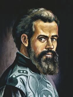
Background imageDissection Collection: Andrea Vesalio. De humani corporis fabrica

Andrea Vesalio. De humani corporis fabrica
Vesalius, Andreas (1514-1564). Brabantian anatomist, physician, and author of one of the most influential books on human anatomy, De humani corporis fabrica. Oil

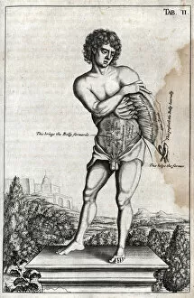
Background imageDissection Collection: Naked male on a plinth with cross section of stomach

Naked male on a plinth with cross section of stomach
Tab II. Naked male on a plinth with cross section of stomach Source: Myographia nova, or, A graphical description of all the muscles in the humane body, as they arise in dissection by Browne, John

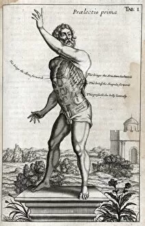
Background imageDissection Collection: Naked man on plinth with cross section of muscles

Naked man on plinth with cross section of muscles
Praelectio secunda - Tab I. Naked man on plinth with cross section of side and shoulder muscles Source: Myographia nova, or, A graphical description of all the muscles in the humane body

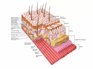
Background imageDissection Collection: Anatomy of the human skin

Anatomy of the human skin

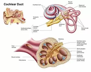
Background imageDissection Collection: Anatomy of the cochlear duct in the human ear

Anatomy of the cochlear duct in the human ear

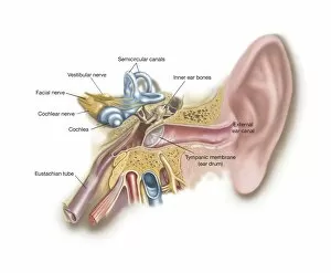
Background imageDissection Collection: Anatomy of human ear

Anatomy of human ear

Background imageDissection Collection: Interior of human heart

Interior of human heart

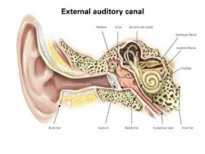
Background imageDissection Collection: External auditory canal of human ear (with labels)

External auditory canal of human ear (with labels)

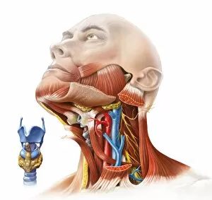
Background imageDissection Collection: Human neck anatomy, artwork C017 / 7259

Human neck anatomy, artwork C017 / 7259
Human neck anatomy, artwork. This dissection of the neck shows the larynx (voicebox) at the upper end of the trachea (windpipe, blue)

Background imageDissection Collection: Anatomical drawing of the human body

Anatomical drawing of the human body
An anatomical drawing of the human body, showing muscles, nerves and internal organs

Background imageDissection Collection: The Anatomy Lesson of Dr. Nicolaes Tulp. An engraving by Johannes Pieter de Frey

The Anatomy Lesson of Dr. Nicolaes Tulp. An engraving by Johannes Pieter de Frey, after the painting by Rembrandt

Background imageDissection Collection: Portrait Thomas Bartholinus Danish physician

Portrait Thomas Bartholinus Danish physician
Portrait of Thomas Bartholinus, Danish physician and anatomist, at the age of 35. At the bottom in the margin a three-line text in Latin

Background imageDissection Collection: Interior pharmacy Title page J Halmaal Dissection

Interior pharmacy Title page J Halmaal Dissection
Interior of a pharmacy Title page for: J. Halmaal, Dissection of Amsterdam pharmacy, 1689, pharmacy, pharmaceutics, Jan Luyken (mentioned on object), Amsterdam, paper, etching, h 142 mm × w 85 mm

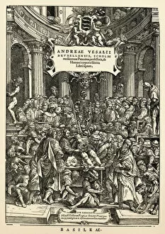
Background imageDissection Collection: Anatomy Lesson On Title Page Of De Humani Corporis Fabrica Libri Septem

Anatomy Lesson On Title Page Of De Humani Corporis Fabrica Libri Septem (On The Fabric Of The Human Body In Seven Books) By Andreas Vesalius, Published Basel, 1543

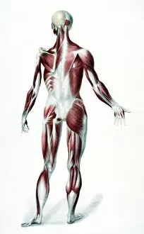
Background imageDissection Collection: Back Of The Male Human Body Showing Muscles Sinews And Bones From The Vessels Of The Human Body

Back Of The Male Human Body Showing Muscles Sinews And Bones From The Vessels Of The Human Body Edited By Jones Quain And William Wilson Published London By Taylor And Watson 1837

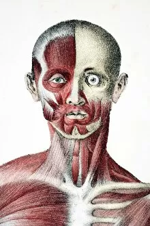
Background imageDissection Collection: Head And Shoulders Of The Male Human Body Showing Muscles Sinews And Bones From The Vessels Of The

Head And Shoulders Of The Male Human Body Showing Muscles Sinews And Bones From The Vessels Of The Human Body Edited By Jones Quain And William Wilson Published London By Taylor And Watson 1837

Background imageDissection Collection: Ecorche head of a man in profile to left, 1660. Creator: Wenceslaus Hollar

Ecorche head of a man in profile to left, 1660. Creator: Wenceslaus Hollar
Ecorche head of a man in profile to left, 1660

Background imageDissection Collection: Drawing from a study of anatomy. Human muscles of the arms

Drawing from a study of anatomy. Human muscles of the arms
History of medicine. Drawing from a study of anatomy. Human muscles of the arms. 18th century

Background imageDissection Collection: Nervous system Leonardo da Vincis drawing. 15 th century

Nervous system Leonardo da Vincis drawing. 15 th century. Leonardo studies the central nervous system in the ox brain an ingenious technique allows him to reproduce the shape of the brain ventricles

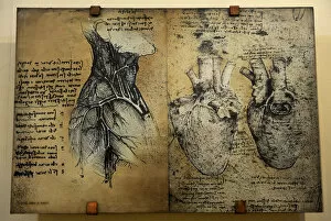
Background imageDissection Collection: Cardiovascular system Leonardo da vincis drawing

Cardiovascular system Leonardo da vincis drawing
Cardiovascular systems drawing. In 1513 Leonardo da Vinci to study the heart and the circulatory system through animal dissections. Windsor, Royal Library 19073, 1510-1513

Background imageDissection Collection: Anatomical drawing of musculature

Anatomical drawing of musculature
Septima Musculorum Tabula (Anatomical drawing of musculature) Source: Andreae Vesalii Bruxellensis, scholae medicorum Patauinae professoris

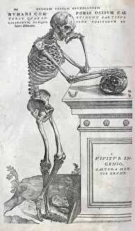
Background imageDissection Collection: Anatomical drawing of a human skeleton

Anatomical drawing of a human skeleton
Humani Corporis ossium caeteris quae sustinent partibus liberorum suaque sede positorum (Anatomical drawing of a human skeleton side view) Source: Andreae Vesalii Bruxellensis

Background imageDissection Collection: Anatomy of human heart, cross section

Anatomy of human heart, cross section with detailed internal structure

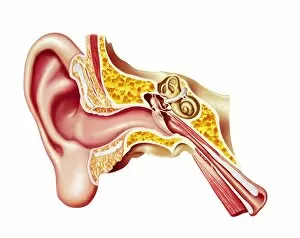
Background imageDissection Collection: Cutaway diagram of human ear

Cutaway diagram of human ear

Background imageDissection Collection: Cross section of internal anatomy of kidney

Cross section of internal anatomy of kidney

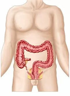
Background imageDissection Collection: Sectional view of large intestine

Sectional view of large intestine

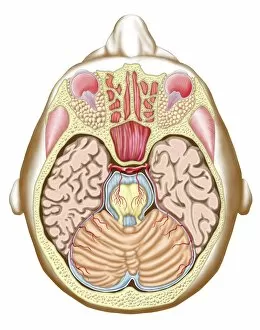
Background imageDissection Collection: Transverse section of the midbrain

Transverse section of the midbrain

Background imageDissection Collection: Face and neck nerves, 1825 artwork

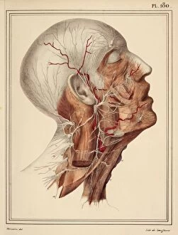
Face and neck nerves, 1825 artwork
Face and neck nerves. Head and neck dissected to show the superficial nerves (white). The parotid gland (normally present below the ear)

Background imageDissection Collection: Macropoma lewesiensis, an extinct coelacanth fish

Macropoma lewesiensis, an extinct coelacanth fish
Restoration of one of the last coelacanth fishes (Macropoma lewesiensis). It lived in the clear water chalk seas of sourthern England 85 mya, and grew to about 60 cm in length

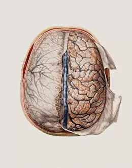
Background imageDissection Collection: Brain meninges

Brain meninges, historical anatomical artwork. This cranial (top) view of the brain (with front at the top) and its surrounding protective meninges, which have been partially dissected away

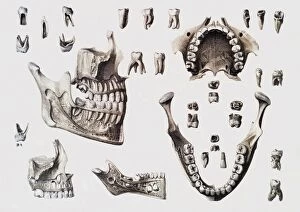
Background imageDissection Collection: Dental anatomy

Dental anatomy. Historical anatomical artwork of healthy and diseased human teeth and jaws. The teeth and jaws are seen from several different directions

Background imageDissection Collection: Blood vessels of chest and neck

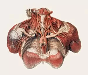
Blood vessels of chest and neck
Blood vessels of the chest and neck, historical anatomical artwork. The thoracic (chest) cavity has been dissected in this anterior (front) view to reveal part of the aorta

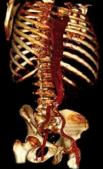
Background imageDissection Collection: Aortic dissection, 3D CT scan

Aortic dissection, 3D CT scan
Aortic dissection. 3D computed tomography (CT) scan of a 67-year-old man with an aortic dissection. The aorta, the bodys main artery, is running vertically down centre (red)

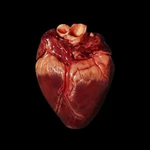
Background imageDissection Collection: Pigs heart

Pigs heart. The heart of a pig is anatomically similar to that of a human. The heart is a hollow muscular organ that pumps blood around the body

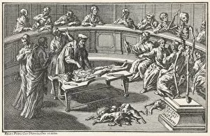
Background imageDissection Collection: Anatomical Theatre C16

Anatomical Theatre C16
Anatomy in the 16th century as depicted in Bartholinus Eustachius works. It looks as though dogs are enjoying the unwanted organs

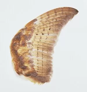
Background imageDissection Collection: Barn Owl wing

Barn Owl wing

Background imageDissection Collection: Title page of Andreas Vesalius De Humani Corporis Fabrica, showing Vesalius dissecting body, 1543

Title page of Andreas Vesalius De Humani Corporis Fabrica, showing Vesalius dissecting body, 1543
Title page of Andreas Vesalius De Humani Corporis Fabrica, showing Vesalius dissecting a body, 1543

Background imageDissection Collection: Late 16th century anatomy theatre, Jacques de Gehyn the Elder, 1633

Late 16th century anatomy theatre, Jacques de Gehyn the Elder, 1633. Artist: Jacques de Gehyn the Elder
Late 16th century anatomy theatre by Jacques de Gehyn the Elder, 1633. From Dutch translation by Vopiscus Plemp of Barthelemy Cabrol Anatomical Alphabet, Amsterdam, 1633. Engraving

Background imageDissection Collection: Eugene-Louis Doyen / Sem

Eugene-Louis Doyen / Sem
EUGENE-LOUIS DOYEN French medical Date: 1859 - 1916

Background imageDissection Collection: VALUE

VALUE
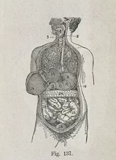
Human Anatomy. Digestive system. Engraving

Background imageDissection Collection: Anatomical drawings; dissection

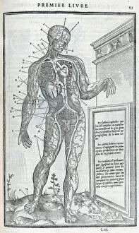
Anatomical drawings; dissection Source: La dissection des parties du corps humain divisee en trois livres / faictz par Charles Estienne

Background imageDissection Collection: Parts of the head

Parts of the head
Tab VII - Praelectio secunda. A head and other diagrams showing parts of the head Source: Myographia nova, or, A graphical description of all the muscles in the humane body

Background imageDissection Collection: Title page - frontispiece - Portrait of John Browne

Title page - frontispiece - Portrait of John Browne
Title page and frontispiece of work - Portrait of John Browne Source: Myographia nova, or, A graphical description of all the muscles in the humane body, as they arise in dissection by Browne, John

Background imageDissection Collection: View of male from side

View of male from side Source: Myographia nova, or, A graphical description of all the muscles in the humane body, as they arise in dissection by Browne, John, 1642-ca. 1700

Background imageDissection Collection: Male on plinth viewed from rear

Male on plinth viewed from rear
Praelectio Quarta - Tab XV. Male on plinth viewed from rear, with cross section of back muscles Source: Myographia nova, or, A graphical description of all the muscles in the humane body

Background imageDissection Collection: Zeiss dissecting microscope

Zeiss dissecting microscope
Antique illustration of a Zeiss dissecting microscope

Background imageDissection Collection: Diagram showing removal of affected head of a femur bone

Diagram showing removal of affected head of a femur bone

Background imageDissection Collection: MEDICAL SCHOOL, c1925. A dissection class at American University in Lebanon, Beirut

MEDICAL SCHOOL, c1925. A dissection class at American University in Lebanon, Beirut. Photograph, c1925


For sale as Licensed Images

Choose your image, Select your licence and Download the media

"Unveiling the Wonders Within: Exploring the Intricacies of Dissection" Step into a world where art and science intertwine as we delve into the captivating realm of dissection. From Leonardo da Vinci's meticulous study of skull anatomy to Rembrandt's haunting depiction of anatomical dissections, these masterpieces offer us a glimpse into the intricate workings of the human body. Witness how Leonardo da Vinci's unrivaled genius comes alive in his Skull Anatomy, revealing every contour and detail with astonishing precision. Marvel at his ability to unravel the mysteries hidden beneath our skin, showcasing not only beauty but also scientific accuracy. Venture further and explore the beating heart, an organ that symbolizes life itself. Discover its complex network of blood vessels intricately depicted by skilled artists throughout history. These illustrations serve as a testament to our relentless pursuit in understanding this vital organ. Continue your journey through time and witness groundbreaking works like De humani corporis fabrica by Andreas Vesalius, which revolutionized anatomical studies during the Renaissance period. Delve deeper into Rembrandt's Dissection series, capturing both fascination and unease surrounding this practice. Feast your eyes on detailed drawings depicting musculature from various angles; each stroke meticulously crafted to showcase every fiber and sinew. These artworks remind us that beneath our skin lies a symphony of muscles working tirelessly together. Immerse yourself in The Anatomy Lesson of Dr Nicolaes Tulp by Rembrandt Harmensz van Rijn—a masterpiece that captures both scientific inquiry and artistic brilliance. Witness how physicians gather around their subject for an educational experience unlike any other. Finally, gaze upon breathtaking artwork illustrating arteries coursing through head, chest, male groin—each stroke telling stories untold until now. These visual narratives transport us back centuries ago when exploration was paramount in unlocking medical knowledge. Join us on this extraordinary journey where art meets science—a celebration of the human body's complexity and our relentless pursuit of knowledge.