Dissection Collection (#5)

"Unveiling the Wonders Within

Background imageDissection Collection: VALUE

VALUE
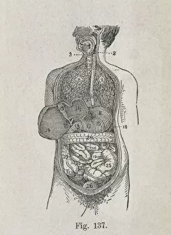
Human Anatomy. Digestive system. Engraving

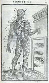
Background imageDissection Collection: Anatomical drawings; dissection

Anatomical drawings; dissection Source: La dissection des parties du corps humain divisee en trois livres / faictz par Charles Estienne

Background imageDissection Collection: Parts of the head

Parts of the head
Tab VII - Praelectio secunda. A head and other diagrams showing parts of the head Source: Myographia nova, or, A graphical description of all the muscles in the humane body

Background imageDissection Collection: Title page - frontispiece - Portrait of John Browne

Title page - frontispiece - Portrait of John Browne
Title page and frontispiece of work - Portrait of John Browne Source: Myographia nova, or, A graphical description of all the muscles in the humane body, as they arise in dissection by Browne, John

Background imageDissection Collection: View of male from side

View of male from side Source: Myographia nova, or, A graphical description of all the muscles in the humane body, as they arise in dissection by Browne, John, 1642-ca. 1700

Background imageDissection Collection: Male on plinth viewed from rear

Male on plinth viewed from rear
Praelectio Quarta - Tab XV. Male on plinth viewed from rear, with cross section of back muscles Source: Myographia nova, or, A graphical description of all the muscles in the humane body

Background imageDissection Collection: Zeiss dissecting microscope

Zeiss dissecting microscope
Antique illustration of a Zeiss dissecting microscope

Background imageDissection Collection: Diagram showing removal of affected head of a femur bone

Diagram showing removal of affected head of a femur bone

Background imageDissection Collection: MEDICAL SCHOOL, c1925. A dissection class at American University in Lebanon, Beirut

MEDICAL SCHOOL, c1925. A dissection class at American University in Lebanon, Beirut. Photograph, c1925

Background imageDissection Collection: KITCHEN, 1518. Interior of a kitchen in the 16th century. Woodcut, German, 1518

KITCHEN, 1518. Interior of a kitchen in the 16th century. Woodcut, German, 1518, from Johannes Stoefflers Calendarium Romanum

Background imageDissection Collection: ANDREAS VESALIUS (1514-1564). Belgian anatomist

ANDREAS VESALIUS (1514-1564). Belgian anatomist. Woodcut from the first edition of De Humani Corporis Fabrica, Basel, 1543

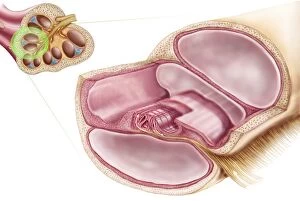
Background imageDissection Collection: Medical illustration of endolymph in the membranous labyrinth of the inner ear

Medical illustration of endolymph in the membranous labyrinth of the inner ear
Medical ilustration showing endolymph in the membranous labyrinth of the inner ear

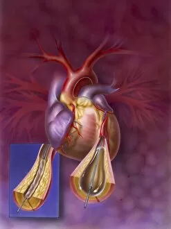
Background imageDissection Collection: Insertion of balloon into atherosclerotic artery

Insertion of balloon into atherosclerotic artery

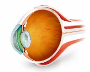
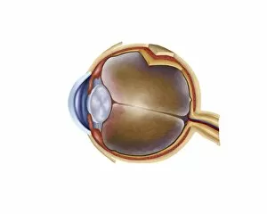
Background imageDissection Collection: Anatomy of human eye, perspective

Anatomy of human eye, perspective

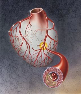
Background imageDissection Collection: Arteries on heart showing atherosclerotic plaque in an artery

Arteries on heart showing atherosclerotic plaque in an artery

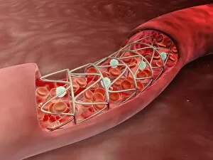
Background imageDissection Collection: Artery cross-section with blood flow and stent deployment

Artery cross-section with blood flow and stent deployment
Microscopic view of an artery cross-section with blood flow and stent deployment

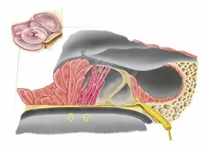
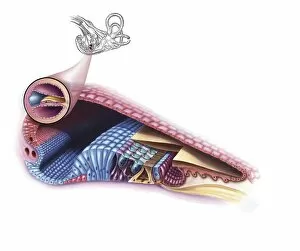
Background imageDissection Collection: Anatomy of the organ of Corti, part of the cochlea of the inner ear

Anatomy of the organ of Corti, part of the cochlea of the inner ear

Background imageDissection Collection: Interior detail of the cochlea

Interior detail of the cochlea

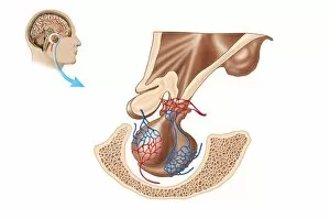
Background imageDissection Collection: Anatomy of pituitary gland

Anatomy of pituitary gland

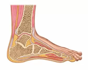
Background imageDissection Collection: Longitudinal section of human foot in a sagittal plane

Longitudinal section of human foot in a sagittal plane

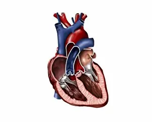
Background imageDissection Collection: Cross section of human heart

Cross section of human heart

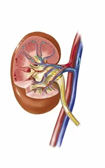
Background imageDissection Collection: Anatomy of human kidney function

Anatomy of human kidney function

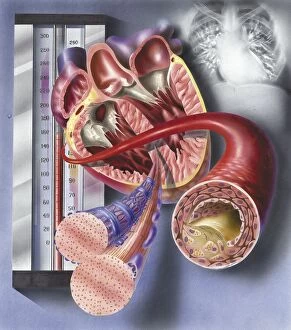
Background imageDissection Collection: Interior view of heart with detail of muscle cells and atherosclerotic artery

Interior view of heart with detail of muscle cells and atherosclerotic artery. Chest x-ray in background

Background imageDissection Collection: Cross section of human kidney on black background

Cross section of human kidney on black background

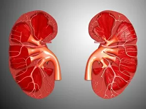
Background imageDissection Collection: Conceptual image of kidneys showing renal pelvis and ureter

Conceptual image of kidneys showing renal pelvis and ureter

Background imageDissection Collection: Heart valves showing pulmonary valve, mitral valve and tricuspid

Heart valves showing pulmonary valve, mitral valve and tricuspid

Background imageDissection Collection: Comparison of normal heart versus heart with a patent foramen ovale

Comparison of normal heart versus heart with a patent foramen ovale

Background imageDissection Collection: Cross section of brain with arteries

Cross section of brain with arteries

Background imageDissection Collection: Internal view of the human heart

Internal view of the human heart

Background imageDissection Collection: Interior of human heart showing atria and ventricles

Interior of human heart showing atria and ventricles

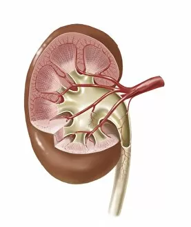
Background imageDissection Collection: Human kidney with artery

Human kidney with artery

Background imageDissection Collection: Cutaway view of the human kidney and adrenal gland

Cutaway view of the human kidney and adrenal gland

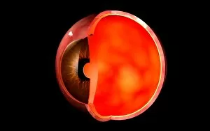
Background imageDissection Collection: Conceptual image of human eye cross section

Conceptual image of human eye cross section

Background imageDissection Collection: Microscopic view of blood flow inside an artery with stent deployment

Microscopic view of blood flow inside an artery with stent deployment

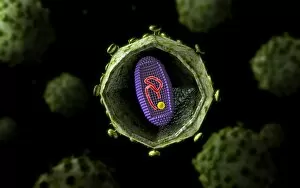
Background imageDissection Collection: Microscopic view of HIV virus, cross section

Microscopic view of HIV virus, cross section

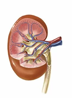
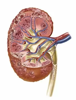
Background imageDissection Collection: Interior detail of human kidney

Interior detail of human kidney

Background imageDissection Collection: Insertion of stent into atherosclerotic artery

Insertion of stent into atherosclerotic artery

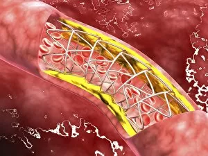
Background imageDissection Collection: Artery cross-section with blood flow, fat plaque and stent deployment

Artery cross-section with blood flow, fat plaque and stent deployment
Microscopic view of an artery cross-section with blood flow, fat plaque and stent deployment

Background imageDissection Collection: Anatomy of human eye

Anatomy of human eye

Background imageDissection Collection: Enlarged left ventricle of the human heart

Enlarged left ventricle of the human heart

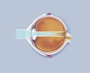
Background imageDissection Collection: Anatomy of human eye showing focal points

Anatomy of human eye showing focal points

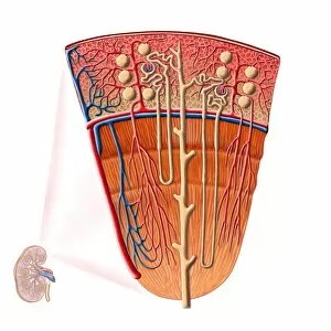
Background imageDissection Collection: Urine production in human kidney

Urine production in human kidney

Background imageDissection Collection: Human kidney

Human kidney

Background imageDissection Collection: Cross-section of human kidney

Cross-section of human kidney

Background imageDissection Collection: A laboratory scientist prepares a human eye for research

A laboratory scientist prepares a human eye for research

Background imageDissection Collection: Polycystic kidney

Polycystic kidney

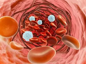
Background imageDissection Collection: Artery cross section with red blood cell flow

Artery cross section with red blood cell flow

Background imageDissection Collection: Anatomy of adrenal gland, transverse section

Anatomy of adrenal gland, transverse section


For sale as Licensed Images

Choose your image, Select your licence and Download the media

"Unveiling the Wonders Within: Exploring the Intricacies of Dissection" Step into a world where art and science intertwine as we delve into the captivating realm of dissection. From Leonardo da Vinci's meticulous study of skull anatomy to Rembrandt's haunting depiction of anatomical dissections, these masterpieces offer us a glimpse into the intricate workings of the human body. Witness how Leonardo da Vinci's unrivaled genius comes alive in his Skull Anatomy, revealing every contour and detail with astonishing precision. Marvel at his ability to unravel the mysteries hidden beneath our skin, showcasing not only beauty but also scientific accuracy. Venture further and explore the beating heart, an organ that symbolizes life itself. Discover its complex network of blood vessels intricately depicted by skilled artists throughout history. These illustrations serve as a testament to our relentless pursuit in understanding this vital organ. Continue your journey through time and witness groundbreaking works like De humani corporis fabrica by Andreas Vesalius, which revolutionized anatomical studies during the Renaissance period. Delve deeper into Rembrandt's Dissection series, capturing both fascination and unease surrounding this practice. Feast your eyes on detailed drawings depicting musculature from various angles; each stroke meticulously crafted to showcase every fiber and sinew. These artworks remind us that beneath our skin lies a symphony of muscles working tirelessly together. Immerse yourself in The Anatomy Lesson of Dr Nicolaes Tulp by Rembrandt Harmensz van Rijn—a masterpiece that captures both scientific inquiry and artistic brilliance. Witness how physicians gather around their subject for an educational experience unlike any other. Finally, gaze upon breathtaking artwork illustrating arteries coursing through head, chest, male groin—each stroke telling stories untold until now. These visual narratives transport us back centuries ago when exploration was paramount in unlocking medical knowledge. Join us on this extraordinary journey where art meets science—a celebration of the human body's complexity and our relentless pursuit of knowledge.