Dissection Collection (#6)

"Unveiling the Wonders Within

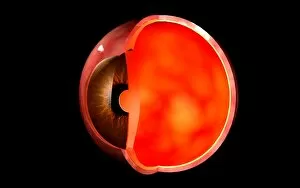
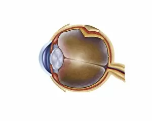
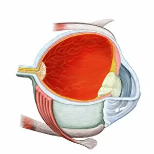
Background imageDissection Collection: Conceptual image of human eye cross section

Conceptual image of human eye cross section

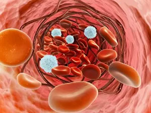
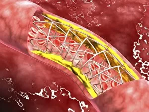
Background imageDissection Collection: Microscopic view of blood flow inside an artery with stent deployment

Microscopic view of blood flow inside an artery with stent deployment

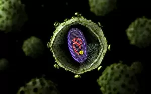
Background imageDissection Collection: Microscopic view of HIV virus, cross section

Microscopic view of HIV virus, cross section

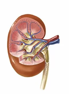
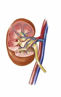
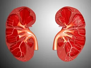
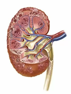
Background imageDissection Collection: Interior detail of human kidney

Interior detail of human kidney

Background imageDissection Collection: Insertion of stent into atherosclerotic artery

Insertion of stent into atherosclerotic artery

Background imageDissection Collection: Artery cross-section with blood flow, fat plaque and stent deployment

Artery cross-section with blood flow, fat plaque and stent deployment
Microscopic view of an artery cross-section with blood flow, fat plaque and stent deployment

Background imageDissection Collection: Anatomy of human eye

Anatomy of human eye

Background imageDissection Collection: Enlarged left ventricle of the human heart

Enlarged left ventricle of the human heart

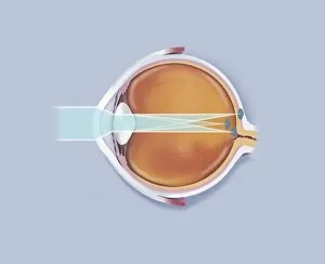
Background imageDissection Collection: Anatomy of human eye showing focal points

Anatomy of human eye showing focal points

Background imageDissection Collection: Urine production in human kidney

Urine production in human kidney

Background imageDissection Collection: Human kidney

Human kidney

Background imageDissection Collection: Cross-section of human kidney

Cross-section of human kidney

Background imageDissection Collection: A laboratory scientist prepares a human eye for research

A laboratory scientist prepares a human eye for research

Background imageDissection Collection: Polycystic kidney

Polycystic kidney

Background imageDissection Collection: Artery cross section with red blood cell flow

Artery cross section with red blood cell flow

Background imageDissection Collection: Anatomy of adrenal gland, transverse section

Anatomy of adrenal gland, transverse section

Background imageDissection Collection: Cross section of human eye

Cross section of human eye

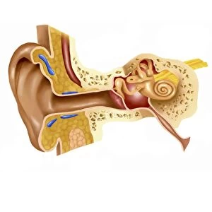
Background imageDissection Collection: Cross section of human ear

Cross section of human ear

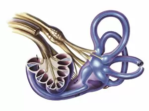
Background imageDissection Collection: Cochlea detail with vestibulocochlear nerve

Cochlea detail with vestibulocochlear nerve

Background imageDissection Collection: Human kidney and adrenal gland

Human kidney and adrenal gland

Background imageDissection Collection: External auditory canal of human ear

External auditory canal of human ear

Background imageDissection Collection: The Manner of Dissecting the Pestilential Body (engraving) (b / w photo)

The Manner of Dissecting the Pestilential Body (engraving) (b / w photo)
XJF135242 The Manner of Dissecting the Pestilential Body (engraving) (b/w photo) by English School, (17th century); Private Collection; English, out of copyright

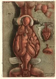
Background imageDissection Collection: Jean-Baptiste Andre Gautier-Dagoty (French, 1740 - 1786). Dissection of a Lizard

Jean-Baptiste Andre Gautier-Dagoty (French, 1740 - 1786). Dissection of a Lizard, Plate 15, 1756. Mezzotint printed in colors on laid paper. Plate: 212 mm x 146 mm (8.35 in. x 5.75 in.)

Background imageDissection Collection: dissection of the third division, medical equipment, surgical instrument, history

dissection of the third division, medical equipment, surgical instrument, history of medicine

Background imageDissection Collection: ANDREAS VESALIUS (1514-1564). Belgian anatomist

ANDREAS VESALIUS (1514-1564). Belgian anatomist. Woodcut from the first edition of Andreas Vesalius De Humani Corporis Fabrica, published in 1543 at Basel, Switzerland

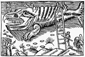
Background imageDissection Collection: WHALE DISSECTION, 1555. The cutting up of a whale

WHALE DISSECTION, 1555. The cutting up of a whale. Woodcut from Olaus Magnus Historia de Gentibus Septentrionalibus, Rome, Italy, 1555

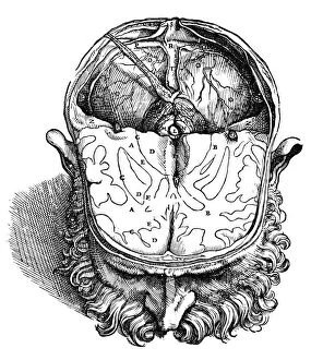
Background imageDissection Collection: Dissection of the brain (fig. 8). Woodcut from the seventh book of Andreas Vesalius De Humani

Dissection of the brain (fig. 8). Woodcut from the seventh book of Andreas Vesalius De Humani Corporis Fabrica
VESALIUS: BRAIN, 1543. Dissection of the brain (fig. 8). Woodcut from the seventh book of Andreas Vesalius De Humani Corporis Fabrica, published in 1543 at Basel, Switzerland

Background imageDissection Collection: Dissection of the brain (fig. 5). Woodcut from the seventh book of Andreas Vesalius De Humani

Dissection of the brain (fig. 5). Woodcut from the seventh book of Andreas Vesalius De Humani Corporis Fabrica
VESALIUS: BRAIN, 1543. Dissection of the brain (fig.5). Woodcut from the seventh book of Andreas Vesalius De Humani Corporis Fabrica, published in 1543 at Basel, Switzerland

Background imageDissection Collection: Dissection of the brain (fig. 6). Woodcut from the seventh book of Andreas Vesalius De Humani

Dissection of the brain (fig. 6). Woodcut from the seventh book of Andreas Vesalius De Humani Corporis Fabrica
VESALIUS: BRAIN, 1543. Dissection of the brain (fig.6). Woodcut from the seventh book of Andreas Vesalius De Humani Corporis Fabrica, published in 1543 at Basel, Switzerland

Background imageDissection Collection: Dissection of the brain (fig. 9). Woodcut from the seventh book of Andreas Vesalius De Humani

Dissection of the brain (fig. 9). Woodcut from the seventh book of Andreas Vesalius De Humani Corporis Fabrica, Basel
VESALIUS: BRAIN, 1543. Dissection of the brain (fig.9). Woodcut from the seventh book of Andreas Vesalius De Humani Corporis Fabrica, Basel, Switzerland, 1543

Background imageDissection Collection: Woodcut bloodletting chart by Johannes Wechtlin, 1517

Woodcut bloodletting chart by Johannes Wechtlin, 1517
BLOODLETTING CHART, 1517. Woodcut bloodletting chart by Johannes Wechtlin, 1517

Background imageDissection Collection: Dissection of the brain (fig. 4). Woodcut from the seventh book of Andreas Vesalius De Humani

Dissection of the brain (fig. 4). Woodcut from the seventh book of Andreas Vesalius De Humani Corporis Fabrica
VESALIUS: BRAIN, 1543. Dissection of the brain (fig.4). Woodcut from the seventh book of Andreas Vesalius De Humani Corporis Fabrica, published in 1543 at Basel, Switzerland

Background imageDissection Collection: Woodcut from the sixth book of Andreas Vesalius De Humani Corporis Fabrica

Woodcut from the sixth book of Andreas Vesalius De Humani Corporis Fabrica, published in 1543 at Basel, Switzerland
VESALIUS: THORACIC CAVITY. Woodcut from the sixth book of Andreas Vesalius De Humani Corporis Fabrica, published in 1543 at Basel, Switzerland

Background imageDissection Collection: Dissection of the brain (fig. 7). Woodcut from the seventh book of Andreas Vesalius De Humani

Dissection of the brain (fig. 7). Woodcut from the seventh book of Andreas Vesalius De Humani Corporis Fabrica
VESALIUS: BRAIN, 1543. Dissection of the brain (fig.7). Woodcut from the seventh book of Andreas Vesalius De Humani Corporis Fabrica published in 1543 at Basel, Switzerland

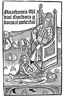
Background imageDissection Collection: Title page of Mondino de Luzzi (Mundinus) Anathomia, published by Martin Pollick

Title page of Mondino de Luzzi (Mundinus) Anathomia, published by Martin Pollick van Mellerstadt, Leipzig, 1493, showing anatomy demonstration

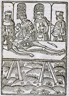
Background imageDissection Collection: Dissection in progress

Dissection in progress
A dissection in progress. From " Le livre de la propriete des choses" (On the Properites of Things) written by the English Franciscan monk Bartholomew Glanville called Bartolomaeus Anglicus

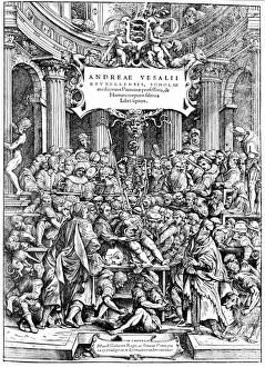
Background imageDissection Collection: Title page of Andreas Vesalius De Humani Corporis Fabrica

Title page of Andreas Vesalius De Humani Corporis Fabrica, Basel, 1543, showing Vesalius dissecting a body

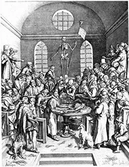
Background imageDissection Collection: Late 16th century anatomy theatre by Jacques de Gehyn the Elder

Late 16th century anatomy theatre by Jacques de Gehyn the Elder. From Dutch translation by Vopiscus Plemp of Barthelemy Cabrol " Anatomical Alphabet", Amsterdam, 1633. Engraving

Background imageDissection Collection: The second plate of the muscles

The second plate of the muscles. From Book II of Andreas Vesalius " De humani corporis fabrica", Basel, 1543. Engraving

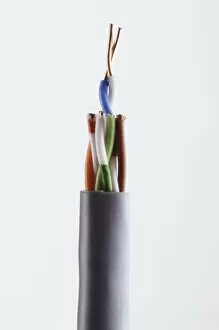
Background imageDissection Collection: Twisted-pair cabling, side view

Twisted-pair cabling, side view

Background imageDissection Collection: Dissected lizard with intestines revealed, view from below

Dissected lizard with intestines revealed, view from below

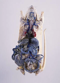
Background imageDissection Collection: Dissected lab rat with intestines revealed, view from below

Dissected lab rat with intestines revealed, view from below

Background imageDissection Collection: Model of Spinner Shark (Carcharhinus brevipinna), side view

Model of Spinner Shark (Carcharhinus brevipinna), side view

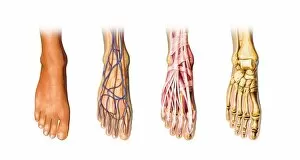
Background imageDissection Collection: Human foot anatomy, artwork

Human foot anatomy, artwork
Human foot anatomy, computer artwork

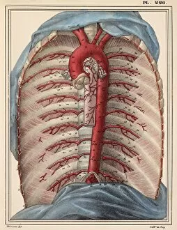
Background imageDissection Collection: Thoracic aorta, 1825 artwork

Thoracic aorta, 1825 artwork
Thoracic aorta. Dissection of the chest cavity with the heart and lungs and other organs removed, showing the thoracic portion of the aorta (red), the main body artery

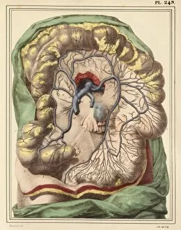
Background imageDissection Collection: Intestinal veins, 1825 artwork

Intestinal veins, 1825 artwork
Intestinal veins. Dissection of the abdominal cavity showing the inferior mesenteric vein (blue) that drains blood from the large intestine

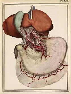
Background imageDissection Collection: Liver and stomach nerves, 1825 artwork

Liver and stomach nerves, 1825 artwork
Liver and stomach nerves. Extensive network of nerves (white) supplying the liver (upper left) and stomach (lower right). These nerves networks (plexuses)

Background imageDissection Collection: Intestinal arteries, 1825 artwork

Intestinal arteries, 1825 artwork
Intestinal arteries. Dissection of the abdomen, showing the inferior mesenteric artery (red) and its branches that supply oxygenated blood to the large intestine


For sale as Licensed Images

Choose your image, Select your licence and Download the media

"Unveiling the Wonders Within: Exploring the Intricacies of Dissection" Step into a world where art and science intertwine as we delve into the captivating realm of dissection. From Leonardo da Vinci's meticulous study of skull anatomy to Rembrandt's haunting depiction of anatomical dissections, these masterpieces offer us a glimpse into the intricate workings of the human body. Witness how Leonardo da Vinci's unrivaled genius comes alive in his Skull Anatomy, revealing every contour and detail with astonishing precision. Marvel at his ability to unravel the mysteries hidden beneath our skin, showcasing not only beauty but also scientific accuracy. Venture further and explore the beating heart, an organ that symbolizes life itself. Discover its complex network of blood vessels intricately depicted by skilled artists throughout history. These illustrations serve as a testament to our relentless pursuit in understanding this vital organ. Continue your journey through time and witness groundbreaking works like De humani corporis fabrica by Andreas Vesalius, which revolutionized anatomical studies during the Renaissance period. Delve deeper into Rembrandt's Dissection series, capturing both fascination and unease surrounding this practice. Feast your eyes on detailed drawings depicting musculature from various angles; each stroke meticulously crafted to showcase every fiber and sinew. These artworks remind us that beneath our skin lies a symphony of muscles working tirelessly together. Immerse yourself in The Anatomy Lesson of Dr Nicolaes Tulp by Rembrandt Harmensz van Rijn—a masterpiece that captures both scientific inquiry and artistic brilliance. Witness how physicians gather around their subject for an educational experience unlike any other. Finally, gaze upon breathtaking artwork illustrating arteries coursing through head, chest, male groin—each stroke telling stories untold until now. These visual narratives transport us back centuries ago when exploration was paramount in unlocking medical knowledge. Join us on this extraordinary journey where art meets science—a celebration of the human body's complexity and our relentless pursuit of knowledge.