Fibres Collection (#4)

"Fibres: Unraveling the Intricacies of the Human Brain and Beyond" Exploring the intricate network of brain fibres through DTI MRI scans (C017 / 7099

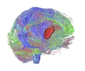
Background imageFibres Collection: Brain tumour, DTI MRI scan C017 / 7057

Brain tumour, DTI MRI scan C017 / 7057
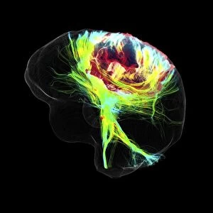
Brain tumour. 3D diffusion tensor imaging (DTI) magnetic resonance imaging (MRI) scan of nerve pathways (coloured) in a brain with a tumour (red, centre right)
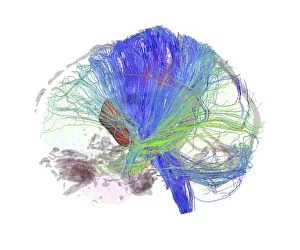
Background imageFibres Collection: Brain fibres, DTI MRI scan C017 / 7100

Brain fibres, DTI MRI scan C017 / 7100
Brain fibres. 3D diffusion tensor imaging (DTI) magnetic resonance imaging (MRI) scan of a selection of nerve pathways (blue, pink, green) in the brain. The front of the brain is at left

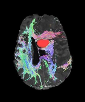
Background imageFibres Collection: Brain tumour, DTI MRI scan C017 / 7059

Brain tumour, DTI MRI scan C017 / 7059
Brain tumour. Axial diffusion tensor imaging (DTI) magnetic resonance imaging (MRI) scan of nerve pathways (coloured) in a brain with a tumour (red, upper centre)

Background imageFibres Collection: Brain tumour, DTI MRI scan C017 / 7058

Brain tumour, DTI MRI scan C017 / 7058
Brain tumour. 3D diffusion tensor imaging (DTI) magnetic resonance imaging (MRI) scan of nerve pathways (coloured) in a brain with a tumour (red, centre left)

Background imageFibres Collection: Sugar uptake in muscles, diagram

Sugar uptake in muscles, diagram
Sugar uptake in muscles. Diagram showing sugar from a drink (upper right) being taken up by the muscles of the human body

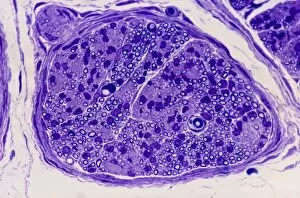
Background imageFibres Collection: Peripheral nerve, light micrograph

Peripheral nerve, light micrograph
Peripheral nerve. Light micrograph of a section through a peripheral nerve. This is a mixed nerve with myelinated axons (dark blue circles)

Background imageFibres Collection: Nerve ganglion, light micrograph

Nerve ganglion, light micrograph
Nerve ganglion. Light micrograph of a section through a dorsal (sensory) spinal root ganglion associated with a sensory nerve root of the spinal cord. Sensory information from peripheral sites e.g

Background imageFibres Collection: Myelinated nerve, light micrograph

Myelinated nerve, light micrograph
Myelinated nerve. Light micrograph of a section through a peripheral myelinated nerve, showing many individual axons each covered with deep blue-staining myelin sheaths

Background imageFibres Collection: Structure of the cochlea, artwork

Structure of the cochlea, artwork
Structure of the cochlea. Computer artwork of a section through the organ of Corti, the auditory sense organ that lines the spiral of the cochlea in the inner ear

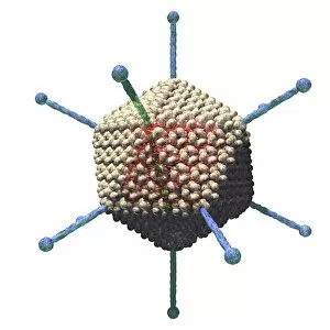
Background imageFibres Collection: Adenovirus, artwork C016 / 8960

Adenovirus, artwork C016 / 8960
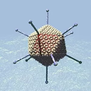
Adenovirus. Computer artwork showing the external protein structure of an adenovirus particle. Adenoviruses are the largest viruses not to have a protein coat covering their capsid

Background imageFibres Collection: Adenovirus, artwork C016 / 8961

Adenovirus, artwork C016 / 8961
Adenovirus. Computer artwork showing the external protein structure of an adenovirus particle. Adenoviruses are the largest viruses not to have a protein coat covering their capsid

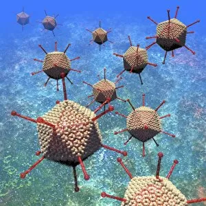
Background imageFibres Collection: Adenovirus particles, artwork C016 / 8963

Adenovirus particles, artwork C016 / 8963
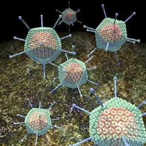
Adenovirus particles. Computer artwork showing the external protein structure of adenovirus particles. Adenoviruses are the largest viruses not to have a protein coat covering their capsid

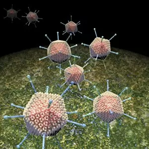
Background imageFibres Collection: Adenovirus particles, artwork C016 / 8965

Adenovirus particles, artwork C016 / 8965
Adenovirus particles. Computer artwork showing the external protein structure of adenovirus particles. Adenoviruses are the largest viruses not to have a protein coat covering their capsid

Background imageFibres Collection: White matter fibres of the human brain C016 / 9017

White matter fibres of the human brain C016 / 9017
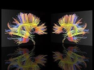
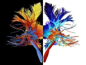
White matter fibres. Coloured 3D diffusion spectral imaging (DSI) scan of the bundles of white matter nerve fibres in the brain, depicting mirror neurons. Mirror neurons are found in the brain

Background imageFibres Collection: Adenovirus, artwork C016 / 8962

Adenovirus, artwork C016 / 8962
Adenovirus. Computer artwork showing the external protein structure of an adenovirus particle. Adenoviruses are the largest viruses not to have a protein coat covering their capsid

Background imageFibres Collection: Adenovirus particles, artwork C016 / 8964

Adenovirus particles, artwork C016 / 8964
Adenovirus particles. Computer artwork showing the external protein structure of adenovirus particles. Adenoviruses are the largest viruses not to have a protein coat covering their capsid

Background imageFibres Collection: White matter fibres of the human brain C016 / 9012

White matter fibres of the human brain C016 / 9012
White matter fibres. Coloured 3D diffusion spectral imaging (DSI) scan of the bundles of white matter nerve fibres in the brain, depicting mirror neurons. Mirror neurons are found in the brain

Background imageFibres Collection: Myenteric nerve plexus, TEM

Myenteric nerve plexus, TEM
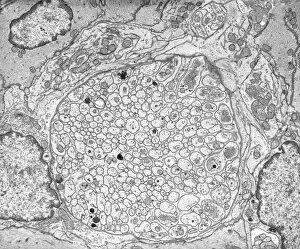
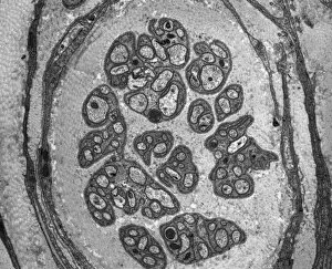
Myenteric nerve plexus. Transmission electron micrograph (TEM) of a section through a bundle of unmyelinated nerve fibres in the myenteric nerve plexus of the intestinal tract

Background imageFibres Collection: Cushings syndrome, light micrograph

Cushings syndrome, light micrograph
Cushings syndrome. Light micrograph of a section through muscle affected by Cushings syndrome, a hormonal condition caused by overproduction of adrenal gland hormones

Background imageFibres Collection: Smooth muscle of gut wall, TEM

Smooth muscle of gut wall, TEM
Smooth muscle of gut wall. Transmission electron micrograph (TEM) of a section through the outermost wall of the intestine

Background imageFibres Collection: Unmyelinated nerve, TEM

Unmyelinated nerve, TEM
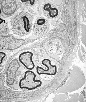
Unmyelinated nerve. Transmission electron micrograph (TEM) of a section through axon (nerve fibre) bundles of unmyelinated nerves

Background imageFibres Collection: Fat tissue, stereoscopic SEM

Fat tissue, stereoscopic SEM
Fat tissue. Coloured stereoscopic (3D) scanning electron micrograph (SEM) of a sample of fat tissue, showing fat cells (adipocytes, round) surrounded by fine strands of supportive connective tissue

Background imageFibres Collection: Silkworm Cocoon Silk Fibres (SEM)

Silkworm Cocoon Silk Fibres (SEM)
Silkworm cocoon silk fibres. Scanning electron micrograph (SEM) of silk fibres. The silkworm is the larva or caterpillar of the silkmoth which has been domesticated from the wild silkmoth Bombyx

Background imageFibres Collection: Myelinated nerve tissue, TEM

Myelinated nerve tissue, TEM
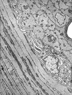
Myelinated nerve tissue. Transmission electron micrograph (TEM) of a section through myelinated nerve fibres (axons). Each axon is coated with many layers of myelin

Background imageFibres Collection: Smooth muscle tissue, TEM

Smooth muscle tissue, TEM
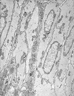
Smooth muscle tissue. Transmission electron micrograph (TEM) of a longitudinal section through smooth muscle cells. Smooth muscle cells are spindle-shaped and often closely packed together

Background imageFibres Collection: Bladder adhesions

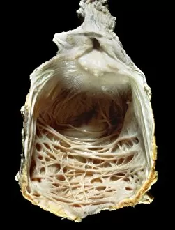
Bladder adhesions. Gross specimen of a urinary bladder with adhesions (string-like) across its inner surface. Adhesions are growths of fibrous (scar)

Background imageFibres Collection: Brain activity white matter fibres C016 / 6296

Brain activity white matter fibres C016 / 6296
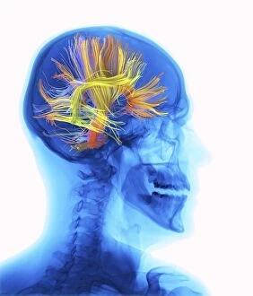
White matter fibres overlaid a human head x-ray. Coloured 3D diffusion spectral imaging (DSI) scan of the bundles of white matter nerve fibres in the brain

Background imageFibres Collection: Brain activity white matter fibres C016 / 6295

Brain activity white matter fibres C016 / 6295
White matter fibres overlaid a human head x-ray. Coloured 3D diffusion spectral imaging (DSI) scan of the bundles of white matter nerve fibres in the brain

Background imageFibres Collection: Connective tissue, SEM C015 / 9929

Connective tissue, SEM C015 / 9929
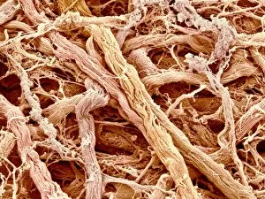
Connective tissue. Coloured scanning electron micrograph (SEM) of connective tissue from the trachea (wind pipe). Connective tissue supports other tissues and organs

Background imageFibres Collection: Connective tissue, SEM C015 / 9933

Connective tissue, SEM C015 / 9933
Connective tissue. Coloured scanning electron micrograph (SEM) of connective tissue from the trachea (wind pipe). Connective tissue supports other tissues and organs

Background imageFibres Collection: Connective tissue, SEM C015 / 9926

Connective tissue, SEM C015 / 9926
Connective tissue. Coloured scanning electron micrograph (SEM) of connective tissue from the trachea (wind pipe). Connective tissue supports other tissues and organs

Background imageFibres Collection: Connective tissue, SEM C015 / 9928

Connective tissue, SEM C015 / 9928
Connective tissue. Coloured scanning electron micrograph (SEM) of connective tissue from the trachea (wind pipe). Connective tissue supports other tissues and organs

Background imageFibres Collection: Lens of the eye, light micrograph C016 / 0526

Lens of the eye, light micrograph C016 / 0526
Lens of the eye. Light micrograph of a section through the lens from a human eye. The lens consists of thousands of precisely aligned stacks of fibres seen here end-on

Background imageFibres Collection: Connective tissue, SEM C015 / 9932

Connective tissue, SEM C015 / 9932
Connective tissue. Coloured scanning electron micrograph (SEM) of connective tissue from the trachea (wind pipe). Connective tissue supports other tissues and organs

Background imageFibres Collection: Cirrhosis of liver, light micrograph C016 / 0530

Cirrhosis of liver, light micrograph C016 / 0530
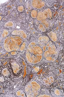
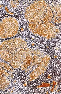
Cirrhosis of liver. Light micrograph of a section through liver tissue, damaged by cirrhosis. Cirrhosis occurs as a result of a range of factors causing damage to liver function

Background imageFibres Collection: Connective tissue, SEM C015 / 9930

Connective tissue, SEM C015 / 9930
Connective tissue. Coloured scanning electron micrograph (SEM) of connective tissue from the trachea (wind pipe). Connective tissue supports other tissues and organs

Background imageFibres Collection: Connective tissue, SEM C015 / 9931

Connective tissue, SEM C015 / 9931
Connective tissue. Coloured scanning electron micrograph (SEM) of connective tissue from the trachea (wind pipe). Connective tissue supports other tissues and organs

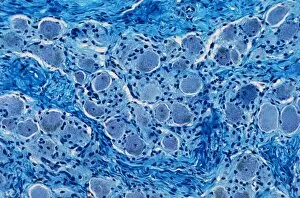
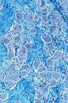
Background imageFibres Collection: Nerve ganglion, light micrograph C016 / 0532

Nerve ganglion, light micrograph C016 / 0532
Nerve ganglion. Light micrograph of a section through a nerve ganglion of the peripheral nervous system, showing clusters of nerve cell bodies

Background imageFibres Collection: Connective tissue, SEM C015 / 9934

Connective tissue, SEM C015 / 9934
Connective tissue. Coloured scanning electron micrograph (SEM) of connective tissue from the trachea (wind pipe). Connective tissue supports other tissues and organs

Background imageFibres Collection: Myelinated nerve fibres, light micrograph C016 / 0531

Myelinated nerve fibres, light micrograph C016 / 0531
Myelinated nerve fibres. Light micrograph of a section through a spinal sensory nerve containing many closely packed axons exhibiting a wave-type appearance

Background imageFibres Collection: Trachea muscle, SEM C015 / 9935

Trachea muscle, SEM C015 / 9935
Trachea muscle. Coloured scanning electron micrograph (SEM) of smooth muscle from the trachea. The trachea (wind pipe) runs from the larynx (voice box) to the lungs

Background imageFibres Collection: Cirrhosis of liver, light micrograph C016 / 0529

Cirrhosis of liver, light micrograph C016 / 0529
Cirrhosis of liver. Light micrograph of a section through liver tissue, damaged by cirrhosis. Cirrhosis occurs as a result of a range of factors causing damage to liver function

Background imageFibres Collection: White matter fibres of the human brain C018 / 6383

White matter fibres of the human brain C018 / 6383
White matter fibres. Computer enhanced 3D diffusion spectral imaging (DSI) scan of the bundles of white matter nerve fibres in the brain

Background imageFibres Collection: White matter fibres, brain mri scan C018 / 6386

White matter fibres, brain mri scan C018 / 6386
White matter fibres overlaid a mri scan of the human head. Computer enhanced 3D diffusion spectral imaging (DSI) scan of the bundles of white matter nerve fibres in the brain

Background imageFibres Collection: White matter fibres of the human brain C018 / 6391

White matter fibres of the human brain C018 / 6391
White matter fibres. Computer enhanced 3D diffusion spectral imaging (DSI) scan of the bundles of white matter nerve fibres in the brain

Background imageFibres Collection: Corpus callosum, DTI MRI scan C017 / 7045

Corpus callosum, DTI MRI scan C017 / 7045
Corpus callosum. Coronal 3D diffusion tensor imaging (DTI) magnetic resonance imaging (MRI) scan of nerve pathways (blue) in and around the brains corpus callosum

Background imageFibres Collection: Glioblastoma brain tumour, DTI MRI scan C017 / 7052

Glioblastoma brain tumour, DTI MRI scan C017 / 7052
Glioblastoma brain tumour. 3D diffusion tensor imaging (DTI) magnetic resonance imaging (MRI) scan of nerve pathways (coloured) around a glioblastoma (upper right) in the brain

Background imageFibres Collection: Sperm tails, TEM C014 / 1463

Sperm tails, TEM C014 / 1463
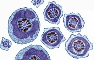
Sperm tails. Transmission electron micrograph (TEM) of sections through the tails of sperm (spermatozoa) cells at various levels


For sale as Licensed Images

Choose your image, Select your licence and Download the media

"Fibres: Unraveling the Intricacies of the Human Brain and Beyond" Exploring the intricate network of brain fibres through DTI MRI scans (C017 / 7099). The mesmerizing artwork depicting white matter fibres and their connection to the brain (C015 / 1930). Delving deeper into understanding brain fibres with DTI MRI scans (C017 / 7035). Illuminating neurons and astrocytes through immunofluorescent LM, revealing the complexity of brain fibres. Peering into the white matter fibres of the human brain, unlocking its mysteries (C014 / 5666). Witnessing nerve cells regenerate under a TEM microscope, showcasing nature's resilience. Marveling at the intricate structure of a lime tree stem captured in a light micrograph. Discovering beauty in unexpected places - castor oil stem captured in a mesmerizing light micrograph. Unveiling myelination's role in enhancing nerve fibre function through TEM imaging. Witnessing cell division unfold under fluorescent micrographs, highlighting life's continuous renewal process. Celebrating nature's ability to heal as we observe regenerating nerve cells under TEM microscopy once again. Exploring further into white matter fibres within our complex human brains (C014/5668). In this captivating journey, we unravel the wonders – from delicate neural connections within our brains to resilient plant stems that provide sustenance for life itself.