Fibres Collection (#5)

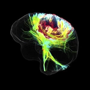
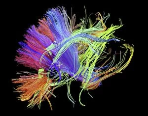
"Fibres: Unraveling the Intricacies of the Human Brain and Beyond" Exploring the intricate network of brain fibres through DTI MRI scans (C017 / 7099

Background imageFibres Collection: Connective tissue, SEM C015 / 9931

Connective tissue, SEM C015 / 9931
Connective tissue. Coloured scanning electron micrograph (SEM) of connective tissue from the trachea (wind pipe). Connective tissue supports other tissues and organs

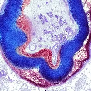
Background imageFibres Collection: Nerve ganglion, light micrograph C016 / 0532

Nerve ganglion, light micrograph C016 / 0532
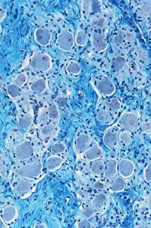
Nerve ganglion. Light micrograph of a section through a nerve ganglion of the peripheral nervous system, showing clusters of nerve cell bodies

Background imageFibres Collection: Connective tissue, SEM C015 / 9934

Connective tissue, SEM C015 / 9934
Connective tissue. Coloured scanning electron micrograph (SEM) of connective tissue from the trachea (wind pipe). Connective tissue supports other tissues and organs

Background imageFibres Collection: Myelinated nerve fibres, light micrograph C016 / 0531

Myelinated nerve fibres, light micrograph C016 / 0531
Myelinated nerve fibres. Light micrograph of a section through a spinal sensory nerve containing many closely packed axons exhibiting a wave-type appearance

Background imageFibres Collection: Trachea muscle, SEM C015 / 9935

Trachea muscle, SEM C015 / 9935
Trachea muscle. Coloured scanning electron micrograph (SEM) of smooth muscle from the trachea. The trachea (wind pipe) runs from the larynx (voice box) to the lungs

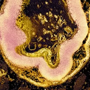
Background imageFibres Collection: Cirrhosis of liver, light micrograph C016 / 0529

Cirrhosis of liver, light micrograph C016 / 0529
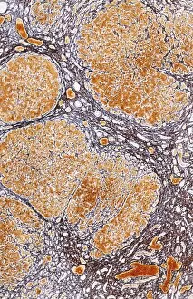
Cirrhosis of liver. Light micrograph of a section through liver tissue, damaged by cirrhosis. Cirrhosis occurs as a result of a range of factors causing damage to liver function

Background imageFibres Collection: White matter fibres of the human brain C018 / 6383

White matter fibres of the human brain C018 / 6383
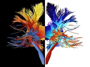
White matter fibres. Computer enhanced 3D diffusion spectral imaging (DSI) scan of the bundles of white matter nerve fibres in the brain

Background imageFibres Collection: White matter fibres, brain mri scan C018 / 6386

White matter fibres, brain mri scan C018 / 6386
White matter fibres overlaid a mri scan of the human head. Computer enhanced 3D diffusion spectral imaging (DSI) scan of the bundles of white matter nerve fibres in the brain

Background imageFibres Collection: White matter fibres of the human brain C018 / 6391

White matter fibres of the human brain C018 / 6391
White matter fibres. Computer enhanced 3D diffusion spectral imaging (DSI) scan of the bundles of white matter nerve fibres in the brain

Background imageFibres Collection: Corpus callosum, DTI MRI scan C017 / 7045

Corpus callosum, DTI MRI scan C017 / 7045
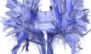
Corpus callosum. Coronal 3D diffusion tensor imaging (DTI) magnetic resonance imaging (MRI) scan of nerve pathways (blue) in and around the brains corpus callosum

Background imageFibres Collection: Glioblastoma brain tumour, DTI MRI scan C017 / 7052

Glioblastoma brain tumour, DTI MRI scan C017 / 7052
Glioblastoma brain tumour. 3D diffusion tensor imaging (DTI) magnetic resonance imaging (MRI) scan of nerve pathways (coloured) around a glioblastoma (upper right) in the brain

Background imageFibres Collection: Sperm tails, TEM C014 / 1463

Sperm tails, TEM C014 / 1463
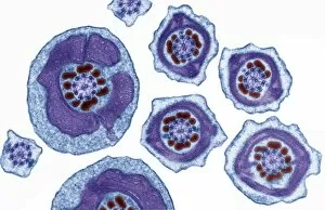
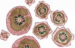
Sperm tails. Transmission electron micrograph (TEM) of sections through the tails of sperm (spermatozoa) cells at various levels

Background imageFibres Collection: Sperm tails, TEM C014 / 1465

Sperm tails, TEM C014 / 1465
Sperm tails. Transmission electron micrograph (TEM) of sections through the tails of sperm (spermatozoa) cells at various levels

Background imageFibres Collection: Silkworm silk fibres (SEM) C014 / 2149

Silkworm silk fibres (SEM) C014 / 2149
Silkworm silk fibres, coloured scanning electron micrograph (SEM). The silkworm is the larva or caterpillar of the silkmoth which has been domesticated from the wild silkmoth Bombyx mandarina

Background imageFibres Collection: Sperm tails, TEM C014 / 1464

Sperm tails, TEM C014 / 1464
Sperm tails. Transmission electron micrograph (TEM) of sections through the tails of sperm (spermatozoa) cells at various levels

Background imageFibres Collection: Nerve fibre node, TEM C014 / 1458

Nerve fibre node, TEM C014 / 1458
Nerve fibre node. Transmission electron micrograph (TEM) of a longitudinal section through a nerve fibre at a node of Ranvier, showing the nerve tissue (red) and the myelin sheath (dark blue)

Background imageFibres Collection: Non-woven textile fibres, SEM C014 / 4739

Non-woven textile fibres, SEM C014 / 4739
Non-woven textile fibres. Coloured scanning electron micrograph (SEM) showing the structure of fibres within a non-woven fabric. Magnification: x125 when printed 10 cm wide

Background imageFibres Collection: White matter fibres of the human brain C014 / 5672

White matter fibres of the human brain C014 / 5672
White matter fibres. Coloured 3D diffusion spectral imaging (DSI) scan of the bundles of white matter nerve fibres in the brain

Background imageFibres Collection: White matter fibres of the human brain C014 / 5664

White matter fibres of the human brain C014 / 5664
White matter fibres. Coloured 3D diffusion spectral imaging (DSI) scan of the bundles of white matter nerve fibres in the brain

Background imageFibres Collection: Human muscle fibres, diagram

Human muscle fibres, diagram
Human muscle fibres. Diagram showing the structure of human muscles from the cellular to the macroscopic level. At lower right, muscle cells contain many myofibrils

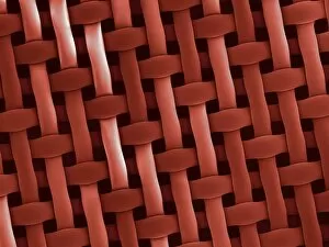
Background imageFibres Collection: Filter, SEM C014 / 4734

Filter, SEM C014 / 4734
Filter. Coloured scanning electron micrograph (SEM) showing the interwoven fibres of a 50 nanometre filter. Magnification: x250 when printed 10 cm wide

Background imageFibres Collection: White matter fibres of the human brain C014 / 5671

White matter fibres of the human brain C014 / 5671
White matter fibres. Coloured 3D diffusion spectral imaging (DSI) scan of the bundles of white matter nerve fibres in the brain

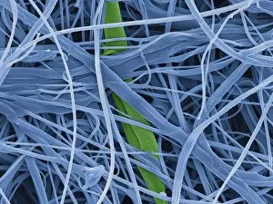
Background imageFibres Collection: Non-woven textile fibres, SEM C014 / 4738

Non-woven textile fibres, SEM C014 / 4738
Non-woven textile fibres. Coloured scanning electron micrograph (SEM) showing the structure of fibres within a non-woven fabric. Magnification: x75 when printed 10 cm wide

Background imageFibres Collection: White matter fibres of the human brain C014 / 5665

White matter fibres of the human brain C014 / 5665
White matter fibres. Coloured 3D diffusion spectral imaging (DSI) scan of the bundles of white matter nerve fibres in the brain

Background imageFibres Collection: White matter fibres of the human brain C014 / 5669

White matter fibres of the human brain C014 / 5669
White matter fibres. Coloured 3D diffusion spectral imaging (DSI) scan of the bundles of white matter nerve fibres in the brain

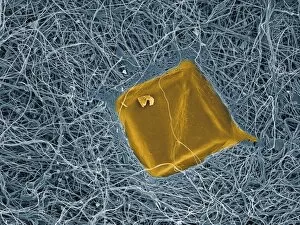
Background imageFibres Collection: MDCK culture cells, SEM C016 / 8028

MDCK culture cells, SEM C016 / 8028
MDCK cells. Coloured scanning electron micrograph (SEM) of cultured MDCK (Madin-Darby canine kidney) epithelial cells on filter paper (fibres)

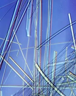
Background imageFibres Collection: Asbestos fibres, light micrograph

Asbestos fibres, light micrograph
Light micrograph with Normarski Differential Contrast (DIC) illumination of asbestos fibres. Asbestos has been used in the building industry for many years as a result of its heat resistance

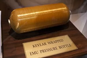
Background imageFibres Collection: Kevlar-wrapped spacesuit pressure bottle

Kevlar-wrapped spacesuit pressure bottle
Kevlar-wrapped pressure bottle for spacesuit Extravehicular Mobility Unit (EMU). Photographed at the International Astronautical Congress, 2011, in Cape Town, South Africa

Background imageFibres Collection: White matter fibres and brain, artwork C015 / 1938

White matter fibres and brain, artwork C015 / 1938
White matter fibres overlaid a 3d model of the human brain in top view. Coloured 3D diffusion spectral imaging (DSI) scan of the bundles of white matter nerve fibres in the brain

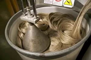
Background imageFibres Collection: Linen manufacturing, France C017 / 7925

Linen manufacturing, France C017 / 7925
Linen manufacturing. Hackled flax fibres coming out the end of the hackling line at the Terre de Lin linen factory, Saint Pierre le Viger, France. These fibres are ready to be made into bales

Background imageFibres Collection: Linen manufacturing, France C017 / 7924

Linen manufacturing, France C017 / 7924
Linen manufacturing. Worker disentangling lines of hackled flax fibres as they come out the end of the hackling line at the Terre de Lin linen factory, Saint Pierre le Viger, France

Background imageFibres Collection: Linen manufacturing, France C017 / 7922

Linen manufacturing, France C017 / 7922
Linen manufacturing. Worker spreading scutched flax fibres before they enter the hackling line of the Terre de Lin linen factory, Saint Pierre le Viger, France

Background imageFibres Collection: Linen manufacturing, France C017 / 7923

Linen manufacturing, France C017 / 7923
Linen manufacturing. Hackled flax fibres coming out the end of the hackling line at the Terre de Lin linen factory, Saint Pierre le Viger, France. These fibres are ready to be made into bales

Background imageFibres Collection: Linen manufacturing, France C017 / 7920

Linen manufacturing, France C017 / 7920
Linen manufacturing. Worker sorting scutched flax fibres at the end of the scutching line of the Terre de Lin linen factory, Saint Pierre le Viger, France

Background imageFibres Collection: Linen manufacturing, France C017 / 7921

Linen manufacturing, France C017 / 7921
Linen manufacturing. Worker spreading scutched flax fibres before they enter the hackling line of the Terre de Lin linen factory, Saint Pierre le Viger, France

Background imageFibres Collection: Myelinated nerve, TEM C016 / 5840

Myelinated nerve, TEM C016 / 5840
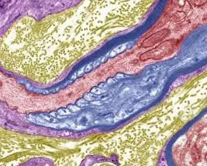
Myelinated nerve. Coloured transmission electron micrograph (TEM) of a section through a myelinated nerve fibre and Schwann cell

Background imageFibres Collection: Myelinated nerve, TEM C016 / 5839

Myelinated nerve, TEM C016 / 5839
Myelinated nerve. Coloured transmission electron micrograph (TEM) of a section through a myelinated nerve fibre and Schwann cell

Background imageFibres Collection: Myelinated nerve, TEM C016 / 5838

Myelinated nerve, TEM C016 / 5838
Myelinated nerve. Coloured transmission electron micrograph (TEM) of a section through a myelinated nerve fibre and Schwann cell

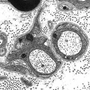
Background imageFibres Collection: Unmyelinated nerve, TEM C016 / 5805

Unmyelinated nerve, TEM C016 / 5805
Unmyelinated nerve. Transmission electron micrograph (TEM) of a section through axon (nerve fibre) bundles of unmyelinated nerves

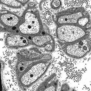
Background imageFibres Collection: Unmyelinated nerve, TEM C016 / 5804

Unmyelinated nerve, TEM C016 / 5804
Unmyelinated nerve. Transmission electron micrograph (TEM) of a section through axon (nerve fibre) bundles of unmyelinated nerves

Background imageFibres Collection: Myelinated nerve, TEM C016 / 5448

Myelinated nerve, TEM C016 / 5448
Myelinated nerve. Transmission electron micrograph (TEM) of a section through a myelinated nerve fibre and Schwann cell. Myelin (black)

Background imageFibres Collection: Myelinated nerve, TEM C016 / 5370

Myelinated nerve, TEM C016 / 5370
Myelinated nerve. Transmission electron micrograph (TEM) of a section through a myelinated nerve fibre and Schwann cell (centre)

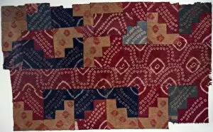
Background imageFibres Collection: Ancient Peruvian fabric C016 / 2369

Ancient Peruvian fabric C016 / 2369
Ancient Peruvian fabric. Section of fabric dating from the from the Paracas Formative Epoch (1250BC-1AD). This textile is made from thread formed from Camelid hair fibres

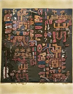
Background imageFibres Collection: Ancient Peruvian fabric C016 / 2368

Ancient Peruvian fabric C016 / 2368
Ancient Peruvian fabric. Section of fabric dating from the Nazca-Huari Fusion Epoch (800AD-1300AD). This textile is decorated with stepped motifs and concentric interlaced rhombuses

Background imageFibres Collection: Fibre optics factory C013 / 9199

Fibre optics factory C013 / 9199
Fibre optics factory. Worker with complete optical patch cables at a factory that produces fibre optic equipment. Optical fibres are made from flexible glass that has a high refractive index

Background imageFibres Collection: Fibre optics factory C013 / 9200

Fibre optics factory C013 / 9200
Fibre optics factory. Worker with complete optical patch cables at a factory that produces fibre optic equipment. Optical fibres are made from flexible glass that has a high refractive index

Background imageFibres Collection: Fibre optics factory C013 / 9193

Fibre optics factory C013 / 9193
Fibre optics factory. Worker using a laser to check the optical contact of an optical cable at a factory that produces fibre optic equipment

Background imageFibres Collection: Myelinated nerves, SEM C013 / 7142

Myelinated nerves, SEM C013 / 7142
Myelinated nerves. Coloured scanning electron micrograph (SEM) of a section through the sciatic nerve, showing the myelinated nerve fibres (axons)


For sale as Licensed Images

Choose your image, Select your licence and Download the media

"Fibres: Unraveling the Intricacies of the Human Brain and Beyond" Exploring the intricate network of brain fibres through DTI MRI scans (C017 / 7099). The mesmerizing artwork depicting white matter fibres and their connection to the brain (C015 / 1930). Delving deeper into understanding brain fibres with DTI MRI scans (C017 / 7035). Illuminating neurons and astrocytes through immunofluorescent LM, revealing the complexity of brain fibres. Peering into the white matter fibres of the human brain, unlocking its mysteries (C014 / 5666). Witnessing nerve cells regenerate under a TEM microscope, showcasing nature's resilience. Marveling at the intricate structure of a lime tree stem captured in a light micrograph. Discovering beauty in unexpected places - castor oil stem captured in a mesmerizing light micrograph. Unveiling myelination's role in enhancing nerve fibre function through TEM imaging. Witnessing cell division unfold under fluorescent micrographs, highlighting life's continuous renewal process. Celebrating nature's ability to heal as we observe regenerating nerve cells under TEM microscopy once again. Exploring further into white matter fibres within our complex human brains (C014/5668). In this captivating journey, we unravel the wonders – from delicate neural connections within our brains to resilient plant stems that provide sustenance for life itself.