
Golgi Apparatus Collection
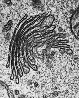
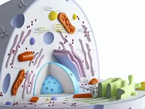
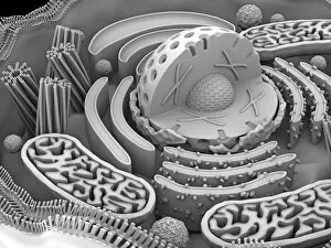
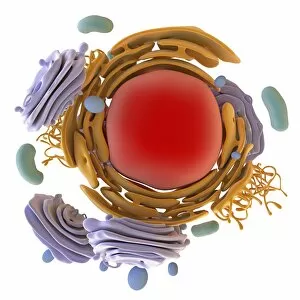
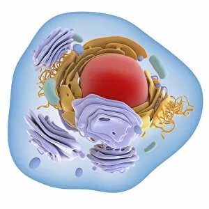
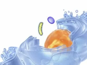
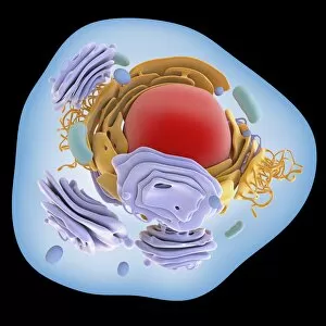
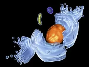
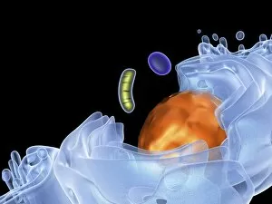
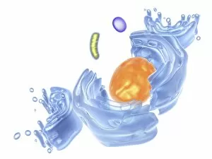
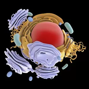
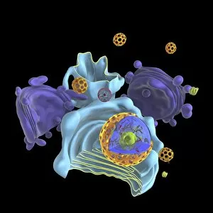
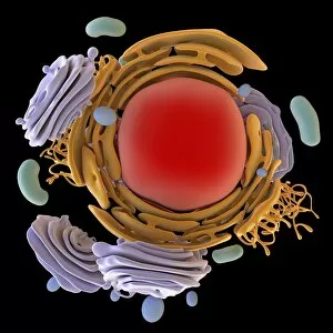
The Golgi apparatus, a vital organelle in animal and plant cells, plays a crucial role in the processing and packaging of proteins


For sale as Licensed Images
Choose your image, Select your licence and Download the media
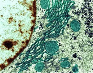
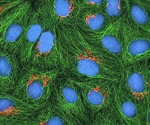
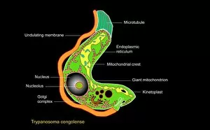
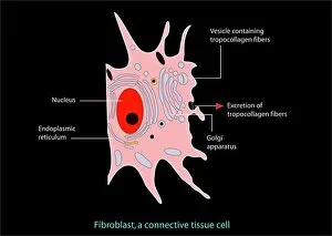
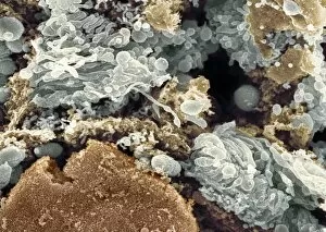
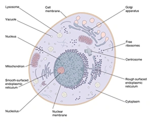
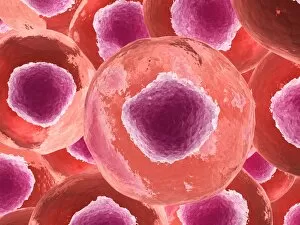
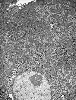
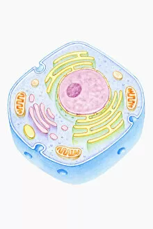
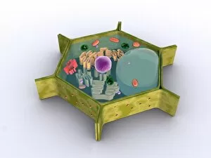
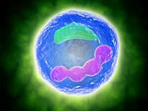
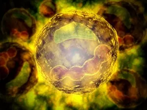
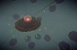
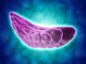
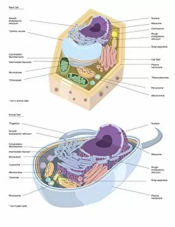
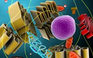
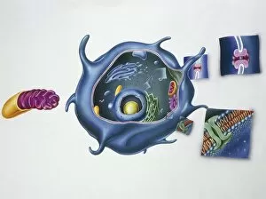
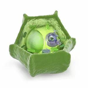
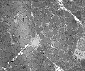
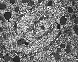
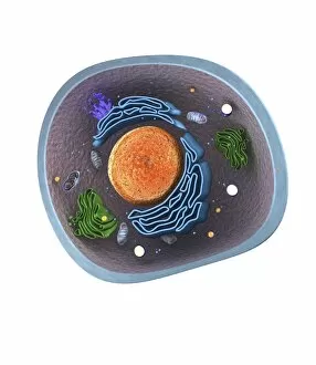
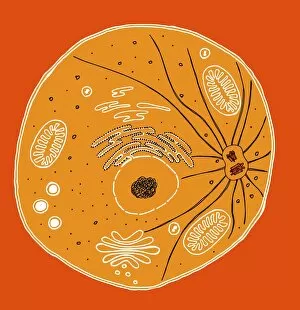
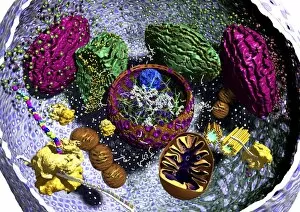
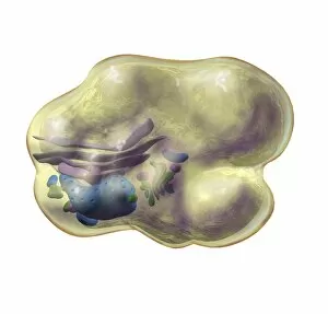
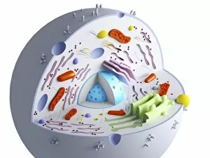
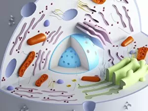
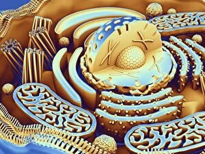
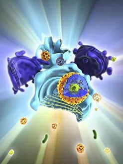
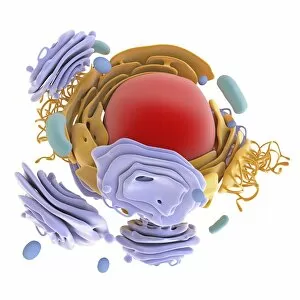
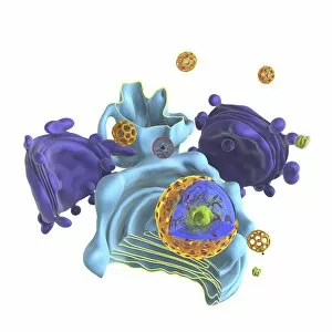
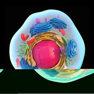
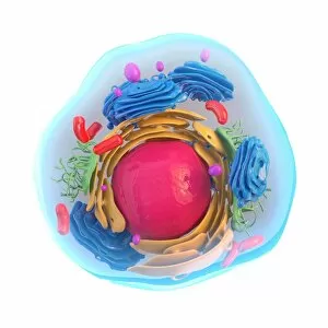
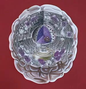
The Golgi apparatus, a vital organelle in animal and plant cells, plays a crucial role in the processing and packaging of proteins. This complex structure is intricately connected to other cellular components like the rough endoplasmic reticulum (ER), as shown in transmission electron microscopy (TEM) images. In HeLa cells, captured through light micrograph C017 / 8298, we can observe the Golgi apparatus's distinctive appearance. Interestingly, this organelle is not limited to animal cells alone; it also exists in trypanosome protozoans as depicted in stunning artwork. Similarly, scanning electron microscopy (SEM) allows us to visualize the intricate details of the Golgi apparatus within fibroblast cells through captivating artwork. Understanding animal cell structures is essential for comprehending their functions fully. Hepatocyte liver cells imaged using TEM provide valuable insights into how the Golgi apparatus operates within these specialized cells. Cross-sectional illustrations of human cells further enhance our understanding of this remarkable organelle's location and function. In plant cells, which possess unique features compared to animal cells, the Golgi apparatus coexists with other essential components such as nuclei, nucleoli, ribosomes, and endoplasmic reticulum. These elements are beautifully illustrated in conceptual images that showcase both plant cell structures and their individual components. Whether it be an artistic representation or a conceptual image capturing its essence within different organisms' cellular contexts—be it animals or plants—the Golgi apparatus remains an intriguing subject for scientific exploration. Its significance cannot be overstated when considering protein synthesis and secretion pathways critical for various biological processes across diverse species.