Histology Collection (#6)

Histology, the study of tissues at a microscopic level, unveils the intricate beauty and complexity of our body's structures

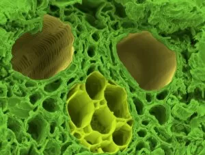
Background imageHistology Collection: Bamboo stem, SEM C015 / 5073

Bamboo stem, SEM C015 / 5073
Bamboo stem. Coloured environmental scanning electron micrograph (ESEM) of a section through the stem of a bamboo plant (family Gramineae), showing a vascular bundle

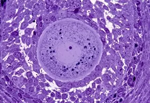
Background imageHistology Collection: Oocyte, light micrograph

Oocyte, light micrograph
Oocyte. Light micrograph of a section through an oocyte within a early antral follicle in an ovary. Oocytes are immature ova, or egg cells

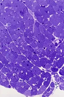
Background imageHistology Collection: Cardiac muscle, light micrograph

Cardiac muscle, light micrograph
Cardiac muscle. Light micrograph of a section through muscle tissue from a heart, showing the cardiomyocyte cells. Heart muscle cells show central nuclei but the dominant feature is the abundance of

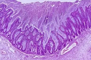
Background imageHistology Collection: Colitis, light micrograph

Colitis, light micrograph
Colitis. Light micrograph of a section through colon (large intestine) tissue affected by bacterial infection, causing injury to the mucosal lining, which has resulted in inflammation (colitis)

Background imageHistology Collection: Hairy scalp skin, light micrograph

Hairy scalp skin, light micrograph
Hairy scalp skin. Light micrograph of a section through healthy skin from a human scalp, showing the numerous hair follicles. The scalp is an example of hairy skin

Background imageHistology Collection: Oxalosis, light micrograph

Oxalosis, light micrograph
Oxalosis. Light micrograph of a section through adult bone tissue affected by oxalosis (primary hyperoxaluria), showing mature bone (green)

Background imageHistology Collection: Peripheral nerve, light micrograph

Peripheral nerve, light micrograph
Peripheral nerve. Light micrograph of a section through a peripheral nerve. This is a mixed nerve with myelinated axons (dark blue circles)

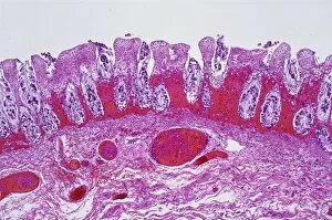
Background imageHistology Collection: Ischaemic bowel, light micrograph

Ischaemic bowel, light micrograph
Ischaemic bowel. Light micrograph of a section through a portion of intestine that has been damaged by an interruption of the blood supply, or ischaemia

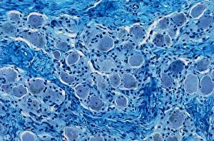
Background imageHistology Collection: Nerve ganglion, light micrograph

Nerve ganglion, light micrograph
Nerve ganglion. Light micrograph of a section through a dorsal (sensory) spinal root ganglion associated with a sensory nerve root of the spinal cord. Sensory information from peripheral sites e.g

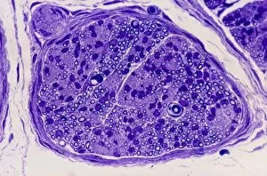
Background imageHistology Collection: Myelinated nerve, light micrograph

Myelinated nerve, light micrograph
Myelinated nerve. Light micrograph of a section through a peripheral myelinated nerve, showing many individual axons each covered with deep blue-staining myelin sheaths

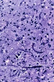
Background imageHistology Collection: Glial cells, light micrograph

Glial cells, light micrograph
Glial cells in the brain. Light micrograph of a section through glial cells (dark) in the grey matter of the brain. Due to their star shape these glial cells are called astrocytes

Background imageHistology Collection: Algae cell wall, SEM

Algae cell wall, SEM
Algae cell wall. Coloured scanning electron micrograph (SEM) of cellulose microfibrils (strands) in a Chaetomorpha sp. algal cell wall

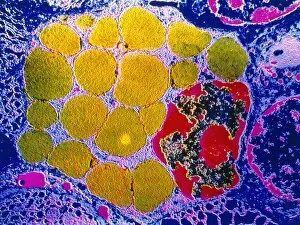
Background imageHistology Collection: Cirrhosis of the liver, light micrograph

Cirrhosis of the liver, light micrograph
Cirrhosis of the liver. Light micrograph of a section through liver tissue affected by cirrhosis, showing a cluster of hepatocyte cells (orange)

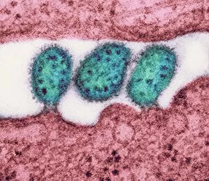
Background imageHistology Collection: Lassa virus particles, TEM C016 / 9409

Lassa virus particles, TEM C016 / 9409
Lassa virus particles. Transmission electron micrograph (TEM) of Lassa virus particles (virions, blue) amongst cell debris. This Arenavirus is the cause of Lassa fever

Background imageHistology Collection: Acute haemorrhagic conjunctivitis, TEM C016 / 9387

Acute haemorrhagic conjunctivitis, TEM C016 / 9387
Acute haemorrhagic conjunctivitis. Transmission electron micrograph (TEM) of a sample of tissue from the conjunctiva of a human eye that is infected by enterovirus particles (purple)

Background imageHistology Collection: Eastern equine encephalitis, TEM C016 / 9390

Eastern equine encephalitis, TEM C016 / 9390
Eastern equine encephalitis. Transmission electron micrograph (TEM) of a section through tissue from a human central nervous system (CNS)

Background imageHistology Collection: Colorado tick fever virus infection, TEM C016 / 9384

Colorado tick fever virus infection, TEM C016 / 9384
Colorado tick fever virus infection. Transmission electron micrograph (TEM) of human tissue infected by Colorado tick fever virus (CTFV) particles (virions, dark circles)

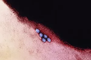
Background imageHistology Collection: Swine flu virus particles, TEM C016 / 9399

Swine flu virus particles, TEM C016 / 9399
Swine flu virus particles. Transmission electron micrograph (TEM) of a section through H3N2 influenza A virus particles (virions)

Background imageHistology Collection: Swine flu virus particles, TEM C016 / 9406

Swine flu virus particles, TEM C016 / 9406
Influenza virus particles. Transmission electron micrograph (TEM) of influenza (flu) virus particles (virions). Each virion consists of ribonucleic acid (RNA, dark patches)

Background imageHistology Collection: HIV virus particles, TEM C016 / 9404

HIV virus particles, TEM C016 / 9404
HIV virus particles. Transmission electron micrograph (TEM) of a section through human immunodeficiency virus (HIV) particles (virions, round)

Background imageHistology Collection: HIV virus particles, TEM C016 / 9405

HIV virus particles, TEM C016 / 9405
HIV virus particles. Transmission electron micrograph (TEM) of a section through human immunodeficiency virus (HIV) particles (virions, round)

Background imageHistology Collection: Swine flu virus particles, TEM C016 / 9400

Swine flu virus particles, TEM C016 / 9400
Swine flu virus particles. Transmission electron micrograph (TEM) of a section through H3N2 influenza A virus particles (virions)

Background imageHistology Collection: Lassa virus particles, TEM C016 / 9408

Lassa virus particles, TEM C016 / 9408
Lassa virus particles. Transmission electron micrograph (TEM) of Lassa virus particles (virions, green) amongst cell debris. This Arenavirus is the cause of Lassa fever

Background imageHistology Collection: Hartmannella vermiformis protozoa cysts C016 / 9402

Hartmannella vermiformis protozoa cysts C016 / 9402
Hartmannella vermiformis protozoa cysts. Transmission electron micrograph (TEM) of a section through cysts (round) containing Hartmannella vermiformis protozoa. H

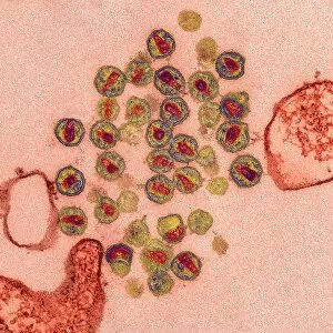
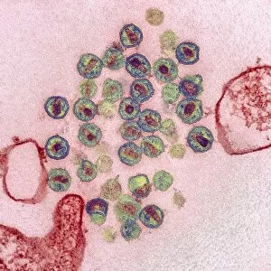
Background imageHistology Collection: Ganjam virus infection, TEM C016 / 9396

Ganjam virus infection, TEM C016 / 9396
Ganjam virus infection. Transmission electron micrograph (TEM) of a section through human tissue infected with ganjam virus particles (virions, blue and yellow)

Background imageHistology Collection: Swine flu virus particles, TEM C016 / 9407

Swine flu virus particles, TEM C016 / 9407
Influenza virus particles. Transmission electron micrograph (TEM) of influenza (flu) virus particles (virions). Each virion consists of ribonucleic acid (RNA, dark patches)

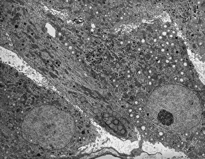
Background imageHistology Collection: Corpus luteum, TEM

Corpus luteum, TEM
Corpus luteum. Transmission electron micrograph (TEM) of a section through the corpus luteum of an ovary, showing several luteal cells

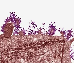
Background imageHistology Collection: Flu virus, TEM

Flu virus, TEM
Flu virus. Transmission electron micrograph (TEM) of a section through influenza (flu) virus particles (virions, purple) budding from a host cell. This is the final stage in viral replication

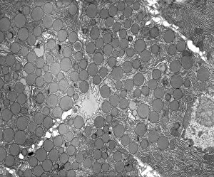
Background imageHistology Collection: Pancreatic exocrine cells, TEM

Pancreatic exocrine cells, TEM
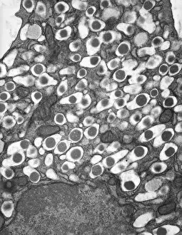
Pancreatic exocrine cells. Transmission electron micrograph (TEM) of a section through exocrine cells in the pancreas, showing numerous zymogen granules (circles), rough endoplasmic reticulum (ER)

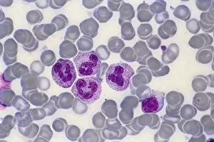
Background imageHistology Collection: Bone marrow blood cells, light micrograph

Bone marrow blood cells, light micrograph

Background imageHistology Collection: Glomerulus, TEM

Glomerulus, TEM
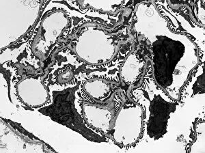
Glomerulus. Transmission electron micrograph (TEM) of a section through a nephron in a human kidney, showing a cluster of capillaries within a renal corpuscle, also known as a glomerulus

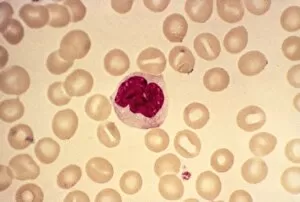
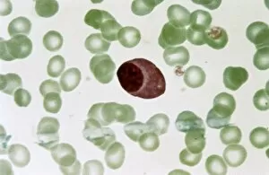
Background imageHistology Collection: Monocyte blood cell, light micrograph

Monocyte blood cell, light micrograph. Monocytes are a type of white blood cell, part of the bodys immune system. Monocytes are the largest white blood cell
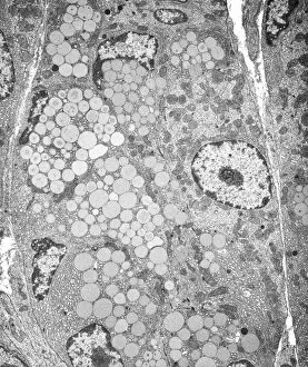
Background imageHistology Collection: Gastric gland, TEM

Gastric gland, TEM
Gastric gland. Transmission electron micrograph (TEM) of a section through the deep region of a gastric (fundus) gland, showing several enzyme-secreting chief cells with many secretory granules

Background imageHistology Collection: Ductal breast cancer, TEM

Ductal breast cancer, TEM
Ductal breast cancer. Transmission electron micrograph (TEM) of a section through breast tissue, showing malignant (cancerous) epithelial cells arising from the milk ducts of the breast

Background imageHistology Collection: Erythroblast blood cell, light micrograph

Erythroblast blood cell, light micrograph
Erythroblast blood cell. Light micrograph of cells from a sample of bone marrow, including a polychromatic erythroblast (upper left). This one has multiple cell nuclei (dark red, four in total)

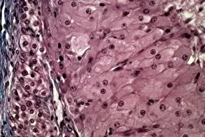
Background imageHistology Collection: Corpus luteum, light micrograph

Corpus luteum, light micrograph
Corpus luteum. Light micrograph of a section through the corpus luteum of an ovary. The corpus luteum is the tissue formed from a mature ovarian follicle after the follicle has released its oocyte
Background imageHistology Collection: Islet of Langerhans beta cell, TEM

Islet of Langerhans beta cell, TEM
Islet of Langerhans beta cell. Transmission electron micrograph (TEM) of a section through a beta cell in an islet of Langerhans in the pancreas

Background imageHistology Collection: Pancreatic acinar cell, TEM

Pancreatic acinar cell, TEM
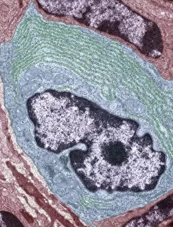
Pancreatic acinar cell. Transmission electron micrograph (TEM) of a section through an enzyme-secreting acinar cell in the human pancreas, showing the nucleus (dark purple, centre)

Background imageHistology Collection: Activated plasma cell, TEM

Activated plasma cell, TEM
Activated plasma cell, coloured transmission electron micrograph (TEM). Plasma cells are derived from stimulated B lymphocytes (white blood cells)

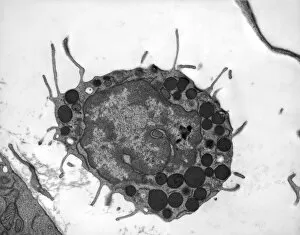
Background imageHistology Collection: Basophil white blood cell, TEM

Basophil white blood cell, TEM
Basophil white blood cell. Transmission electron micrograph (TEM) of section through a circulating basophil cell. Basophils are the smallest and least common of the white blood cells

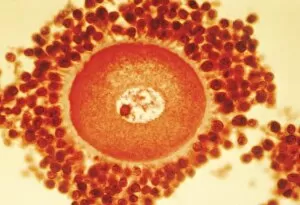
Background imageHistology Collection: Oocyte before ovulation, light micrograph

Oocyte before ovulation, light micrograph
Oocyte before ovulation. Light micrograph of a section through tissue from an ovary, showing an oocyte two days before ovulation

Background imageHistology Collection: Plasmocyte blood cell, light micrograph

Plasmocyte blood cell, light micrograph. Plasmocytes, also called plasma cells, are derived from stimulated B lymphocytes (white blood cells)

Background imageHistology Collection: Myenteric nerve plexus, TEM

Myenteric nerve plexus, TEM
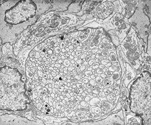
Myenteric nerve plexus. Transmission electron micrograph (TEM) of a section through a bundle of unmyelinated nerve fibres in the myenteric nerve plexus of the intestinal tract

Background imageHistology Collection: Golgi membranes, TEM

Golgi membranes, TEM
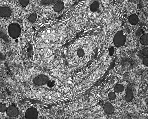
Golgi membranes. Transmission electron micrograph (TEM) of a section through a cell, showing the membranes (dark lines) of the Golgi apparatus

Background imageHistology Collection: Ovarian follicle tissue, light micrograph

Ovarian follicle tissue, light micrograph
Ovarian follicle tissue. Light micrograph of a section through tissue from an ovarian follicle, showing a boundary between two layers

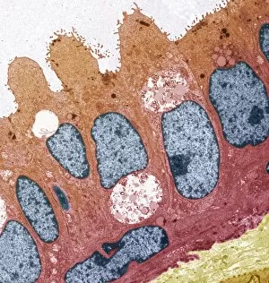
Background imageHistology Collection: Goblet cell, TEM

Goblet cell, TEM
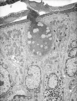
Goblet cell. Transmission electron micrograph (TEM) of a section through a goblet cell in the lining (epithelium, top) of the small intestine

Background imageHistology Collection: Mast cells, light micrograph

Mast cells, light micrograph
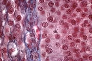
Mast cells. Light micrograph of several mast cells (pink rounded objects). These leucocytes (white blood cells) contain the chemical mediators histamine, serotonin and heparin


For sale as Licensed Images

Choose your image, Select your licence and Download the media

Histology, the study of tissues at a microscopic level, unveils the intricate beauty and complexity of our body's structures. Through techniques like light micrography and transmission electron microscopy (TEM), scientists have been able to explore various tissues and unravel their secrets. One such tissue is the cerebellum, which plays a crucial role in coordinating movement and balance. By examining cerebellum tissue under a light microscope, we can observe its distinct layers and cell types. The synapse nerve junctions captured through TEM reveal the precise connections between neurons that allow for seamless communication. In 1894, Spanish histologist Santiago Ramon y Cajal created an exquisite drawing showcasing different cell types within the mammalian cerebellum. His meticulous work laid the foundation for understanding neural networks. Moving beyond just one region of the brain, histologists also delve into other fascinating areas like the hippocampus. Microscopic examination of hippocampus brain tissue provides insights into memory formation and spatial navigation. Purkinje nerve cells found within the cerebellum are particularly captivating under scrutiny. Their elaborate branching patterns give rise to their unique appearance when observed through a microscope slide. Histological studies extend beyond neurological tissues; they encompass organs throughout our body systems as well. For instance, kidney tubules in section offer glimpses into renal function while highlighting their structural organization. The human brain itself holds countless mysteries waiting to be unraveled by histologists examining microscope slides containing delicate slices of this complex organ. These slides provide glimpses into both healthy brains and those affected by diseases like Alzheimer's - offering valuable insights into neurodegenerative disorders. Exploring deeper with TEM reveals cellular components such as rough endoplasmic reticulum - an organelle involved in protein synthesis - providing detailed views at nanoscale resolution. Histology not only focuses on neurons but also encompasses glial cells that support neuronal functions. Light micrographs capturing glial stem cell cultures showcase the potential for regeneration and repair within the nervous system.