Histopathology Collection (#3)

Histopathology is a fascinating field that delves into the intricate details of diseases and their impact on our bodies

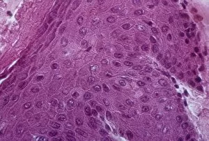
Background imageHistopathology Collection: Cervical thickening, light micrograph C015 / 6166

Cervical thickening, light micrograph C015 / 6166
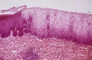
Cervical thickening. Light micrograph of a section through the thickened mucous membrane of a cervix (neck of the womb). This condition, known as leukoplakia, occurs in 1% of elderly women

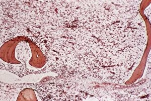
Background imageHistopathology Collection: Chronic myeloid leukaemia, micrograph C015 / 6227

Chronic myeloid leukaemia, micrograph C015 / 6227
Chronic myeloid leukaemia. Light micrograph of bone marrow from a patient with chronic myeloid leukaemia (CML). This cancer affects the myeloid tissue (bone marrow)

Background imageHistopathology Collection: Acute myeloid leukaemia C015 / 6225

Acute myeloid leukaemia C015 / 6225
Acute myeloid leukaemia. Light micrograph of bone marrow from a patient with acute myeloid leukaemia (AML). This cancer affects the myeloid tissue (bone marrow)

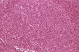
Background imageHistopathology Collection: Lymph node hyperplasia, light micrograph C015 / 6224

Lymph node hyperplasia, light micrograph C015 / 6224
Lymph node hyperplasia. Light micrograph of a section through a lymph node showing follicular hyperplasia. Hyperplasia is the abnormal proliferation of cells

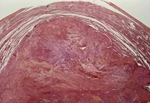
Background imageHistopathology Collection: Uterine fibroid, light micrograph C015 / 6413

Uterine fibroid, light micrograph C015 / 6413
Uterine fibroid. Light micrograph of a section through a uterine fibroid. A fibroid is a fibrous benign tumour originating from muscular tissue

Background imageHistopathology Collection: Uterine fibroid, light micrograph C015 / 6411

Uterine fibroid, light micrograph C015 / 6411
Uterine fibroid. Light micrograph of a section through a uterine fibroid (centre). A fibroid is a fibrous benign tumour originating from muscular tissue

Background imageHistopathology Collection: Uterine fibroid, light micrograph C015 / 6412

Uterine fibroid, light micrograph C015 / 6412
Uterine fibroid. Light micrograph of a section through a uterine fibroid (oval). A fibroid is a fibrous benign tumour originating from muscular tissue

Background imageHistopathology Collection: Paratubal cysts, light micrograph C015 / 6410

Paratubal cysts, light micrograph C015 / 6410
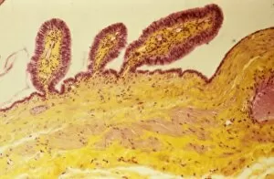
Paratubal cysts. Light micrograph of a section through a fallopian tube (oviduct) with three paratubal cysts (top). Cysts are fluid-filled sacs

Background imageHistopathology Collection: Cervical cyst, light micrograph C015 / 6408

Cervical cyst, light micrograph C015 / 6408
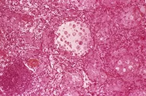
Cervical cyst. Light micrograph of a section through tissue from the cervix (neck of the womb) showing a mucous cyst (pale, centre)

Background imageHistopathology Collection: Listeriosis, light micrograph C015 / 6405

Listeriosis, light micrograph C015 / 6405
Listeriosis. Light micrograph of a section through a placenta in a case of listeriosis. This rare infection by the Listeria monocytogenes bacteria is usually due to food poisoning

Background imageHistopathology Collection: Endometrial squamous metaplasia C015 / 6404

Endometrial squamous metaplasia C015 / 6404
Endometrial squamous metaplasia. Light micrograph of a section through the endometrium (lining of the womb) showing squamous metaplasia

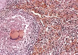
Background imageHistopathology Collection: Tuberculosis, light micrograph C015 / 6400

Tuberculosis, light micrograph C015 / 6400
Tuberculosis. Light micrograph of a section through a lymph node showing a Langhans cell (centre) caused by tuberculosis (TB)

Background imageHistopathology Collection: Dermoid ovarian cyst C015 / 6402

Dermoid ovarian cyst C015 / 6402
Dermoid ovarian cyst. Light micrograph of a section through an ovary with a dermoid, or teratoma, ovarian cyst. This is a benign (non-cancerous) cyst that arises from germ (sex) cells

Background imageHistopathology Collection: Tumour embolism, light micrograph C015 / 6397

Tumour embolism, light micrograph C015 / 6397
Tumour embolism. Light micrograph of a section through an embolism (blood clot) caused by cells from a cervical cancer. The cells have left the primary tumour and are metastasising (spreading)

Background imageHistopathology Collection: Excessive iron absorption C015 / 6394

Excessive iron absorption C015 / 6394
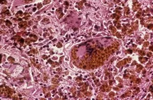
Excessive iron absorption. Light micrograph of a section through tissue showing haemochromatosis, a physiological disorder where excessive amounts of iron are absorbed by the body

Background imageHistopathology Collection: Gauchers disease, light micrograph

Gauchers disease, light micrograph
Gauchers disease. Light micrograph of a section through the spleen of a patient with Gauchers disease. This is an inherited metabolic disorder in which a fatty substance called glucocerebroside

Background imageHistopathology Collection: Cervical thickening, light micrograph C015 / 6167

Cervical thickening, light micrograph C015 / 6167
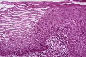
Cervical thickening. Light micrograph of a section through the thickened mucous membrane (dark purple, left) of a cervix (neck of the womb). Normal epithelium is at right

Background imageHistopathology Collection: Cervical thickening, light micrograph C015 / 6168

Cervical thickening, light micrograph C015 / 6168
Cervical thickening. Light micrograph of a section through the thickened mucous membrane (dark purple, right) of a cervix (neck of the womb). Normal epithelium is at left

Background imageHistopathology Collection: Glomerulosclerosis, TEM C016 / 5836

Glomerulosclerosis, TEM C016 / 5836
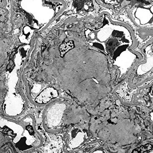
Glomerulosclerosis. Transmission electron micrograph (TEM) of a section through through the glomerulus of a kidney affected by glomerulosclerosis

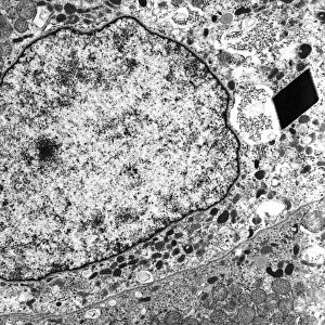
Background imageHistopathology Collection: Alveolar soft part sarcoma, TEM C016 / 5826

Alveolar soft part sarcoma, TEM C016 / 5826
Alveolar soft part sarcoma. Transmission electron micrograph (TEM) of a section through an alveolar soft part sarcoma (ASPS) showing a characteristic rhomboid crystal (black, centre)

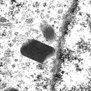
Background imageHistopathology Collection: Alveolar soft part sarcoma, TEM C016 / 5816

Alveolar soft part sarcoma, TEM C016 / 5816
Alveolar soft part sarcoma. Transmission electron micrograph (TEM) of a section through an alveolar soft part sarcoma (ASPS) showing a characteristic rhomboid crystal (black)

Background imageHistopathology Collection: Mitochondrial inclusions, TEM C016 / 5807

Mitochondrial inclusions, TEM C016 / 5807
Mitochondrial inclusions. Transmission electron micrograph (TEM) of a section through a mitochondrion showing crystalline inclusions (thick parallel rods) in the intermembrane spaces

Background imageHistopathology Collection: Mitochondrial inclusions, TEM C016 / 5806

Mitochondrial inclusions, TEM C016 / 5806
Mitochondrial inclusions. Transmission electron micrograph (TEM) of a section through a mitochondrion showing crystalline inclusions (thick parallel rods) in the intermembrane spaces

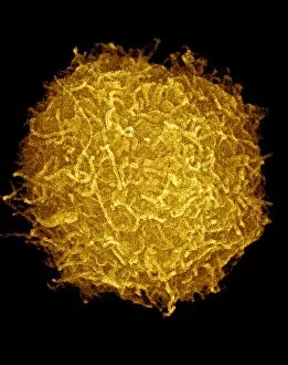
Background imageHistopathology Collection: Prostate cancer cell, SEM C013 / 5012

Prostate cancer cell, SEM C013 / 5012
Prostate cancer cell, coloured scanning electron micrograph (SEM). The uneven surface of this cell is typical of cancer cells

Background imageHistopathology Collection: Prostate cancer cell, SEM C013 / 5011

Prostate cancer cell, SEM C013 / 5011
Prostate cancer cell, coloured scanning electron micrograph (SEM). The uneven surface of this cell is typical of cancer cells

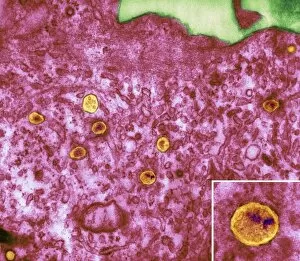
Background imageHistopathology Collection: HIV particles in infected cell, TEM

HIV particles in infected cell, TEM
HIV particles. Coloured transmission electron micrograph (TEM) of human immunodeficiency virus (HIV) particles (orange) in a host cell. HIV causes the disease AIDS

Background imageHistopathology Collection: Crystals in seminal fluid

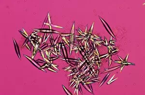
Crystals in seminal fluid. Polarised light micrograph of a sample of abnormal seminal fluid. The sample contains crystals, which may indicate a problem with the prostate

Background imageHistopathology Collection: Rickets, light micrograph

Rickets, light micrograph
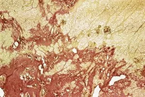
Rickets. Light micrograph of a section through bone of a patient with rickets, a softening of the bones. The light orange areas are unmineralised (unhardened) cartilage
Background imageHistopathology Collection: Bacterial infection of nail, SEM

Bacterial infection of nail, SEM
Bacterial infection of a nail. Coloured scanning electron micrograph (SEM) of a nail with a bacterial infection (paronychia)

Background imageHistopathology Collection: Neutrophils and Shigella bacteria, SEM

Neutrophils and Shigella bacteria, SEM
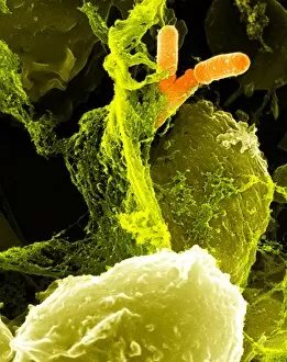
Neutrophils and Shigella bacteria. Coloured scanning electron micrograph (SEM) of neutrophil white blood cells (large, yellow-green) and Shigella bacteria (orange, upper right)

Background imageHistopathology Collection: Psoriasis, light micrograph

Psoriasis, light micrograph
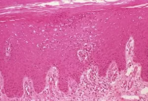
Psoriasis. Light micrograph of a section through a psoriasis pustule. Psoriasis is a chronic skin disease causing inflammation and scaly lesions

Background imageHistopathology Collection: Bacterial pneumonia infection

Bacterial pneumonia infection. Light micrograph of pneumonia bacteria (Klebsiella pneumoniae, rod- shaped, pink) among cells in a sample of pus from a pneumonia patient. K

Background imageHistopathology Collection: Typhoid nodules, light micrograph

Typhoid nodules, light micrograph
Typhoid nodules, coloured light micrograph. Section through a lymph node of a patient with typhoid, showing macrophages (large cells, pink) forming typhoid nodules

Background imageHistopathology Collection: Liver tuberculosis, light micrograph

Liver tuberculosis, light micrograph
Liver tuberculosis. Coloured light micrograph of a section through the liver of a patient with miliary tuberculosis (TB). A tubercle, a nodular lesion of infected dead tissue, is seen at left

Background imageHistopathology Collection: Miliary tuberculosis, light micrograph

Miliary tuberculosis, light micrograph
Miliary tuberculosis. Light micrograph of a section through a single miliary tubercle (left) in a patients lung. Tubercles are nodular lesions of infected dead tissue that arise from tuberculosis (TB)

Background imageHistopathology Collection: Thyroiditis

Thyroiditis (Hashimotos disease). Light micrograph of a section through a thyroid gland with the autoimmune disorder Hashimotos disease

Background imageHistopathology Collection: Alcoholic hepatitis, light micrograph

Alcoholic hepatitis, light micrograph
Alcoholic hepatitis. Coloured light micrograph of a section through the liver of a patient with alcoholic hepatitis, inflammation of the liver due to prolonged heavy alcohol consumption

Background imageHistopathology Collection: Systemic lupus erythematosus

Systemic lupus erythematosus. Light micrograph of a section through a glomerulus (blood filtration structure) from kidney tissue in a case of SLE

Background imageHistopathology Collection: Coronary thrombosis, light micrograph

Coronary thrombosis, light micrograph
Coronary thrombosis. Light micrograph of a section through a thrombus (blood clot) in the coronary artery of the heart. The clot is packed full of red blood cells (red)

Background imageHistopathology Collection: Gauchers disease

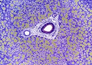
Gauchers disease. Light micrograph of a section through a human liver with Gauchers disease. It is a genetic disorder (autosomal recessive)

Background imageHistopathology Collection: Inflamed gall bladder, light micrograph

Inflamed gall bladder, light micrograph
Inflamed gall bladder. Light micrograph of a section through a gall bladder with cholecystitis (inflammation). The mucosa lining (purple) is thickened

Background imageHistopathology Collection: Skin inflammation, light micrograph

Skin inflammation, light micrograph
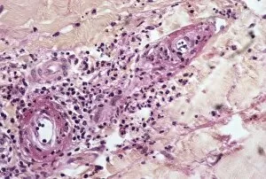
Skin inflammation. Light micrograph of a section through skin showing inflamed capillaries (lower left and upper right) caused by capillaritis, also known as pigmented purpurea

Background imageHistopathology Collection: Ulcerative colitis, SEM

Ulcerative colitis, SEM
Ulcerative colitis. Coloured scanning electron micrograph (SEM) of freeze-fractured bowel tissue from a patient suffering from ulcerative colitis

Background imageHistopathology Collection: AIDS pneumonia infection, TEM

AIDS pneumonia infection, TEM
AIDS pneumonia infection. Coloured transmission electron micrograph (TEM) of a section through Pneumocystis carinii protozoa causing pneumonia. The protozoa are crescent-shaped (red/yellow)

Background imageHistopathology Collection: Bilharzia infection, ureter tissue

Bilharzia infection, ureter tissue
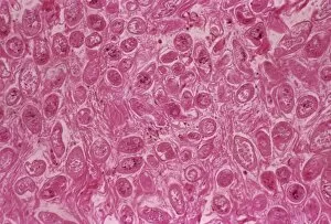
Bilharzia infection. Light micrograph of human ureter tissue that contains numerous eggs from Schistosoma flukes. The ureters are tubes that carry urine from the kidneys to the bladder

Background imageHistopathology Collection: Skin pigmentation in Addisons disease

Skin pigmentation in Addisons disease
Skin pigmentation from Addisons disease. Light micrograph of a section through the skin of a patient with Addisons disease

Background imageHistopathology Collection: Alzheimers disease brain cell, TEM

Alzheimers disease brain cell, TEM
Alzheimers disease brain cell. Coloured transmission electron micrograph (TEM) of a neurofibrillary tangle in a nerve cell from the brain of a patient with Alzheimers disease

Background imageHistopathology Collection: Alzheimers disease brain tissue

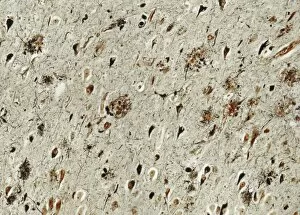
Alzheimers disease brain tissue, light micrograph. Two characteristic features of Alzheimers disease are seen here; neurofibrillary tangles (dark teardrop shapes)


For sale as Licensed Images

Choose your image, Select your licence and Download the media

Histopathology is a fascinating field that delves into the intricate details of diseases and their impact on our bodies. From Salmonella bacteria to Dohle bodies in blood cells, histopathologists use various techniques like SEM (scanning electron microscopy) and micrographs to unravel the mysteries within. In acute promyelocytic leukemia, abnormal cells can be observed through micrographs, providing crucial insights for diagnosis and treatment. Similarly, ovarian cancer reveals its telltale signs under light micrograph C015 / 7103, aiding in early detection. Liver tissue cirrhosis showcases the damaging effects of certain conditions on this vital organ. Through light micrographs, pathologists can identify key features that aid in understanding disease progression. Cystic fibrosis affects multiple organs including the lungs and pancreas. Histopathological analysis helps uncover cellular changes associated with this genetic disorder. Osteoporotic bone exhibits reduced density and weakened structure due to bone loss over time. By studying these samples closely, researchers gain valuable knowledge about prevention strategies and potential treatments. Malaria parasites are intricately studied using TEM (transmission electron microscopy), revealing their distinct characteristics necessary for accurate diagnosis and effective interventions against this deadly disease. Herpes simplex viruses leave their mark through TEM images showcasing their unique structures. Understanding these viral particles aids in developing antiviral therapies to combat outbreaks effectively. Diabetic foot ulcers present complex challenges due to impaired wound healing mechanisms caused by diabetes-related complications. Histopathology plays a pivotal role in unraveling the underlying pathophysiology behind these ulcers for better management strategies. Gout crystals provide visual evidence of joint inflammation caused by excess uric acid deposition. Studying these crystals enables clinicians to tailor treatment plans accordingly for patients suffering from gout attacks. HIV particles reveal themselves under TEM examination as we strive towards understanding this global health crisis better. This microscopic exploration contributes significantly towards developing effective antiretroviral therapies and potential vaccines.