
Histopathology Collection (#2)
Histopathology is a fascinating field that delves into the intricate details of diseases and their impact on our bodies


For sale as Licensed Images
Choose your image, Select your licence and Download the media
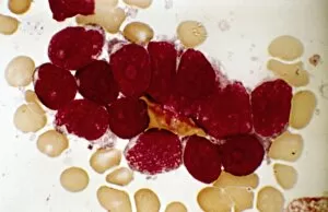
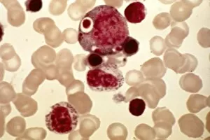
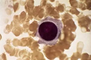
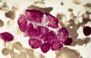
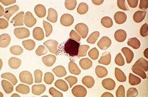
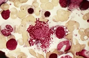
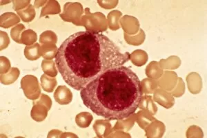
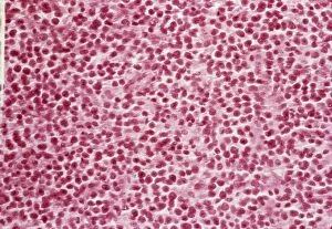
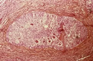
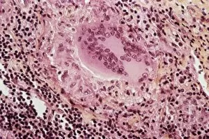
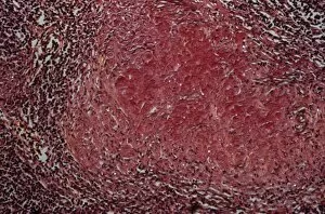
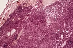
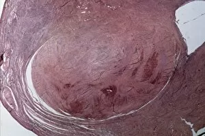
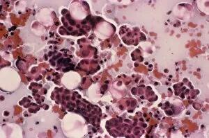
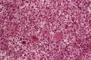
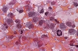
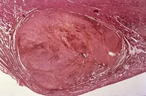
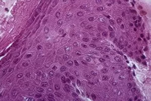
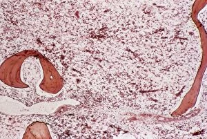
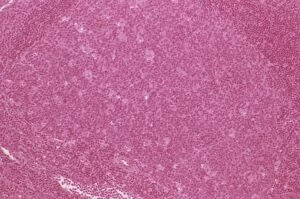
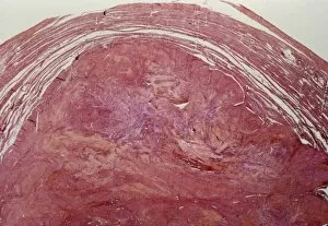
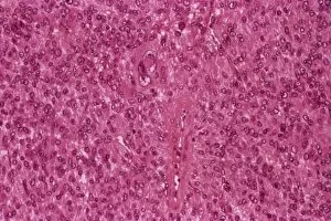
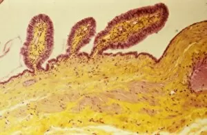
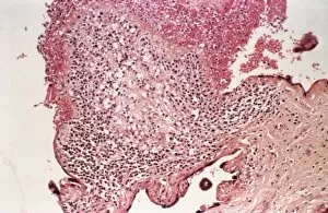
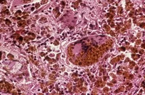
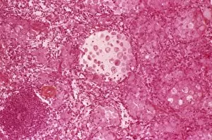
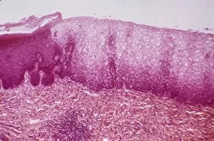
Histopathology is a fascinating field that delves into the intricate details of diseases and their impact on our bodies. From Salmonella bacteria to Dohle bodies in blood cells, histopathologists use various techniques like SEM (scanning electron microscopy) and micrographs to unravel the mysteries within. In acute promyelocytic leukemia, abnormal cells can be observed through micrographs, providing crucial insights for diagnosis and treatment. Similarly, ovarian cancer reveals its telltale signs under light micrograph C015 / 7103, aiding in early detection. Liver tissue cirrhosis showcases the damaging effects of certain conditions on this vital organ. Through light micrographs, pathologists can identify key features that aid in understanding disease progression. Cystic fibrosis affects multiple organs including the lungs and pancreas. Histopathological analysis helps uncover cellular changes associated with this genetic disorder. Osteoporotic bone exhibits reduced density and weakened structure due to bone loss over time. By studying these samples closely, researchers gain valuable knowledge about prevention strategies and potential treatments. Malaria parasites are intricately studied using TEM (transmission electron microscopy), revealing their distinct characteristics necessary for accurate diagnosis and effective interventions against this deadly disease. Herpes simplex viruses leave their mark through TEM images showcasing their unique structures. Understanding these viral particles aids in developing antiviral therapies to combat outbreaks effectively. Diabetic foot ulcers present complex challenges due to impaired wound healing mechanisms caused by diabetes-related complications. Histopathology plays a pivotal role in unraveling the underlying pathophysiology behind these ulcers for better management strategies. Gout crystals provide visual evidence of joint inflammation caused by excess uric acid deposition. Studying these crystals enables clinicians to tailor treatment plans accordingly for patients suffering from gout attacks. HIV particles reveal themselves under TEM examination as we strive towards understanding this global health crisis better. This microscopic exploration contributes significantly towards developing effective antiretroviral therapies and potential vaccines.