
Immune Cell Collection
"Exploring the Mighty Immune Cell: Unveiling its Intricate World" In the vast realm of our immune system, lies a group of remarkable cells known as immune cells


For sale as Licensed Images
Choose your image, Select your licence and Download the media
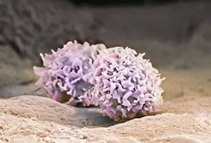
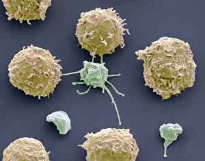
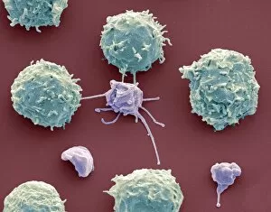
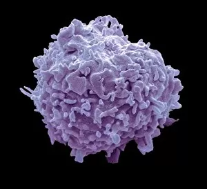
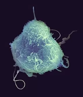
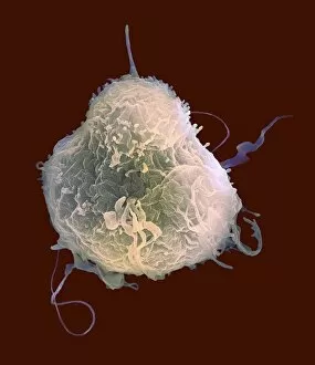
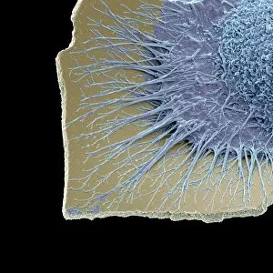
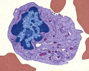
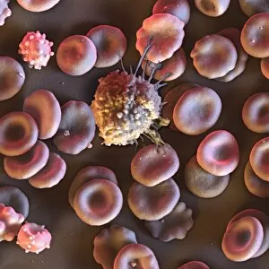
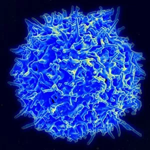
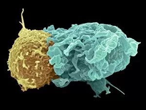
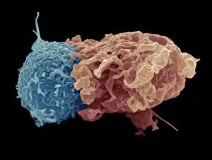
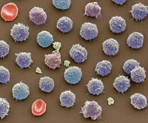
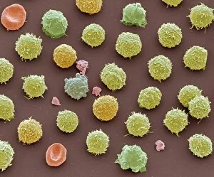
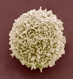
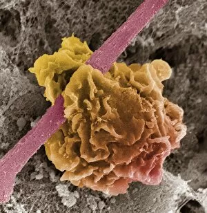
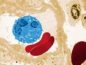
"Exploring the Mighty Immune Cell: Unveiling its Intricate World" In the vast realm of our immune system, lies a group of remarkable cells known as immune cells. Among them, macrophage cells stand tall as the guardians of our body's defense mechanism. Through the lens of a transmission electron microscope (TEM), we witness their intricate structure and marvel at their ability to engulf and destroy foreign invaders. Moving on to another chapter in this captivating story, we encounter white blood cells and platelets under the scanning electron microscope (SEM). In SEM C016 / 3099 and SEM C016 / 3098 images, these brave warriors are captured in action, ready to combat any threat that comes their way. Their resilience is truly awe-inspiring. As we delve deeper into this microscopic world, monocyte white blood cell takes center stage in SEM C016 / 3089 image. This versatile cell plays a crucial role in initiating an immune response by transforming into various specialized defenders when needed. Shifting our focus towards lymphocyte white blood cells showcased in SEM C016 / 9414, SEM C016 / 9415, SEM C016 / 9140, and SEM C016 / 9138 images; we witness their diversity and adaptability. These resilient soldiers tirelessly patrol our body to identify potential threats while orchestrating an orchestrated attack against invading pathogens. Lastly but not leastly, microglial white blood cell emerges from obscurity through stunning images like SEM C019/0247, SEM C019/0246, and SEMC019/0245. These unique defenders reside within our central nervous system acting as vigilant sentinels safeguarding against any harm or inflammation that may arise. Through TEM imaging techniques even monocyte white blood cell reveals its true form - a key player behind-the-scenes orchestrating vital immune responses with precision. The captivating journey through these mesmerizing images provides us with a glimpse into the extraordinary world of immune cells.