Immunology Collection (page 9)

"Unleashing the Power of Immunology: Exploring the Intricate World of Immune Responses" Immunology

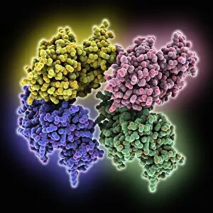
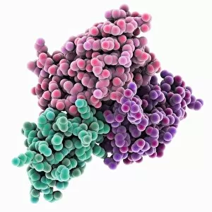
Background imageImmunology Collection: C3-degrading proteinase enzyme C016 / 1363

C3-degrading proteinase enzyme C016 / 1363
C3-degrading proteinase enzyme. Molecular model of a proteinase enzyme from the TIGR4 strain of the bacterium Streptococcus pneumoniae

Background imageImmunology Collection: Human interleukin-28B C016 / 1364

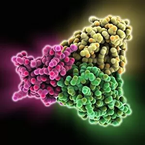
Human interleukin-28B C016 / 1364
Human interleukin-28B (IL-28B), molecular model. Interleukins a group of secreted proteins and signalling molecules that play a range of roles in cellular communication

Background imageImmunology Collection: Human interleukin-28B C016 / 1365

Human interleukin-28B C016 / 1365
Human interleukin-28B (IL-28B), molecular model. Interleukins a group of secreted proteins and signalling molecules that play a range of roles in cellular communication

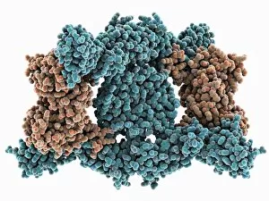
Background imageImmunology Collection: IBDV subviral particle, molecular model

IBDV subviral particle, molecular model. IBDV (infectious bursal disease virus) is an avian virus that infects the bursa of Fabricius (specialised bird immune organ) in young chickens

Background imageImmunology Collection: C3-degrading proteinase enzyme C016 / 1362

C3-degrading proteinase enzyme C016 / 1362
C3-degrading proteinase enzyme. Molecular model of a proteinase enzyme from the TIGR4 strain of the bacterium Streptococcus pneumoniae

Background imageImmunology Collection: Macrophage engulfing TB bacteria, SEM C014 / 0596

Macrophage engulfing TB bacteria, SEM C014 / 0596
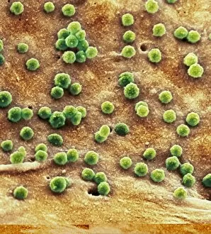
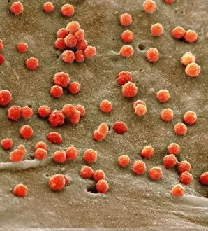
Macrophage engulfing TB bacteria. Coloured scanning electron micrograph (SEM) of a macrophage white blood cell engulfing a tuberculosis (Mycobacterium tuberculosis) bacterium (orange)

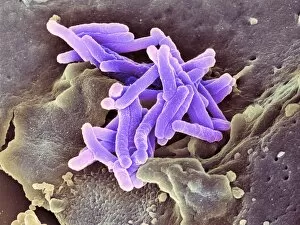
Background imageImmunology Collection: Macrophage engulfing TB bacteria, SEM C014 / 0598

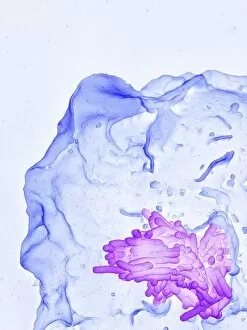
Macrophage engulfing TB bacteria, SEM C014 / 0598
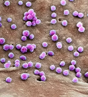
Macrophage engulfing TB bacteria. Coloured scanning electron micrograph (SEM) of a macrophage white blood cell engulfing a tuberculosis (Mycobacterium tuberculosis) bacterium (purple)

Background imageImmunology Collection: Macrophage engulfing TB bacteria, SEM C014 / 0595

Macrophage engulfing TB bacteria, SEM C014 / 0595
Macrophage engulfing TB bacteria. Coloured scanning electron micrograph (SEM) of a macrophage white blood cell engulfing a tuberculosis (Mycobacterium tuberculosis) bacterium (pink)

Background imageImmunology Collection: Macrophage engulfing TB bacteria, SEM C014 / 0594

Macrophage engulfing TB bacteria, SEM C014 / 0594
Macrophage engulfing TB bacteria. Coloured scanning electron micrograph (SEM) of a macrophage white blood cell engulfing a tuberculosis (Mycobacterium tuberculosis) bacterium (yellow)

Background imageImmunology Collection: Macrophage engulfing TB bacteria, SEM C014 / 0593

Macrophage engulfing TB bacteria, SEM C014 / 0593
Macrophage engulfing TB bacteria. Coloured scanning electron micrograph (SEM) of a macrophage white blood cell engulfing a tuberculosis (Mycobacterium tuberculosis) bacterium (purple)

Background imageImmunology Collection: Macrophage engulfing TB bacteria, SEM C014 / 0592

Macrophage engulfing TB bacteria, SEM C014 / 0592
Macrophage engulfing TB bacteria. Coloured scanning electron micrograph (SEM) of a macrophage white blood cell engulfing a tuberculosis (Mycobacterium tuberculosis) bacterium (orange)

Background imageImmunology Collection: Macrophage engulfing TB bacteria, SEM C014 / 0590

Macrophage engulfing TB bacteria, SEM C014 / 0590
Macrophage engulfing TB bacteria. Coloured scanning electron micrograph (SEM) of a macrophage white blood cell engulfing a tuberculosis (Mycobacterium tuberculosis) bacterium (blue)

Background imageImmunology Collection: Macrophage engulfing TB bacteria, SEM C014 / 0589

Macrophage engulfing TB bacteria, SEM C014 / 0589
Macrophage engulfing TB bacteria. Coloured scanning electron micrograph (SEM) of a macrophage white blood cell engulfing a tuberculosis (Mycobacterium tuberculosis) bacterium (yellow)

Background imageImmunology Collection: Macrophage engulfing TB bacteria, SEM C014 / 0591

Macrophage engulfing TB bacteria, SEM C014 / 0591
Macrophage engulfing TB bacteria. Coloured scanning electron micrograph (SEM) of a macrophage white blood cell engulfing a tuberculosis (Mycobacterium tuberculosis) bacterium (bright green)

Background imageImmunology Collection: Cell infected with HIV, SEM C014 / 0581

Cell infected with HIV, SEM C014 / 0581
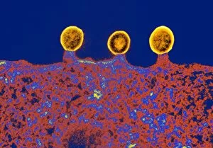
Cell infected with HIV. Coloured scanning electron micrograph (SEM) of HIV particles (round) budding from the membrane of a host cell

Background imageImmunology Collection: Cell infected with HIV, SEM C014 / 0580

Cell infected with HIV, SEM C014 / 0580
Cell infected with HIV. Coloured scanning electron micrograph (SEM) of HIV particles (round) budding from the membrane of a host cell

Background imageImmunology Collection: Cell infected with HIV, SEM C014 / 0579

Cell infected with HIV, SEM C014 / 0579
Cell infected with HIV. Coloured scanning electron micrograph (SEM) of HIV particles (round) budding from the membrane of a host cell

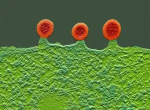
Background imageImmunology Collection: Cell infected with HIV, SEM C017 / 8338

Cell infected with HIV, SEM C017 / 8338
Cell infected with HIV. Coloured scanning electron micrograph (SEM) of HIV particles (orange) budding from the membrane of a host cell

Background imageImmunology Collection: Cell infected with HIV, SEM C017 / 8339

Cell infected with HIV, SEM C017 / 8339
Cell infected with HIV. Coloured scanning electron micrograph (SEM) of HIV particles (orange) budding from the membrane of a host cell

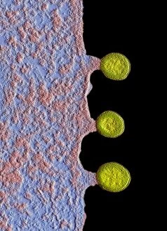
Background imageImmunology Collection: Cell infected with HIV, SEM C017 / 8337

Cell infected with HIV, SEM C017 / 8337
Cell infected with HIV. Coloured scanning electron micrograph (SEM) of HIV particles (yellow) budding from the membrane of a host cell

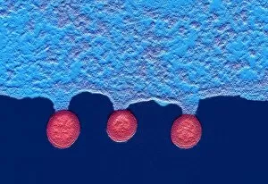
Background imageImmunology Collection: Cell infected with HIV, SEM C017 / 8336

Cell infected with HIV, SEM C017 / 8336
Cell infected with HIV. Coloured scanning electron micrograph (SEM) of HIV particles (pink) budding from the membrane of a host cell

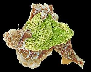
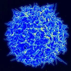
Background imageImmunology Collection: T-lymphocyte, SEM C017 / 8306

T-lymphocyte, SEM C017 / 8306
T-Lymphocyte, coloured scanning electron micrograph (SEM). T-lymphocytes, or T-cells, are a type of white blood cell and components of the bodys immune system. They mature in the thymus

Background imageImmunology Collection: Macrophage engulfing TB bacteria, SEM C017 / 8309

Macrophage engulfing TB bacteria, SEM C017 / 8309
Macrophage engulfing TB bacteria. Coloured scanning electron micrograph (SEM) of a macrophage white blood cell engulfing tuberculosis (Mycobacterium tuberculosis) bacteria (pink)

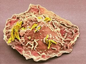
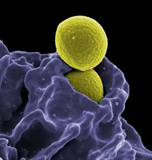
Background imageImmunology Collection: Neutrophil engulfing MRSA, SEM C017 / 8301

Neutrophil engulfing MRSA, SEM C017 / 8301
Neutrophil engulfing MRSA. Coloured scanning electron micrograph (SEM) of a neutrophil white blood cell (purple) engulfing methicillin-resistant Staphylococcus aureus bacteria (MRSA, yellow)

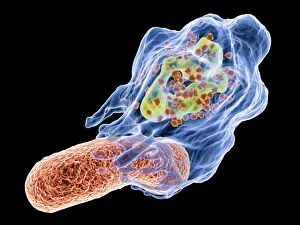
Background imageImmunology Collection: Macrophage feeding bacteria, artwork C017 / 8247

Macrophage feeding bacteria, artwork C017 / 8247
Computer artwork of a macrophage (blue), ingesting a bacterium (red), a process known as phagocytosis. The nucleus is depicted in green
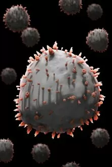
Background imageImmunology Collection: Interferon antagonism by viral protein C015 / 5421

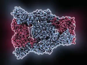
Interferon antagonism by viral protein C015 / 5421
Interferon (IFN) antagonism by viral protein. Molecular model of an orthopoxvirus IFN-gamma-binding protein tetramer bound to an IFN-gamma dimer
Background imageImmunology Collection: Interferon antagonism by viral protein C015 / 5420

Interferon antagonism by viral protein C015 / 5420
Interferon (IFN) antagonism by viral protein. Molecular model of an orthopoxvirus IFN-gamma-binding protein tetramer bound to an IFN-gamma dimer

Background imageImmunology Collection: Lymph node, light micrograph C015 / 6223

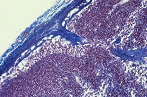
Lymph node, light micrograph C015 / 6223
Lymph node. Light micrograph of a section through a lymph node. The blue tissue at right is the fibrous outer capsule of the node. Beneath it is the subcapsular sinus (white)

Background imageImmunology Collection: Lymph node, light micrograph C015 / 6222

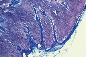
Lymph node, light micrograph C015 / 6222
Lymph node. Light micrograph of a section through a lymph node. The blue tissue at left is the fibrous outer capsule of the node. Beneath it is the subcapsular sinus (white)

Background imageImmunology Collection: Scorpion toxin bound to antibody C015 / 5158

Scorpion toxin bound to antibody C015 / 5158
Scorpion toxin bound to antibody, molecular model. The toxin is Cn2 from a Centruroides noxius scorpion. The antibodies are human single-chain antibody fragments (scFv) called 9004G

Background imageImmunology Collection: Scorpion toxin bound to antibody C015 / 5157

Scorpion toxin bound to antibody C015 / 5157
Scorpion toxin bound to antibody, molecular model. The toxin is Cn2 from a Centruroides noxius scorpion. The antibodies are human single-chain antibody fragments (scFv) called 9004G

Background imageImmunology Collection: Multiple sclerosis protein complex C015 / 3496

Multiple sclerosis protein complex C015 / 3496
Multiple sclerosis protein complex, molecular model. The proteins forming this complex are a T-cell receptor (TCR), a peptide antigen (myelin basic protein, MBP)

Background imageImmunology Collection: Multiple sclerosis protein complex

Multiple sclerosis protein complex, molecular model. The proteins forming this complex are a T-cell receptor (TCR), a peptide antigen (myelin basic protein, MBP)

Background imageImmunology Collection: Dendritic cell, SEM C016 / 4763

Dendritic cell, SEM C016 / 4763
Dendritic cell. Coloured ion-abrasion scanning electron micrograph (IA-SEM) showing sheet-like cellular extensions on a dendritic cell

Background imageImmunology Collection: Red and white blood cells, artwork C016 / 4629

Red and white blood cells, artwork C016 / 4629
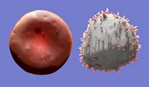
Red and white blood cells, computer artwork. Red blood cells are biconcave, giving them a large surface area for gas exchange, and highly elastic

Background imageImmunology Collection: Red and white blood cells, artwork C016 / 4630

Red and white blood cells, artwork C016 / 4630
Red and white blood cells, computer artwork. Red blood cells are biconcave, giving them a large surface area for gas exchange, and highly elastic

Background imageImmunology Collection: White blood cell, artwork C016 / 4628

White blood cell, artwork C016 / 4628
White blood cell, computer artwork. White blood cells are part of the bodys immune system

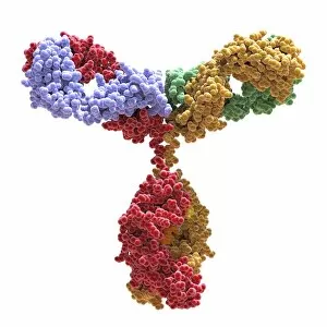
Background imageImmunology Collection: Immunoglobulin G antibody molecule C016 / 4456

Immunoglobulin G antibody molecule C016 / 4456
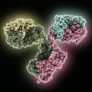
Immunoglobulin G antibody molecule. Computer artwork of a model of the secondary structure of immunoglobulin G (IgG). This is the most abundant immunoglobulin and is found in all body fluids

Background imageImmunology Collection: Immunoglobulin G antibody molecule C016 / 3452

Immunoglobulin G antibody molecule C016 / 3452
Immunoglobulin G antibody molecule. Computer model of the secondary structure of immunoglobulin G (IgG). This is the most abundant immunoglobulin and is found in all body fluids

Background imageImmunology Collection: Antigen presentation, SEM C016 / 3105

Antigen presentation, SEM C016 / 3105
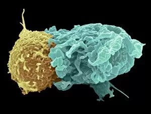
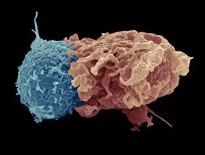
Antigen presentation. Coloured scanning electron micrograph (SEM) showing the interaction between a macrophage (blue) and a T helper lymphocyte (Th cell, yellow)

Background imageImmunology Collection: Antigen presentation, SEM C016 / 3104

Antigen presentation, SEM C016 / 3104
Antigen presentation. Coloured scanning electron micrograph (SEM) showing the interaction between a macrophage (brown) and a T helper lymphocyte (Th cell, red)

Background imageImmunology Collection: White blood cells, SEM C016 / 3102

White blood cells, SEM C016 / 3102
White blood cells. Coloured scanning electron micrograph (SEM) of white blood cells (leucocytes) from a sample of peripheral blood

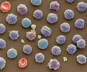
Background imageImmunology Collection: Blood cells, SEM C016 / 3101

Blood cells, SEM C016 / 3101
Blood cells. Coloured scanning electron micrograph (SEM) of a variety of different white blood cells (leucocytes, blue/purple) with two red blood cells (erythrocytes)

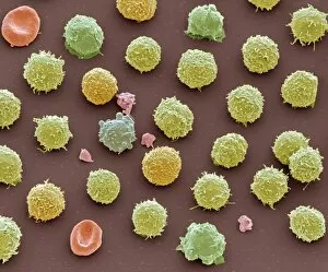
Background imageImmunology Collection: Blood cells, SEM C016 / 3100

Blood cells, SEM C016 / 3100
Blood cells. Coloured scanning electron micrograph (SEM) of a variety of different white blood cells (leucocytes, yellow/green) with two red blood cells (erythrocytes)

Background imageImmunology Collection: Blood cells, SEM C016 / 3096

Blood cells, SEM C016 / 3096
Blood cells. Coloured scanning electron micrograph of a lymphocyte white blood cell (left) and a red blood cell (erythrocyte, right)

Background imageImmunology Collection: Macrophage and platelets, SEM C016 / 3094

Macrophage and platelets, SEM C016 / 3094
Macrophage and platelets. Coloured scanning electron micrograph (SEM) of platelets (green) attached to a macrophage white blood cell (turquoise)

Background imageImmunology Collection: Platelets, SEM C016 / 3091

Platelets, SEM C016 / 3091
Platelets, coloured scanning electron micrograph (SEM). Platelets, or thrombocytes, are fragments of white blood cells. When blood clots

Background imageImmunology Collection: Lymphocyte white blood cell, SEM C016 / 3087

Lymphocyte white blood cell, SEM C016 / 3087
Lymphocyte white blood cell. Coloured scanning electron micrograph (SEM) of a lymphocyte white blood cell. Lymphocytes are involved in the immune systems defence mechanisms, lymph system


For sale as Licensed Images

Choose your image, Select your licence and Download the media

"Unleashing the Power of Immunology: Exploring the Intricate World of Immune Responses" Immunology, a fascinating field that delves into the complex mechanisms of our immune system, holds immense potential in combating diseases. T lymphocytes and cancer cells engage in a constant battle for supremacy, as depicted by SEM C001 / 1679. These tiny warriors play a crucial role in identifying and eliminating abnormal cells. The Immunoglobulin G antibody molecule (F007 / 9894) stands tall as one of our body's most powerful defenders against pathogens. Its remarkable structure enables it to neutralize harmful invaders with precision and efficiency. Neutrophils, exemplified by SEM C018 / 8596, showcase their extraordinary ability to engulf MRSA bacteria—an awe-inspiring sight indeed. Meanwhile, dendritic cells (artwork) act as vigilant sentinels, capturing antigens and presenting them to other immune cells for recognition. TEM reveals an up-close view of human white blood cells bearing HLA antigens—a key component in distinguishing self from non-self. Antibodies (artwork), resembling elegant warriors on a mission, bind specifically to foreign substances to mark them for destruction. HIV reverse transcription enzyme serves as a reminder of the challenges faced by immunologists worldwide. This relentless virus exploits our own cellular machinery but continues to be targeted through innovative research efforts. Human macrophages (TEM) demonstrate their exceptional phagocytic abilities while basophil white blood cells stand ready at the frontlines—both integral players in mounting effective immune responses against invading pathogens. Intriguingly captured by SEM imagery is bacteria infecting a macrophage—a visual representation highlighting how these microscopic organisms can exploit host defenses while also serving as valuable tools for studying infection dynamics. Lastly, Dohle bodies within blood cells offer insights into various pathological conditions affecting neutrophils—an essential clue guiding immunologists towards understanding and treating immune disorders.