
Lens Collection (#17)
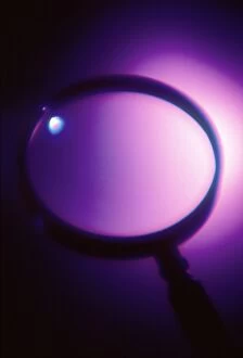
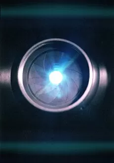
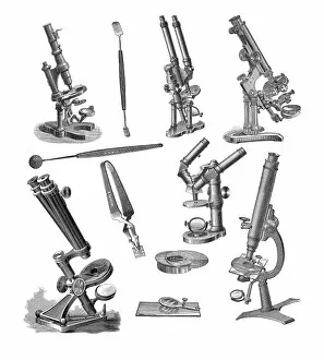
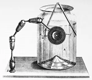
"Lens: Unveiling the World through Optics and Vision" Step into a world of wonder as we explore the captivating realm of lenses


For sale as Licensed Images
Choose your image, Select your licence and Download the media
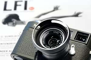
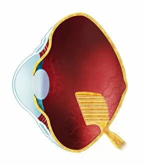
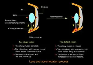
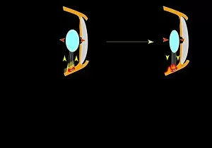
"Lens: Unveiling the World through Optics and Vision" Step into a world of wonder as we explore the captivating realm of lenses. From Sir Isaac Newton's groundbreaking work in his masterpiece "Opticks" to unraveling the mesmerizing color spectrum, lenses have been instrumental in our understanding of light. Delve into the intricate anatomy of the human eye, where lenses play a vital role in focusing incoming light onto our retinas. Witness how artists throughout history have skillfully depicted this remarkable feature, capturing its essence on canvas for eternity. Travel back to the 17th century and discover René Descartes' revolutionary optics theory that laid the foundation for modern lens technology. Marvel at camera lenses, which enable us to freeze moments in time with unparalleled clarity and precision. Take a closer look at nature's ingenious creation - the compound eye of a fly - magnified under an SEM Z340/0698 microscope. Appreciate its complexity and marvel at how it inspired advancements in lens design. Learn about The Royal Society's endorsement of skilled lens-grinders during the 1600s, recognizing their invaluable contributions to science and innovation. Explore Kilauea Lighthouse perched majestically on Kilauea Point, Hawaii – guiding ships safely with its powerful lens since 1913. Reflect upon history as you visit Vimy Canadian Memorial near Lens, Nord Pas de Calais – a poignant tribute honoring those who fought valiantly during World War I. Discover how cameras equipped with lenses captured these significant moments forever etched in our collective memory. Transport yourself to 1894 when hand-held stereoscopic cameras revolutionized photography; witness an advertisement from Stereoscopic Company showcasing their innovative products that brought images to life like never before. Witness firsthand how television cameras capture live events while skilled cameramen navigate every angle effortlessly. Celebrate your own roots by exploring your family tree through generations captured by various lenses throughout time. Finally, embrace the nostalgia of film cameras and their timeless charm.