Light Micrograph Collection (#4)

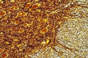
Capturing the intricate beauty of biological structures, a light micrograph reveals the mesmerizing complexity of cerebellum tissue

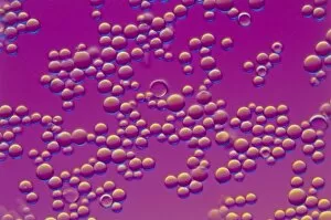
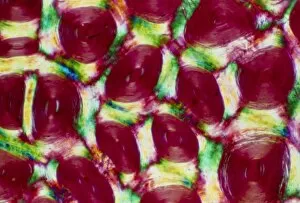
Background imageLight Micrograph Collection: LM of HPLC column packing

LM of HPLC column packing
Light micrograph of resin beads used in High Pressure Liquid Chromatography (HPLC), a technique for separating closely-related components from a sample solution

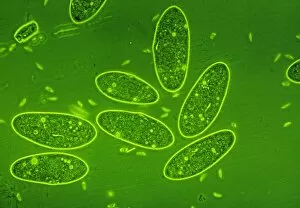
Background imageLight Micrograph Collection: LM of the protozoan Paramecium sp

LM of the protozoan Paramecium sp
Light micrograph of the ciliate protozoan, Paramecium sp. They are one of the most highly- evolved of all unicellular animals, and are plentiful in fresh water ponds

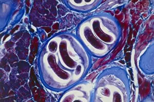
Background imageLight Micrograph Collection: LM of Trichina larvae encysted in muscle tissue

LM of Trichina larvae encysted in muscle tissue
Light micrograph of Trichina spiralis (formerly called Trichinella spiralis) larvae encysted in human muscle tissue. Each cyst (blue)

Background imageLight Micrograph Collection: LM of a field of red and white blood cells

LM of a field of red and white blood cells
Light micrograph of a sample of normal human blood, showing numerous erythrocytes (red blood cells) and four leucocytes (white blood cells) with purple-stained, lobed nuclei

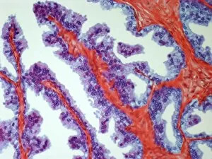
Background imageLight Micrograph Collection: LM of mammary gland

LM of mammary gland
Light micrograph of normal female breast (mammary gland) tissue from a non-pregnant woman, showing aggregations of glandular lobules

Background imageLight Micrograph Collection: Human skin section, light micrograph

Human skin section, light micrograph
Human skin. Light micrograph of a section through healthy human skin. The outer surface of the skin is at top. The uppermost surface is the epidermis

Background imageLight Micrograph Collection: LM of of a single Paramecium sp

LM of of a single Paramecium sp
Light micrograph of the ciliate protozoan Paramecium sp. The internal structure of the organism is particularly clear. The large dark area toward the top of the frame is the meganucleus

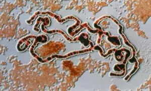
Background imageLight Micrograph Collection: LM of Drosophila giant (polytene) chromosome

LM of Drosophila giant (polytene) chromosome
Giant chromosome. Coloured computer-enhanced light micrograph of a giant (polytene) chromosome in a salivary gland cell of the fruit fly Drosophila funebris

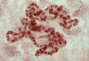
Background imageLight Micrograph Collection: LM of giant polytene chromosomes, Drosophila

LM of giant polytene chromosomes, Drosophila
Light micrograph of stained giant polytene chromosomes of Drosophila funebris. These chromosomes are found in the salivary glands of D.funebris and many other fly species

Background imageLight Micrograph Collection: LM of paramecium sp

LM of paramecium sp
Light micrograph in phase contrast illumination of the ciliate protozoan, Paramecium sp. The internal structure of Paramecium is visible here with many food and water vacuoles seen as bright spots

Background imageLight Micrograph Collection: LM of rhino horn

LM of rhino horn
Polarised light micrograph of a cross-section of horn from the Indian rhinoceros. The horn consists throughout of keratin, and does not have a bony pith (unlike those of cattle)

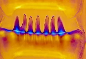
Background imageLight Micrograph Collection: Euro bank note hologram

Euro bank note hologram. Light micrograph (LM) of part of the security hologram on a euro bank note. The hologram is of the euro symbol (blue/green) & the bank note denomination (five, orange)

Background imageLight Micrograph Collection: LM of live human blood cells

LM of live human blood cells
Light micrograph in interference contrast illumination of human blood cells. The majority of the cells seen are red cells, carriers of oxygen throughout the body

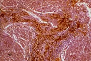
Background imageLight Micrograph Collection: LM of human liver showing alcoholic cirrhosis

LM of human liver showing alcoholic cirrhosis
Cirrhosis. Light micrograph of a section of human liver tissue showing cirrhosis due to excessive alcohol consumption. The liver responds to the injury or death of some of its cells by producing

Background imageLight Micrograph Collection: Blastocyst embryo, light micrograph F008 / 3573

Blastocyst embryo, light micrograph F008 / 3573
Blastocyst embryo, light micrograph. This embryo is five to six days old

Background imageLight Micrograph Collection: LM of a section through the human spinal cord

LM of a section through the human spinal cord
Light micrograph of a transverse section through the human spinal cord. Grey matter, consisting of unmyelinated nerve cells

Background imageLight Micrograph Collection: Chilodonella ciliate protozoa, LM

Chilodonella ciliate protozoa, LM
Chilodonella protozoa. Light micrograph of several Chilodonella sp. ciliate protozoa. Chilodonella is a single-celled animal that is found in freshwater

Background imageLight Micrograph Collection: LM of Waved Whelk tooth and radula

LM of Waved Whelk tooth and radula
Light micrograph in interference contrast illumination of a tooth plate on the radula of the Waved Whelk (Buccinum undatum). The radula is a strip-like organ studded with rows of horny tooth plates

Background imageLight Micrograph Collection: LM of radula of slug

LM of radula of slug
Light micrograph in interference contrast illumination of the radula of the Black Slug (Arion ater). The radula is a horny strip of tooth plates found in most molluscs

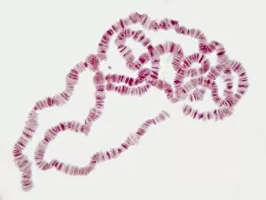
Background imageLight Micrograph Collection: Giant chromosomes, light micrograph P657 / 0038

Giant chromosomes, light micrograph P657 / 0038
Giant chromosomes. Light micrograph of giant (polytene) chromosomes from a fruit fly (Drosophila busckii). These chromosomes are formed from repeated rounds of DNA (deoxyribonucleic acid)

Background imageLight Micrograph Collection: Human skin section, light micrograph P710 / 0472

Human skin section, light micrograph P710 / 0472
Human skin. Light micrograph of a section through healthy human skin. The outer surface of the skin is at top. The uppermost surface is the epidermis

Background imageLight Micrograph Collection: LM of Kodachrome 64 film emulsion

LM of Kodachrome 64 film emulsion
Light micrograph of the emulsion from Kodachrome 64 colour transparency film. This film works on a subtractive process, a method of colour synthesis whereby two or more superimposed colourants are

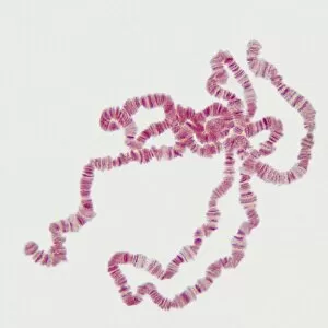
Background imageLight Micrograph Collection: Giant chromosomes, light micrograph P657 / 0037

Giant chromosomes, light micrograph P657 / 0037
Giant chromosomes. Light micrograph of giant (polytene) chromosomes from a fruit fly (Drosophila busckii). These chromosomes are formed from repeated rounds of DNA (deoxyribonucleic acid)
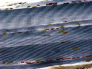
Background imageLight Micrograph Collection: Skeletal muscle fibres, light micrograph

Skeletal muscle fibres, light micrograph
Skeletal muscle fibres. Light micrograph of skeletal muscle fibres showing the characteristic banding. Cell nuclei are yellow and red blood cells, in capillaries in the endomycium, are red

Background imageLight Micrograph Collection: Coccidiosis, light micrograph F005 / 6082

Coccidiosis, light micrograph F005 / 6082
Coccidiosis. Light micrograph of a section through an hepatic bile duct infected with cocidian protozoa (green). Magnification: x300 when printed at 10 centimetres wide

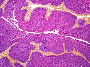
Background imageLight Micrograph Collection: Bladder cancer, light micrograph F005 / 6066

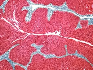
Bladder cancer, light micrograph F005 / 6066
Bladder cancer. Light micrograph of a section through a transitional cell carcinoma (TCC, cancer) of the bladder wall. TCC is the most common form of bladder cancer

Background imageLight Micrograph Collection: Nerve bundle, light micrograph

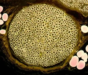
Nerve bundle, light micrograph
Nerve bundle. Light micrograph of a section through a nerve bundle from the sciatic nerve. Myelin sheaths (dark blue circles) can be seen surrounding the axons (light blue dots)

Background imageLight Micrograph Collection: Secondary lung cancer, light micrograph F005 / 6072

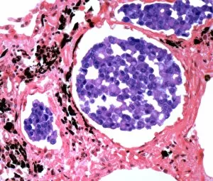
Secondary lung cancer, light micrograph F005 / 6072
Secondary lung cancer. Light micrograph of a section through a secondary carcinoma of the lung. This cancer has metastasised (spread) from a primary tumour of the breast

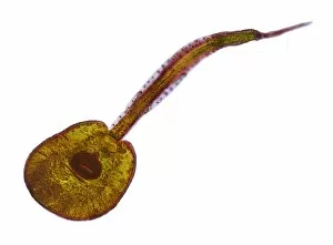
Background imageLight Micrograph Collection: Immature fluke, light micrograph

Immature fluke, light micrograph
Fluke. Light micrograph of an immature parasitic trematode (fluke). Magnification: x15 when printed at 10 centimetres wide

Background imageLight Micrograph Collection: Trachea, light micrograph

Trachea, light micrograph. The trachea is lined with ciliated columnar epithelium. Goblet cells (green), which secrete mucus, are seen in the epithelium

Background imageLight Micrograph Collection: Testicular cancer, light micrograph F005 / 6062

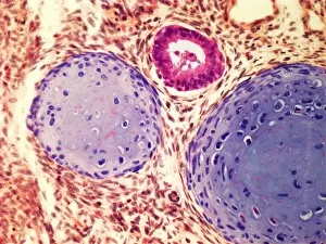
Testicular cancer, light micrograph F005 / 6062
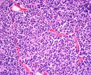
Testicular cancer. Light micrograph of a section through a teratoma of the testis, a rare type of cancer. Teratomas are encapsulated malignant (cancerous)

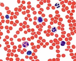
Background imageLight Micrograph Collection: Blood smear, light micrograph F005 / 6090

Blood smear, light micrograph F005 / 6090
Blood smear. Light micrograph showing normal red and white blood cells. At upper centre is monocyte, at top right and centre right are neutrophils, at bottom right is a basoophil

Background imageLight Micrograph Collection: Prostate cancer, light micrograph F005 / 6093

Prostate cancer, light micrograph F005 / 6093
Prostate cancer. Light micrograph of a section through a carcinoma (cancer) of the prostate. Magnification: x200 when printed at 10 centimetres wide

Background imageLight Micrograph Collection: Bladder cancer, light micrograph F005 / 6065

Bladder cancer, light micrograph F005 / 6065
Bladder cancer. Light micrograph of a section through a transitional cell carcinoma (TCC, cancer) of the bladder wall. TCC is the most common form of bladder cancer

Background imageLight Micrograph Collection: Secondary liver cancer, light micrograph F005 / 6100

Secondary liver cancer, light micrograph F005 / 6100
Secondary liver cancer. Light micrograph of a section through liver tissue showing a secondary cancer that had metastasised (spread) from a primary small cell lung cancer

Background imageLight Micrograph Collection: Skin cancer, light micrograph F005 / 6061

Skin cancer, light micrograph F005 / 6061
Skin cancer. Light micrograph of a section through a basal cell carcinoma (rodent ulcer) a skin cancer. Magnification: x80 when printed at 10 centimetres wide

Background imageLight Micrograph Collection: Secondary heart cancer, light micrograph F005 / 6069

Secondary heart cancer, light micrograph F005 / 6069
Secondary heart cancer. Light micrograph of a section through cardiac muscle showing a metastatic seminoma. This is a germ cell tumour that originated in the testis

Background imageLight Micrograph Collection: Scalp skin showing hair follicles, LM

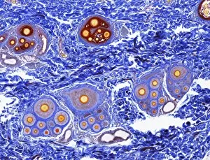
Scalp skin showing hair follicles, LM
Scalp skin. Light micrograph of a section through skin from the scalp. Numerous hair shafts are seen (purple rings), which are surrounded by connective tissue (dark blue)

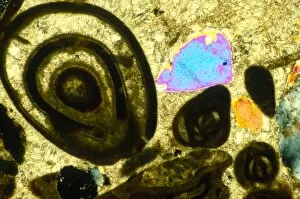
Background imageLight Micrograph Collection: Polarised LM of Bracklesham limestone, thin section

Polarised LM of Bracklesham limestone, thin section
Polarised light micrograph of Bracklesham limestone in thin section. The limestone takes its name from the town on the south coast of England where the limestone beds are found

Background imageLight Micrograph Collection: Fluke larva, light micrograph

Fluke larva, light micrograph
Fluke larva. Light micrograph of a parasitic trematode (fluke) larva. Magnification: x75 when printed at 10 centimetres wide

Background imageLight Micrograph Collection: Kidney cancer, light micrograph F005 / 6071

Kidney cancer, light micrograph F005 / 6071
Kidney cancer. Light micrograph of a section through a Wilms tumour of the kidney. This solid tumour generally affects children, with most cases occurring between 3 and 4 years of age

Background imageLight Micrograph Collection: Uterine cancer, light micrograph F005 / 6074

Uterine cancer, light micrograph F005 / 6074
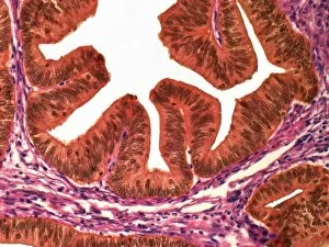
Uterine cancer. Light micrograph of a section through an adenocarcinoma (cancer) of the endometrium (womb lining). Magnification: x300 when printed at 10 centimetres

Background imageLight Micrograph Collection: Coccidiosis, light micrograph F005 / 6081

Coccidiosis, light micrograph F005 / 6081
Coccidiosis. Light micrograph of a section through an hepatic bile duct infected with cocidian protozoa (purple). Magnification: x300 when printed at 10 centimetres wide

Background imageLight Micrograph Collection: Uterine cancer, light micrograph F005 / 6073

Uterine cancer, light micrograph F005 / 6073
Uterine cancer. Light micrograph of a section through an adenocarcinoma (cancer) of the endometrium (womb lining). Magnification: x300 when printed at 10 centimetres

Background imageLight Micrograph Collection: Embryonic smooth muscle cell C018 / 8595

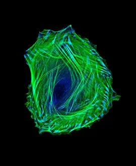
Embryonic smooth muscle cell C018 / 8595
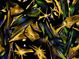
Embryonic smooth muscle cell, immunofluorescence micrograph. Actin filaments are green, the protein vinculin is light blue. Actin is part of the cytoskeleton

Background imageLight Micrograph Collection: Neural progenitor cell differentiation C018 / 8758

Neural progenitor cell differentiation C018 / 8758
Neural progenitor cell differentiation. Fluorescence light micrograph of neural progenitor cells that have been grown in a medium that is selective for astrocytes for three weeks

Background imageLight Micrograph Collection: Mouse embryo, light micrograph C018 / 8600

Mouse embryo, light micrograph C018 / 8600
Mouse embryo head. Light micrograph of the head of a mouse (Mus sp.) embryo 13 days after fertilisation

Background imageLight Micrograph Collection: Graves disease, light micrograph

Graves disease, light micrograph
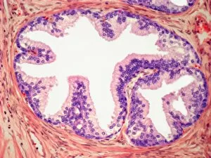
Graves disease. Light micrograph of a section through follicle from a thyroid gland affected by Graves disease. This is an autoimmune disease, where the thyroid is overactive


For sale as Licensed Images

Choose your image, Select your licence and Download the media

Capturing the intricate beauty of biological structures, a light micrograph reveals the mesmerizing complexity of cerebellum tissue. Delicate nerve and glial cells intertwine like a finely woven tapestry, forming the foundation for neurological function. In another stunning image, copper and magnesium sulphate crystals shimmer under the lens of a light microscope, showcasing their crystalline elegance. Moving beyond human anatomy, an awe-inspiring glimpse into early development is unveiled through a light micrograph of a human blastocyst. This embryonic stage brims with promise and potential as it prepares to embark on its journey towards life. Immunofluorescent LM unveils vibrant hues that illuminate neurons and astrocytes in breathtaking detail. These vital components of our nervous system come alive under fluorescent markers, revealing their interconnectedness in supporting brain function. Stepping away from biology but not lacking in fascination, caffeine crystals take center stage in yet another captivating light micrograph. Their jagged edges and distinct patterns mirror the stimulating effects they have on our bodies. Venturing deeper into brain tissue exploration, hippocampus tissue emerges as an enchanting subject for study. Its convoluted structure houses memories and emotions while providing insight into cognitive processes that shape who we are. Glial cells take on an ethereal quality when observed through confocal light microscopy. The interplay between these supportive cells becomes apparent as they weave together like delicate threads within neural networks. HeLa cells become protagonists under the gaze of a light microscope C017/8299 - immortalized cell lines that have revolutionized medical research since their discovery over half a century ago. Their unique characteristics continue to unlock mysteries about cancer and other diseases plaguing humanity. Intricacy extends beyond living organisms; even plant stems hold secrets waiting to be revealed by science's lens. A dicotyledon stem showcases its vascular bundles with precision while hinting at nature's ingenious design principles.