Light Micrograph Collection (#6)

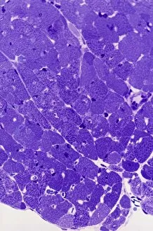
Capturing the intricate beauty of biological structures, a light micrograph reveals the mesmerizing complexity of cerebellum tissue

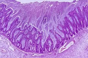
Background imageLight Micrograph Collection: Gall bladder lining, light micrograph F006 / 9814

Gall bladder lining, light micrograph F006 / 9814
Gall bladder lining. Light micrograph of a section through the gall bladder showing the folds of tall columnar epithelium (purple) overlying a connective tissue (brown)

Background imageLight Micrograph Collection: Bone cancer, light micrograph F006 / 9810

Bone cancer, light micrograph F006 / 9810
Bone cancer. Light micrograph of a section through an undifferentiated pleomorphic sarcoma of bone. This is a malignant (cancerous) tumour. Magnification: x200 when printed at 10 centimetres wide

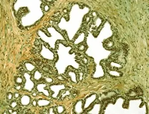
Background imageLight Micrograph Collection: Seminal vesicle, light micrograph F006 / 9809

Seminal vesicle, light micrograph F006 / 9809
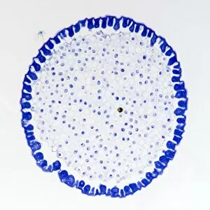
Seminal vesicle. Light micrograph of a section through the seminal vesicle showing the complex mucosal folding. The seminal vesicles are a pair of glands situated on each side of the male urinary

Background imageLight Micrograph Collection: Rectal papilloma, light micrograph F006 / 9807

Rectal papilloma, light micrograph F006 / 9807
Rectal papilloma, light micrograph. Magnification: x100 when printed at 10 centimetres wide

Background imageLight Micrograph Collection: Seminal vesicle, light micrograph F006 / 9808

Seminal vesicle, light micrograph F006 / 9808
Seminal vesicle. Light micrograph of a section through the seminal vesicle showing the complex mucosal folding. The seminal vesicles are a pair of glands situated on each side of the male urinary

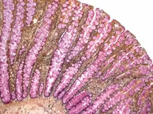
Background imageLight Micrograph Collection: Colon, light micrograph F006 / 9806

Colon, light micrograph F006 / 9806
Colon, light micrograph. Goblet cells, which secrete mucous to lubricate the epithelium, are red. Magnification: x150 when printed at 10 centimetres wide

Background imageLight Micrograph Collection: Metastatic breast cancer F006 / 9802

Metastatic breast cancer F006 / 9802
Metastatic breast cancer. Light micrograph of a section through a lymph node showing a malignant (cancerous) tumour (purple) that originated in the breast

Background imageLight Micrograph Collection: Acute bronchitis, light micrograph F006 / 9803

Acute bronchitis, light micrograph F006 / 9803
Acute bronchitis. Light micrograph of a section through a bronchus (airway) affected by acute bronchitis. The lumen is filled with pus (purple) consisting mainly of neutrophil white blood cells

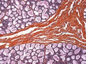
Background imageLight Micrograph Collection: Lactating breast tissue, light micrograph F006 / 9797

Lactating breast tissue, light micrograph F006 / 9797
Lactating breast tissue, light micrograph. Magnification: x150 when printed at 10 centimetres wide

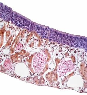
Background imageLight Micrograph Collection: Nasal mucosa, light micrograph F006 / 9798

Nasal mucosa, light micrograph F006 / 9798
Nasal mucosa. Light micrograph of a section through the nasal mucosa, the tissue that lines the airways of the nose. The epithelium is purple, Bowmans glands, which secrete mucus

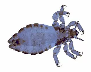
Background imageLight Micrograph Collection: Head louse, light micrograph F006 / 9796

Head louse, light micrograph F006 / 9796
Head louse, light micrograph

Background imageLight Micrograph Collection: Head louse, light micrograph F006 / 9795

Head louse, light micrograph F006 / 9795
Human head louse (Pediculus humanus capitis), light micrograph. Magnification: x20 when printed at 10 centimetres wide

Background imageLight Micrograph Collection: Giant chromosomes, light micrograph F006 / 9794

Giant chromosomes, light micrograph F006 / 9794
Giant chromosomes, light micrograph. Polytene chromosomes are over-sized chromosomes that have developed from standard chromosomes

Background imageLight Micrograph Collection: Giant chromosomes, light micrograph F006 / 9793

Giant chromosomes, light micrograph F006 / 9793
Giant chromosomes, light micrograph. Polytene chromosomes are over-sized chromosomes that have developed from standard chromosomes

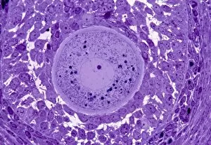
Background imageLight Micrograph Collection: Oocyte, light micrograph

Oocyte, light micrograph
Oocyte. Light micrograph of a section through an oocyte within a early antral follicle in an ovary. Oocytes are immature ova, or egg cells

Background imageLight Micrograph Collection: Cardiac muscle, light micrograph

Cardiac muscle, light micrograph
Cardiac muscle. Light micrograph of a section through muscle tissue from a heart, showing the cardiomyocyte cells. Heart muscle cells show central nuclei but the dominant feature is the abundance of

Background imageLight Micrograph Collection: Colitis, light micrograph

Colitis, light micrograph
Colitis. Light micrograph of a section through colon (large intestine) tissue affected by bacterial infection, causing injury to the mucosal lining, which has resulted in inflammation (colitis)

Background imageLight Micrograph Collection: Hairy scalp skin, light micrograph

Hairy scalp skin, light micrograph
Hairy scalp skin. Light micrograph of a section through healthy skin from a human scalp, showing the numerous hair follicles. The scalp is an example of hairy skin

Background imageLight Micrograph Collection: Oxalosis, light micrograph

Oxalosis, light micrograph
Oxalosis. Light micrograph of a section through adult bone tissue affected by oxalosis (primary hyperoxaluria), showing mature bone (green)

Background imageLight Micrograph Collection: Peripheral nerve, light micrograph

Peripheral nerve, light micrograph
Peripheral nerve. Light micrograph of a section through a peripheral nerve. This is a mixed nerve with myelinated axons (dark blue circles)

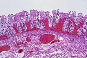
Background imageLight Micrograph Collection: Ischaemic bowel, light micrograph

Ischaemic bowel, light micrograph
Ischaemic bowel. Light micrograph of a section through a portion of intestine that has been damaged by an interruption of the blood supply, or ischaemia

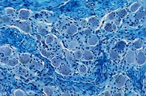
Background imageLight Micrograph Collection: Nerve ganglion, light micrograph

Nerve ganglion, light micrograph
Nerve ganglion. Light micrograph of a section through a dorsal (sensory) spinal root ganglion associated with a sensory nerve root of the spinal cord. Sensory information from peripheral sites e.g

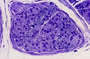
Background imageLight Micrograph Collection: Myelinated nerve, light micrograph

Myelinated nerve, light micrograph
Myelinated nerve. Light micrograph of a section through a peripheral myelinated nerve, showing many individual axons each covered with deep blue-staining myelin sheaths

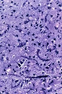
Background imageLight Micrograph Collection: Glial cells, light micrograph

Glial cells, light micrograph
Glial cells in the brain. Light micrograph of a section through glial cells (dark) in the grey matter of the brain. Due to their star shape these glial cells are called astrocytes

Background imageLight Micrograph Collection: Copper sulphate crystals light micrograph

Copper sulphate crystals light micrograph
Copper sulphate crystals, polarised light micrograph. Magnification: x2.5, when printed 10 centimetres wide

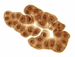
Background imageLight Micrograph Collection: Cirrhosis of the liver, light micrograph

Cirrhosis of the liver, light micrograph
Cirrhosis of the liver. Light micrograph of a section through liver tissue affected by cirrhosis, showing a cluster of hepatocyte cells (orange)

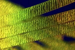
Background imageLight Micrograph Collection: Spirogyra algae, light micrograph C016 / 9592

Spirogyra algae, light micrograph C016 / 9592
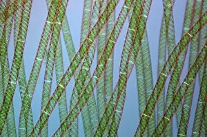
Spirogyra algae. Polarised light micrograph of Spirogyra sp. algae. This filamentous green algae is named for the spiral arrangement of its chloroplasts (green)

Background imageLight Micrograph Collection: Spirogyra algae, light micrograph C016 / 9594

Spirogyra algae, light micrograph C016 / 9594
Spirogyra algae. Rheinberg illuminated light micrograph of Spirogyra sp. algae. This filamentous green algae is named for the spiral arrangement of its chloroplasts (green)

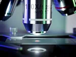
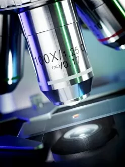
Background imageLight Micrograph Collection: Light microscopy C016 / 9700

Light microscopy C016 / 9700
Light microscopy. Light microscope being used to magnify a blood sample

Background imageLight Micrograph Collection: Desmids and spirogyra, light micrograph C016 / 9595

Desmids and spirogyra, light micrograph C016 / 9595
Desmids and spirogyra. Polarised light micrograph of two Micrasterias rotata desmids (round) and a single filament of Spirogyra sp. green alga (lower right)

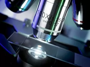
Background imageLight Micrograph Collection: Light microscopy C016 / 9703

Light microscopy C016 / 9703
Light microscopy. Light microscope being used to magnify a blood sample

Background imageLight Micrograph Collection: Fossil diatom, light micrograph C016 / 8603

Fossil diatom, light micrograph C016 / 8603
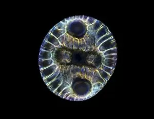
Fossil diatom. Darkfield illuminated light micrograph of a fossilised diatom. Diatoms are a group of photosynthetic, single-celled algae containing about 10, 000 species

Background imageLight Micrograph Collection: IVF treatment, light microscope C016 / 6536

IVF treatment, light microscope C016 / 6536
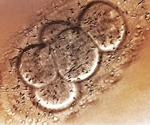
IVF treatment. Light microscope of human egg cells (large, round) and sperm (small dark ovals with tails) in a petri dish during in vitro fertilisation (IVF)

Background imageLight Micrograph Collection: Roundworm germ cells, light micrograph C016 / 9538

Roundworm germ cells, light micrograph C016 / 9538
Roundworm germ cells. Light micrograph of germ cells from a roundworm (Ascaris sp.), undergoing mitosis (nuclear division)

Background imageLight Micrograph Collection: IVF treatment, light microscope C016 / 6535

IVF treatment, light microscope C016 / 6535
IVF treatment. Light microscope of human egg cells (large, round) and sperm (small dark ovals with tails) in a petri dish during in vitro fertilisation (IVF)

Background imageLight Micrograph Collection: IVF treatment, light microscope C016 / 6512

IVF treatment, light microscope C016 / 6512
IVF treatment. Light microscope of human egg cells (large, round) and sperm (small dark ovals with tails) in a petri dish during in vitro fertilisation (IVF)

Background imageLight Micrograph Collection: Light microscopy C016 / 9702

Light microscopy C016 / 9702
Light microscopy. Light microscope being used to magnify a blood sample

Background imageLight Micrograph Collection: Spirogyra algae, light micrograph C016 / 9593

Spirogyra algae, light micrograph C016 / 9593
Spirogyra algae. Polarised light micrograph of Spirogyra sp. algae. This filamentous green algae is named for the spiral arrangement of its chloroplasts (green)

Background imageLight Micrograph Collection: Micrasterias desmids, light micrograph C016 / 9596

Micrasterias desmids, light micrograph C016 / 9596
Micrasterias desmids. Rheinberg illuminated light micrograph of Micrasterias rotata desmid green algae. Desmids are a common group of freshwater single-celled algae that have intricate cell walls

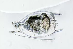
Background imageLight Micrograph Collection: Rotifer, light micrograph C016 / 9550

Rotifer, light micrograph C016 / 9550
Rotifer. Light micrograph of a freshwater rotifer (Brachionus calyciflorus). Rotifers are microscopic aquatic animals that are related to roundworms

Background imageLight Micrograph Collection: Deep brain stimulation and nerve cells C016 / 7200

Deep brain stimulation and nerve cells C016 / 7200
Deep brain stimulation (DBS). Computer artwork showing an electrode sending electrical impulses to nerve cells. DBS was developed for the treatment of Parkinsons disease

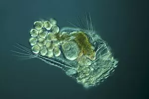
Background imageLight Micrograph Collection: Rotifer with eggs, light micrograph C016 / 8587

Rotifer with eggs, light micrograph C016 / 8587
Rotifer with eggs. Differential interference contrast (DIC) light micrograph of a rotifer (phylum Rotifera) carrying eggs (small, round)

Background imageLight Micrograph Collection: Light microscopy C016 / 9701

Light microscopy C016 / 9701
Light microscopy. Light microscope being used to magnify a blood sample

Background imageLight Micrograph Collection: Primordial egg follicles in an ovary

Primordial egg follicles in an ovary
Light micrograph of three primordial follicles (at centre) surrounded by flattened stroma cells in the human ovary. These follicles will form into oocytes which become eggs

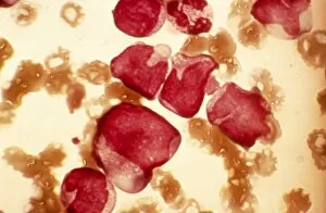
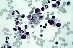
Background imageLight Micrograph Collection: Acute myeloid leukaemia, micrograph

Acute myeloid leukaemia, micrograph
Acute myeloid leukaemia. Light micrograph of blood cells from bone marrow in a case of acute myeloid leukaemia. These cells include a monocytoid myeloblast stem cell

Background imageLight Micrograph Collection: Elliptocytosis, light micrograph

Elliptocytosis, light micrograph
Elliptocytosis. Light micrograph of red blood cells in a case of elliptocytosis. Red blood cells (erythrocytes) carry oxygen and carbon dioxide to and from body tissues

Background imageLight Micrograph Collection: Cystine in bone marrow, light micrograph

Cystine in bone marrow, light micrograph
Cystine in bone marrow. Light micrograph of crystals of cystine among blood cells in a sample of bone marrow. Cystine is an amino acid that can form crystals in urine

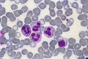
Background imageLight Micrograph Collection: Bone marrow blood cells, light micrograph

Bone marrow blood cells, light micrograph


For sale as Licensed Images

Choose your image, Select your licence and Download the media

Capturing the intricate beauty of biological structures, a light micrograph reveals the mesmerizing complexity of cerebellum tissue. Delicate nerve and glial cells intertwine like a finely woven tapestry, forming the foundation for neurological function. In another stunning image, copper and magnesium sulphate crystals shimmer under the lens of a light microscope, showcasing their crystalline elegance. Moving beyond human anatomy, an awe-inspiring glimpse into early development is unveiled through a light micrograph of a human blastocyst. This embryonic stage brims with promise and potential as it prepares to embark on its journey towards life. Immunofluorescent LM unveils vibrant hues that illuminate neurons and astrocytes in breathtaking detail. These vital components of our nervous system come alive under fluorescent markers, revealing their interconnectedness in supporting brain function. Stepping away from biology but not lacking in fascination, caffeine crystals take center stage in yet another captivating light micrograph. Their jagged edges and distinct patterns mirror the stimulating effects they have on our bodies. Venturing deeper into brain tissue exploration, hippocampus tissue emerges as an enchanting subject for study. Its convoluted structure houses memories and emotions while providing insight into cognitive processes that shape who we are. Glial cells take on an ethereal quality when observed through confocal light microscopy. The interplay between these supportive cells becomes apparent as they weave together like delicate threads within neural networks. HeLa cells become protagonists under the gaze of a light microscope C017/8299 - immortalized cell lines that have revolutionized medical research since their discovery over half a century ago. Their unique characteristics continue to unlock mysteries about cancer and other diseases plaguing humanity. Intricacy extends beyond living organisms; even plant stems hold secrets waiting to be revealed by science's lens. A dicotyledon stem showcases its vascular bundles with precision while hinting at nature's ingenious design principles.