
Light Micrograph Collection (#2)
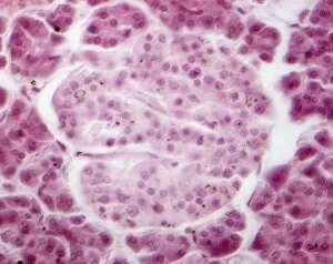
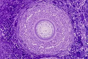
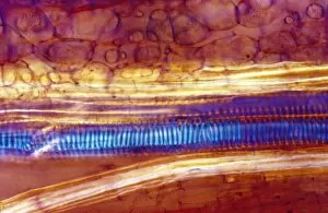
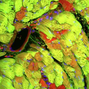
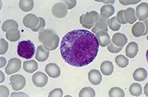
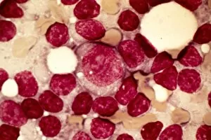
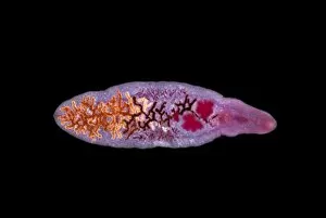
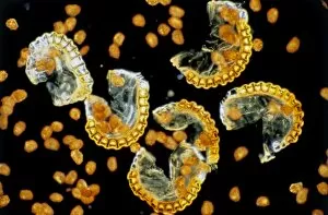
Capturing the intricate beauty of biological structures, a light micrograph reveals the mesmerizing complexity of cerebellum tissue


For sale as Licensed Images
Choose your image, Select your licence and Download the media
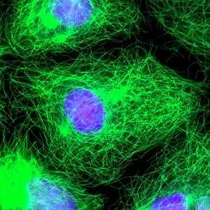
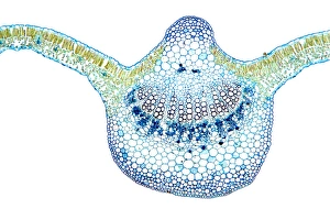
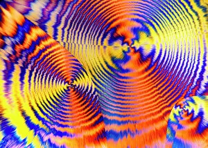
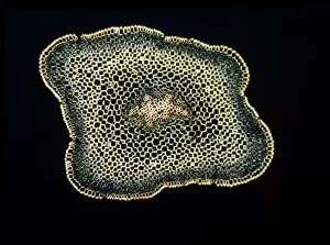
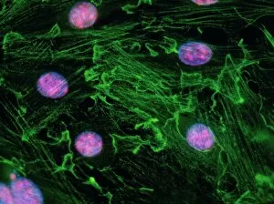
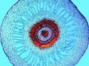
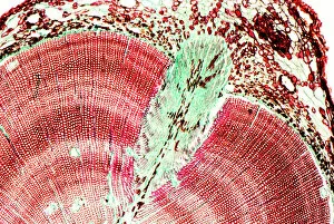
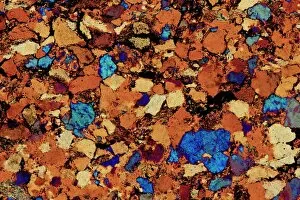
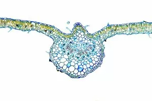
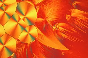
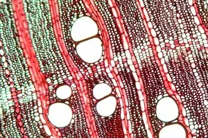
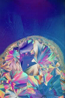
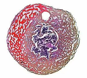
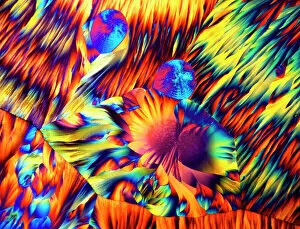
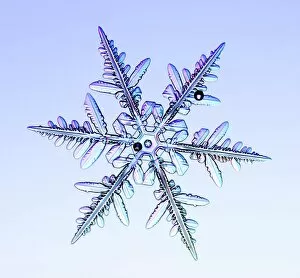
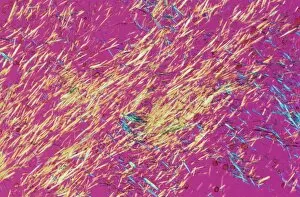
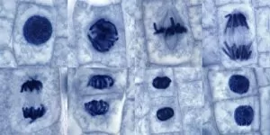
Capturing the intricate beauty of biological structures, a light micrograph reveals the mesmerizing complexity of cerebellum tissue. Delicate nerve and glial cells intertwine like a finely woven tapestry, forming the foundation for neurological function. In another stunning image, copper and magnesium sulphate crystals shimmer under the lens of a light microscope, showcasing their crystalline elegance. Moving beyond human anatomy, an awe-inspiring glimpse into early development is unveiled through a light micrograph of a human blastocyst. This embryonic stage brims with promise and potential as it prepares to embark on its journey towards life. Immunofluorescent LM unveils vibrant hues that illuminate neurons and astrocytes in breathtaking detail. These vital components of our nervous system come alive under fluorescent markers, revealing their interconnectedness in supporting brain function. Stepping away from biology but not lacking in fascination, caffeine crystals take center stage in yet another captivating light micrograph. Their jagged edges and distinct patterns mirror the stimulating effects they have on our bodies. Venturing deeper into brain tissue exploration, hippocampus tissue emerges as an enchanting subject for study. Its convoluted structure houses memories and emotions while providing insight into cognitive processes that shape who we are. Glial cells take on an ethereal quality when observed through confocal light microscopy. The interplay between these supportive cells becomes apparent as they weave together like delicate threads within neural networks. HeLa cells become protagonists under the gaze of a light microscope C017/8299 - immortalized cell lines that have revolutionized medical research since their discovery over half a century ago. Their unique characteristics continue to unlock mysteries about cancer and other diseases plaguing humanity. Intricacy extends beyond living organisms; even plant stems hold secrets waiting to be revealed by science's lens. A dicotyledon stem showcases its vascular bundles with precision while hinting at nature's ingenious design principles.