Magnified Image Collection (page 2)

"Exploring the unseen wonders of the microscopic world: from particle tracks to intricate tissues and beyond

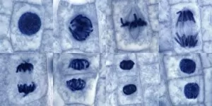
Background imageMagnified Image Collection: Plant cell mitosis, light micrograph

Plant cell mitosis, light micrograph
Plant cell mitosis. Light micrograph of root tip cells from an onion (Allium sp.) undergoing cell division (mitosis). From top left to bottom right

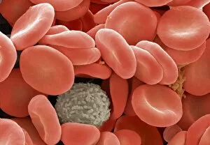
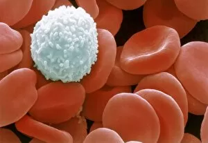
Background imageMagnified Image Collection: Red and white blood cells

Red and white blood cells. Coloured scanning electron micrograph (SEM) showing two types of human blood cells. Red blood cells (or erythrocytes, red) and a white blood cell (or leucocyte)

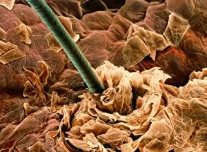
Background imageMagnified Image Collection: Coloured SEM of a human hair on the skin

Coloured SEM of a human hair on the skin
Hair on skin. Coloured Scanning Electron Micro- graph (SEM) of a hair on the surface of human skin. At centre is a shaft of hair (dark grey) which has emerged from a follicle in the skin

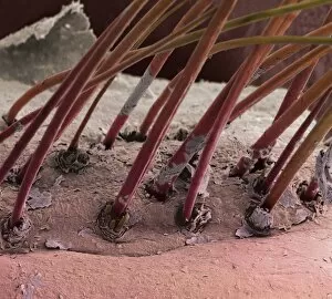
Background imageMagnified Image Collection: Eyelash hairs, SEM

Eyelash hairs, SEM
Eyelash hairs and skin. Coloured scanning electron micrograph (SEM) of eyelash hairs growing from the surface of human skin

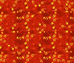
Background imageMagnified Image Collection: Human sperm cells

Human sperm cells
Sperm. Computer artwork of human sperm (spermatozoa), the sex cells of the male. Each sperm is about 0.065 millimetres in length

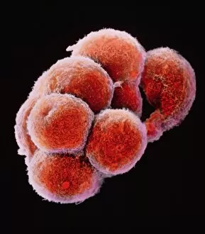
Background imageMagnified Image Collection: False-colour SEM of embryo at the morula stage

False-colour SEM of embryo at the morula stage
Embryo development. False-colour scanning electron micrograph of an embryo at the early stage known as the morula. The egg reaches this phase about 4 days after fertilisation after a series of

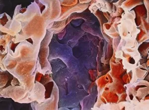
Background imageMagnified Image Collection: Alveolus in human lung

Alveolus in human lung
Alveolus. False-colour scanning electron micrograph (SEM) of an alveolus (blue cavity), one of the microscopic air sacs of the lungs

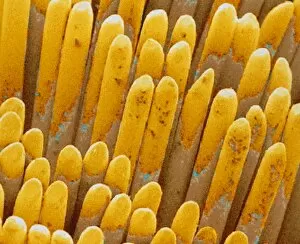
Background imageMagnified Image Collection: SEM of rod & cone cell of the retina

SEM of rod & cone cell of the retina
False-colour scanning electron micrograph (SEM) of rod and cone cells of the eye retina. Cone cells (blue) and more numerous rod cells (pink, purple) are specialized light-sensitive cells

Background imageMagnified Image Collection: Starch grains, SEM

Starch grains, SEM
Starch grains. Coloured scanning electron micrograph (SEM) of starch grains (orange) in a black-eyed pea (Vigna sp.). Starch is synthesised from sucrose

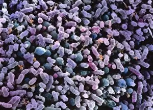
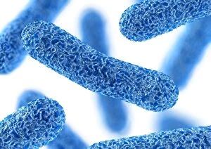
Background imageMagnified Image Collection: Rod-shaped bacteria

Rod-shaped bacteria. Computer artwork of rod- shaped bacteria (bacilli). Typical bacilli are E. coli and Salmonella bacteria, though there are many others as well

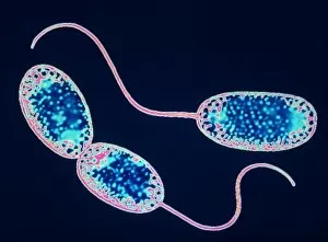
Background imageMagnified Image Collection: Nitrobacter sp bacteria, TEM

Nitrobacter sp bacteria, TEM

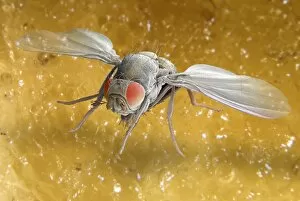
Background imageMagnified Image Collection: Fruit fly, SEM Z340 / 0662

Fruit fly, SEM Z340 / 0662
Fruit fly. Coloured scanning electron micrograph (SEM) of a fruit fly (Drosophila busckii) on a banana. Its compound eyes (red) are seen and its wings are outstretched

Background imageMagnified Image Collection: Fruit fly, SEM Z340 / 0663

Fruit fly, SEM Z340 / 0663
Fruit fly. Coloured scanning electron micrograph (SEM) of a fruit fly (Drosophila busckii) on a banana. Its compound eyes (red) are seen and its wings are outstretched

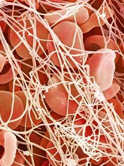
Background imageMagnified Image Collection: Blood clot, SEM P260 / 0123

Blood clot, SEM P260 / 0123
Blood clot. Coloured scanning electron micrograph (SEM) of a blood clot. The red blood cells (erythrocytes) are trapped in filaments of fibrin protein (pink)

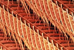
Background imageMagnified Image Collection: Coloured SEM of the feather of a wren

Coloured SEM of the feather of a wren
Feather. Coloured scanning electron micrograph of barbs, barbules and barbicels on the feather of a wren, Troglodytes troglodytes

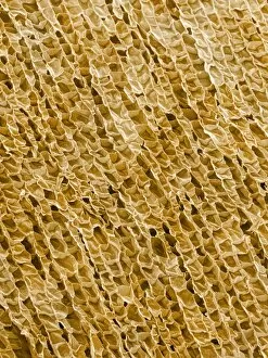
Background imageMagnified Image Collection: Wine cork, SEM

Wine cork, SEM
Wine cork. Coloured scanning electron micrograph (SEM) of a section through a wine cork. The structure of the cells can be seen. Cork is taken from the bark of the cork oak tree (Quercus suber)

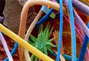
Background imageMagnified Image Collection: Col. SEM of filaments of a tooth brush

Col. SEM of filaments of a tooth brush
Toothbrush. Coloured scanning electron micrograph of the filaments of a toothbrush. Magnification: x37 at 6x7cm size. x74 at 8x6ins, x40 at 9x7cm master size

Background imageMagnified Image Collection: Light bulb filament, SEM

Light bulb filament, SEM
Tungsten light bulb filament, coloured scanning electron micrograph (SEM). The heat generated by an electrical current flowing through the filament makes it glow white hot

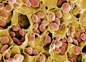
Background imageMagnified Image Collection: Colour SEM of red & white blood cells

Colour SEM of red & white blood cells
Blood cells. Coloured scanning electron micrograph (SEM) of human blood showing red and white cells. Red blood cells (erythrocytes) have a characteristic biconcave-disc shape and are numerous

Background imageMagnified Image Collection: Coloured SEM of household dust (allergens)

Coloured SEM of household dust (allergens)
Household dust. Coloured Scanning Electron Micro- graph (SEM) of a sample of household dust. Dust contains an assortment of substances which can cause asthma or other allergic reactions

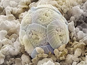
Background imageMagnified Image Collection: Calcareous phytoplankton fossil, SEM Z100 / 0212

Calcareous phytoplankton fossil, SEM Z100 / 0212
Calcareous phytoplankton fossil. Coloured scanning electron micrograph (SEM) of a fossil of the skeleton (coccosphere) of a coccolithotrope, a small marine algal organism

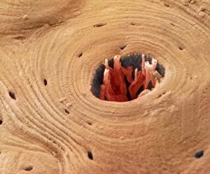
Background imageMagnified Image Collection: Coloured SEM of transverse section of compact bone

Coloured SEM of transverse section of compact bone
Compact bone. Coloured scanning electronmicrograph (SEM) of a transverse section ofcompact bone. The Haversian canal (dark centralarea) contains blood vessels

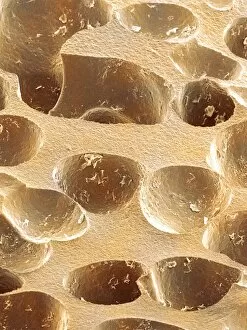
Background imageMagnified Image Collection: Synthetic cork, SEM

Synthetic cork, SEM
Synthetic cork. Coloured scanning electron micrograph (SEM) of a section through a plastic wine bottle cork. The air holes within the cork allow it to shrink and expand to fit the bottle neck

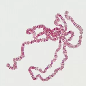
Background imageMagnified Image Collection: Giant chromosomes, light micrograph P657 / 0037

Giant chromosomes, light micrograph P657 / 0037
Giant chromosomes. Light micrograph of giant (polytene) chromosomes from a fruit fly (Drosophila busckii). These chromosomes are formed from repeated rounds of DNA (deoxyribonucleic acid)

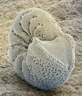
Background imageMagnified Image Collection: Foraminiferan fossil, SEM Z110 / 0239

Foraminiferan fossil, SEM Z110 / 0239
Foraminiferan fossil. Coloured scanning electron micrograph (SEM) of the fossilised shell (test) of a foraminiferan. Foraminifera are single-celled marine protozoa that construct

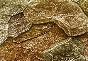
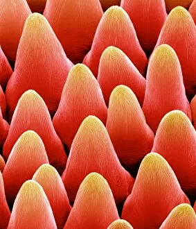
Background imageMagnified Image Collection: Coloured SEM of the surface of human skin

Coloured SEM of the surface of human skin
Human skin. Coloured scanning electron micrograph (SEM) of the surface of healthy human skin. The scaly appearance is due to the dead outer cells being constantly shed (sloughed off) and replaced

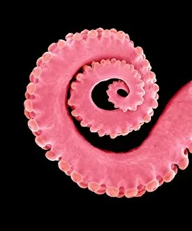
Background imageMagnified Image Collection: Octopus suckers, SEM C016 / 9672

Octopus suckers, SEM C016 / 9672
Octopus suckers. Coloured scanning electron micrograph (SEM) of suckers on an octopus (order Octopoda) arm. The suckers are used to grasp prey when hunting

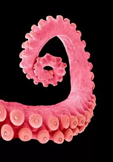
Background imageMagnified Image Collection: Octopus suckers, SEM C016 / 9669

Octopus suckers, SEM C016 / 9669
Octopus suckers. Coloured scanning electron micrograph (SEM) of suckers on an octopus (order Octopoda) arm. The suckers are used to grasp prey when hunting

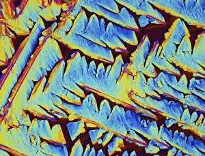
Background imageMagnified Image Collection: Oxytocin hormone crystals, PLM C016 / 7199

Oxytocin hormone crystals, PLM C016 / 7199
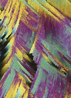
Oxytocin. Polarised light micrograph (PLM) of crystals of the female hormone oxytocin. In women this hormone is secreted naturally by the pituitary gland

Background imageMagnified Image Collection: Octopus suckers, SEM C016 / 9671

Octopus suckers, SEM C016 / 9671
Octopus suckers. Coloured scanning electron micrograph (SEM) of suckers on an octopus (order Octopoda) arm. The suckers are used to grasp prey when hunting

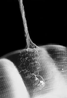
Background imageMagnified Image Collection: Synapse, SEM C018 / 0122

Synapse, SEM C018 / 0122
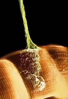
Synapse. Scanning electron micrograph (SEM) of a neuromuscular junction showing a motor neurone (vertical line) terminating on skeletal muscle fibres (across bottom frame)

Background imageMagnified Image Collection: Synapse, SEM

Synapse, SEM
Synapse. Coloured scanning electron micrograph (SEM) of a neuromuscular junction showing a motor neurone (green) terminating on skeletal muscle fibres (orange)

Background imageMagnified Image Collection: Salt crystal, light micrograph

Salt crystal, light micrograph
Salt. Polarised light micrograph of a crystal of sodium chloride, the common salt. This is a white crystalline compound widespread in nature as a solid (rock salt)

Background imageMagnified Image Collection: Testosterone hormone, light micrograph C015 / 6787

Testosterone hormone, light micrograph C015 / 6787
Testosterone hormone. Polarised light micrograph of crystals of the male sex hormone testosterone. Testosterone is the main human androgen hormone

Background imageMagnified Image Collection: Insulin crystals, light micrograph C017 / 8246

Insulin crystals, light micrograph C017 / 8246
Insulin. Polarised light micrograph (PLM) of crystals of the hormone insulin. The insulin molecule is made up of two chains of amino acids (A & B chains)

Background imageMagnified Image Collection: Blue green algae stigonema, LM C013 / 5286

Blue green algae stigonema, LM C013 / 5286
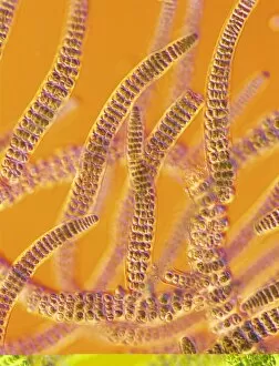
Light micrograph using differential interference contrast microscopy (DIC) of the filamentous blue-green algae (group name Cyanophycophyta), called stigonema

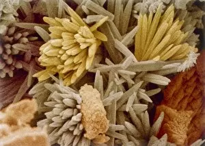
Background imageMagnified Image Collection: Bog moss, SEM

Bog moss, SEM
Bog moss. Coloured scanning electron micrograph (SEM) of bog moss (Sphagnum sp.). Magnification: x350 when printed at 10 centimetres wide

Background imageMagnified Image Collection: SEM of daisy pollen B786 / 0524

SEM of daisy pollen B786 / 0524
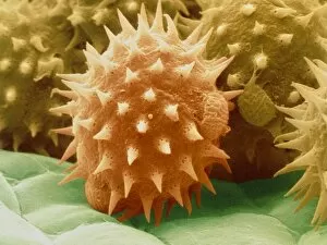
Daisy pollen. Coloured scanning electron micrograph (SEM) of pollen grains of a common Daisy (Bellis perennis). The outer wall (exine) has many small spiky projections

Background imageMagnified Image Collection: Coloured SEM of sunflower pollen grains

Coloured SEM of sunflower pollen grains
Pollen grains. Coloured Scanning Electron Micro- graph of pollen grains of the sunflower, Helianthus annuus. Pollen grain size, shape and surface texture differ from one plant species to another

Background imageMagnified Image Collection: Petal papillae, SEM

Petal papillae, SEM
Petal papillae. Coloured scanning electron micrograph (SEM) of papillae on the surface of a cultivated primrose (Primula obconica) petal. Papillae are projections from epidermal cells

Background imageMagnified Image Collection: Reflective belt material

Reflective belt material
Light micrograph in interference contrast illumination of a reflective material used in safety clothing. The material is retro-reflective

Background imageMagnified Image Collection: Crystals of silver on copper wire

Crystals of silver on copper wire
Light micrograph in incident illumination of crystalline silver deposited on a copper wire. Copper wire is suspended in a solution of silver nitrate

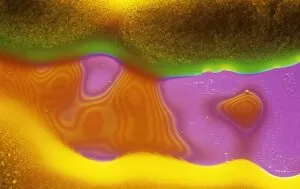
Background imageMagnified Image Collection: LM of melted arsenic disulphide

LM of melted arsenic disulphide
Light micrograph of molten arsenic disulphide. Interference contrast. Magnification: x200 at 35mm size
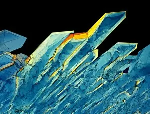
Background imageMagnified Image Collection: Kettle fur limescale

Kettle fur limescale
Kettle fur". Coloured scanning electron micrograph (SEM) of fur from a domestic kettle, showing the crystals that form in a hard water area

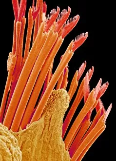
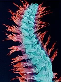
Background imageMagnified Image Collection: Fireworm bristles, SEM

Fireworm bristles, SEM
Fireworm bristles. Coloured scanning electron micrograph (SEM) of a fireworms bristles (setae). Fireworms (family Amphinomidae) are polychaete (meaning many-bristled) worms

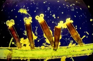
Background imageMagnified Image Collection: Sessile rotifers

Sessile rotifers. Light micrograph of five rotifers (Floscularia ringens) attached to vegetation. Each rotifer is encased in a sheath (brown) constructed from faecal pellets

Background imageMagnified Image Collection: Fireworm, SEM

Fireworm, SEM
Fireworm. Coloured scanning electron micrograph (SEM) of a fireworm (family Amphinomidae). This is a polychaete (meaning many-bristled) and segmented worm

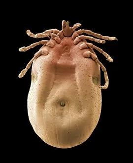
Background imageMagnified Image Collection: Brown dog tick, SEM

Brown dog tick, SEM
Brown dog tick. Coloured scanning electron micrograph (SEM) of a brown dog tick (Rhipicephalus sanguineus) engorged with blood. Ticks are parasitic arachnids


For sale as Licensed Images

Choose your image, Select your licence and Download the media

"Exploring the unseen wonders of the microscopic world: from particle tracks to intricate tissues and beyond. " "Unveiling the hidden beauty within: a closer look at cerebellum tissue through a mesmerizing light micrograph. " "A captivating glimpse into the mysteries of subatomic particles: behold the bubble chamber photo capturing sigma particle decay. " "The groundbreaking moment in science history: witness the first observation of omega-minus particle, forever changing our understanding of matter. " "Awe-inspiring beginnings: marvel at the intricate structure of a human blastocyst, where life takes its first steps. " "Diving deep into neural networks: an illuminating immunofluorescent LM reveals stunning neurons and astrocytes in all their glory. " "Unraveling nature's cycle within us: witness the uterus lining during menstruation, as seen through SEM imaging - a fascinating display of renewal and change. " "Peering into viral threats that loom above us all: explore avian flu virus up close, revealing its complex structure and potential dangers it holds. " "The crystalline beauty behind love and bonding: discover oxytocin hormone crystals under PLM C016 / 7196 microscopy - nature's secret ingredient for connection. " "A tapestry woven by nature itself: delve into intricately detailed fabric structures captured through SEM imaging - artistry on a microscopic scale. " "Glimpsing into windows to our souls with breathtaking precision: explore the iris of an eye like never before, revealing unique patterns that make each person truly one-of-a-kind. " "Revealing fragility beneath strength: examine osteoporotic bone under SEM, shedding light on this silent disease affecting millions worldwide.