Mandible Collection (page 11)

The mandible, also known as the jawbone, is a fascinating and crucial component of our skull anatomy

Background imageMandible Collection: Broken jawbone, X-ray

Broken jawbone, X-ray
Broken jawbone. X-ray of the jaw of a young female patient with a fractured mandible (lower jawbone). Two cracks can be seen in the bone, at right and bottom left

Background imageMandible Collection: Broken chin, 3D CT scan

Broken chin, 3D CT scan
Broken chin. Three-quarter side view 3D computed tomography (CT) scan of a patient with a broken chin. The lower jaw is fractured just below the bottom front incisor teeth

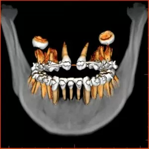
Background imageMandible Collection: Teeth, 3D CT scan

Teeth, 3D CT scan. Frontal view of a persons jaw and teeth with braces on the upper teeth. The wisdom teeth (upper left and upper right) have not yet penetrated the gums

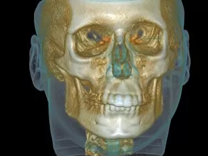
Background imageMandible Collection: Normal head, cone beam CT scan

Normal head, cone beam CT scan. Front view of a dental patients head. The scan was produced using a cone beam x-ray computed tomography (CT) scanner

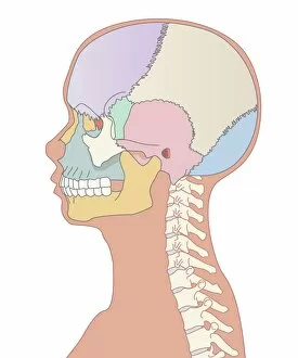
Background imageMandible Collection: Skull bones, artwork

Skull bones, artwork
Skull bones. Computer artwork showing the major bones of the human skull. These are: frontal (purple, upper left), parietal (green/beige, upper right), occipital (blue, lower right), temporal (pink)

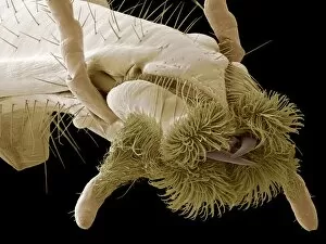
Background imageMandible Collection: Scorpion fly mouth, SEM

Scorpion fly mouth, SEM
Scorpion fly mouth. Coloured scanning electron micrograph (SEM) of a close-up of the mouth of a scorpion fly (Panorpa sp.)

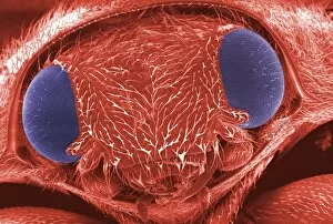
Background imageMandible Collection: Ladybird head, SEM

Ladybird head, SEM
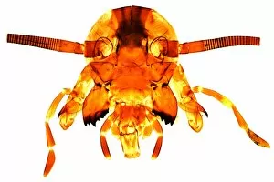
Ladybird head. Coloured scanning electron micrograph (SEM) of the head of a ladybird (family Coccinellidae). The compound eyes of this beetle are at left and right (blue)

Background imageMandible Collection: Skeletons head, artwork

Skeletons head, artwork
Skeletons head. Computer artwork showing the bones of the head, neck and shoulders of a human skeleton that is lying down

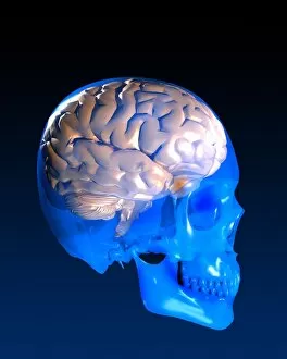
Background imageMandible Collection: Human brain and skull, artwork

Human brain and skull, artwork
Human brain and skull, computer artwork. The human brain occupies a relatively large volume within the skull compared to other primates

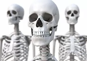
Background imageMandible Collection: Skeletons, artwork

Skeletons, artwork
Skeletons. Computer artwork of the skull and neck of a human skeleton, flanked by two other skeletons

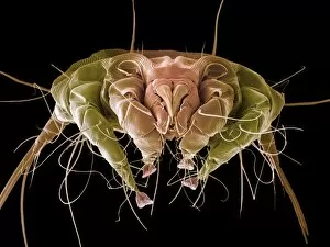
Background imageMandible Collection: Hedgehog mite, SEM

Hedgehog mite, SEM
Hedgehog mite (Caparinia sp.), coloured scanning electron micrograph (SEM). View of the mites specialised mouthparts with blunt chewing chelicerae (fang-like protrusions, centre)

Background imageMandible Collection: Human skull, artwork

Human skull, artwork. The skull consists of the cranium (upper area) which encases and protects the brain, and the facial bones

Background imageMandible Collection: Daddy long legs spider, SEM

Daddy long legs spider, SEM
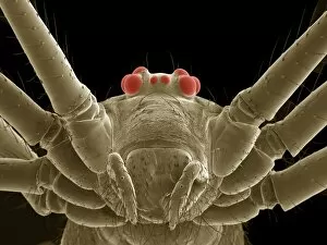
Daddy long legs spider. Coloured scanning electron micrograph (SEM) of the head of a daddy long legs, showing its eight eyes (red) above its mouth-parts (lower centre)

Background imageMandible Collection: Wasp head

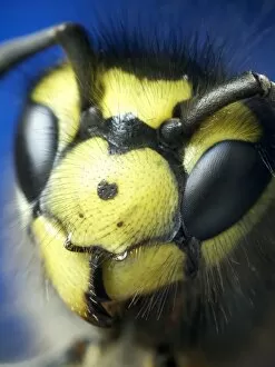
Wasp head. Close-up of the head of a European wasp (Vespula germanica), showing its two large compound eyes, and its antennae (black) between them

Background imageMandible Collection: Daddy long legs spider, SEM

Daddy long legs spider, SEM
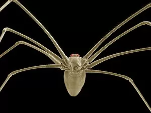
Daddy long legs spider. Coloured scanning electron micrograph (SEM) of a daddy long legs, showing its characteristic long legs

Background imageMandible Collection: Ant head, SEM

Ant head, SEM
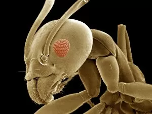
Ant (family Formicidae) head, coloured scanning electron micrograph (SEM). One of its compound eyes (red) is seen, as well as the two antennae that are mounted on the head between the eyes

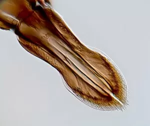
Background imageMandible Collection: Black fly proboscis, light micrograph

Black fly proboscis, light micrograph
Black fly proboscis. Differential interference contrast micrograph of the proboscis of a black fly (Simulium sp.). Female black flies feed on blood, while the males feed on nectar

Background imageMandible Collection: Black ant carrying grain seed

Black ant carrying grain seed
Black ant. Close up of a black ant (family Formicidae) carrying a grain seed in its large jaws (mandibles). Photographed in Israel

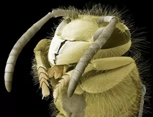
Background imageMandible Collection: Common wasp head, SEM

Common wasp head, SEM
Common wasp (Vespula vulgaris) head, coloured scanning electron micrograph (SEM). The antennae are seen on the top of the head, between the two large compound eyes

Background imageMandible Collection: Bee proboscis, light micrograph

Bee proboscis, light micrograph
Bee proboscis. Polarised light micrograph of the proboscis of the common honey bee (Apis mellifera). The bees proboscis (centre) consists of two outer angled parts (the galea)

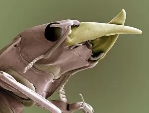
Background imageMandible Collection: Ground beetle head, SEM

Ground beetle head, SEM
Ground beetle. Coloured scanning electron micrograph (SEM) of the head of a ground beetle (family Carabidae). The beetles large jaws (mandibles) can be seen at upper right (yellow)

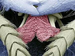
Background imageMandible Collection: Wasp tongue, SEM

Wasp tongue, SEM
Wasp tongue. Coloured scanning electron micrograph (SEM) of the tongue (red) of a wasp. Unlike the equivalent structure in bees (the proboscis)

Background imageMandible Collection: Gnat head, light micrograph

Gnat head, light micrograph
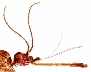
Gnat head. Light micrograph of the head (lower left), antennae (upper left), proboscis (lower right) and other mouthparts of a gnat (Culex pipiens)

Background imageMandible Collection: Cockroachs head, light micrograph

Cockroachs head, light micrograph
Cockroachs head. Light micrograph of the head and mouthparts of the American cockroach (Periplaneta orientalis). The head consists of two hemispherical compound eyes (top centre)

Background imageMandible Collection: Bulldog ant - mandibles, used with tenacity to seize and hold prey before stinging it

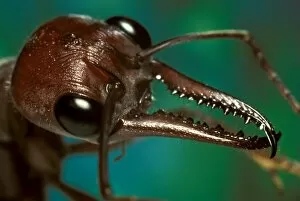
Bulldog ant - mandibles, used with tenacity to seize and hold prey before stinging it, hence the common name
AUS-504 Bulldog ant - mandibles, used with tenacity to seize and hold prey before stinging it, hence the common name. Australia Myrmecia brevinoda Jim Frazier-Densey Clyne / Auscape / ardea.com

Background imageMandible Collection: A snout-nosed katydid - with powerful mandibles used to crack seeds or give a painful nip if

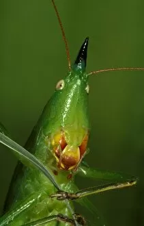
A snout-nosed katydid - with powerful mandibles used to crack seeds or give a painful nip if the insect is mishandled
AUS-463 A snout-nosed katydid - with powerful mandibles used to crack seeds or give a painful nip if the insect is mishandled. Australia Euconocephalus sp


For sale as Licensed Images

Choose your image, Select your licence and Download the media

The mandible, also known as the jawbone, is a fascinating and crucial component of our skull anatomy. Leonardo da Vinci's detailed sketches in his "Skull Anatomy" showcase the intricacies of this bone. A panoramic dental X-ray allows us to examine the mandible's structure and its relationship with teeth. Looking back in time, the Paranthropus boisei (Zinjanthropus) cranium (OH5) provides valuable insights into our evolutionary history. The study of human skulls through X-rays reveals not only the mandible but also other vital structures within our head. Nature never fails to amaze us; even tiny creatures like the red-barbed ant can be observed under a scanning electron microscope (SEM), revealing their unique mandibles adapted for various tasks. Meanwhile, a cross-section diagram illustrates how our mouth and jaw work together harmoniously. Insects are no exception when it comes to showcasing intriguing mandibular adaptations. Witnessing a red flour beetle in flight or examining an artwork depicting a Black Death rat flea reminds us of nature's diversity and complexity. However, sometimes unfortunate incidents occur that affect this essential bone. Fractured jawbones captured by X-rays remind us of both medical challenges and advancements in treating such injuries. Zooming further into nature's wonders, we encounter spiny spiders under SEMs - their menacing-looking yet functional jaws ready for capturing prey. Ultimately, whether studying ancient fossils or observing modern-day organisms, understanding the mandible plays an integral role in comprehending head and neck anatomy as depicted beautifully through artworks dedicated to this subject matter.