Mandible Collection (page 8)

The mandible, also known as the jawbone, is a fascinating and crucial component of our skull anatomy

Background imageMandible Collection: Sarcophilus laniarius, Tasmanian devil

Sarcophilus laniarius, Tasmanian devil
Jaw of a Tasmanian devil from the collections at the Natural History Museum

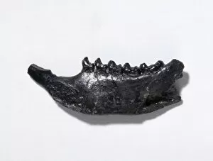
Background imageMandible Collection: Plesiadapis fodinatus, primate

Plesiadapis fodinatus, primate
Left section of a mandible of the primate, Plesiadapis fondinatus. Specimen dates from the Late Paleocene, silver coulee Quarry, Park County, Wyoming, USA

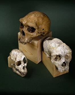
Background imageMandible Collection: Proconsul, Homo heildebergensis & Homo neanderthalensis cran

Proconsul, Homo heildebergensis & Homo neanderthalensis cran
L to R: Cranium of Proconsul, an extinct primate that lived 18 million years ago; Homo heildebergensis (Broken Hill 1) discovered in Zambia; and Homo neanderthalensis (Gibraltar 1)

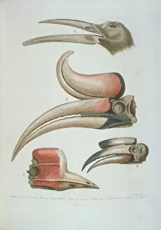
Background imageMandible Collection: Detail of bird beaks

Detail of bird beaks
Plate 281, hand coloured copperplate etching from George Edwards The Gleanings of Natural History, Vol. 2 (1760). Annotated Four curious beaks of birds but little known

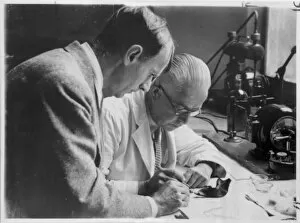
Background imageMandible Collection: Preliminary flourine test of the Piltdown remains in 1949

Preliminary flourine test of the Piltdown remains in 1949
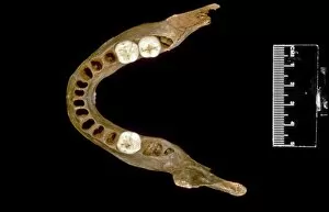
Dr Kenneth Oakley discussing with Mr L. E. Parsons (right) where the mandible of Piltdown man could be sampled with the least risk of damage

Background imageMandible Collection: Homo sapiens (Goughs Cave 49) mandible

Homo sapiens (Goughs Cave 49) mandible
Adolescent modern human mandible found during excavations in Goughs Cave, Cheddar, Somerset, dates back to around 14, 000 - 12, 000 year ago, (Creswellian)

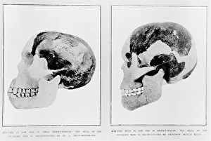
Background imageMandible Collection: Cranium and mandible (Piltdown 1 & 2)

Cranium and mandible (Piltdown 1 & 2)
Piltdown man cranium and mandible as reconstructed on the left by Dr. A Smith Woodward and on the right by Professor Arthur Keith

Background imageMandible Collection: Lateral view of mandible

Lateral view of mandible

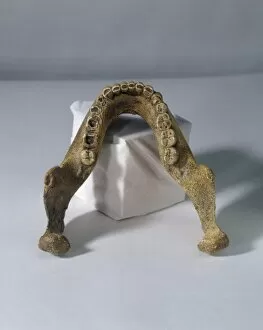
Background imageMandible Collection: Homo heidelbergensis mandible (Mauer 1)

Homo heidelbergensis mandible (Mauer 1)

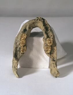
Background imageMandible Collection: Homo erectus mandible (SK 15)

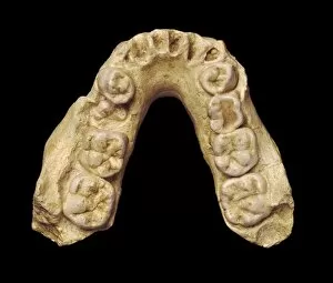
Homo erectus mandible (SK 15)
A cast of a mandible with six teeth belonging to Homo erectus. The original, known as SK 15 Member 2 was found at Swartkrans, South Africa
Background imageMandible Collection: Homo sapiens, human mandible

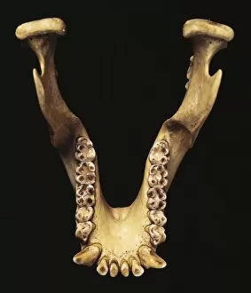
Homo sapiens, human mandible
A specimen of a modern human mandible, or jaw bone with a complete set of teeth. Specimen held at the Natural History Museum, London

Background imageMandible Collection: Australopithecus africanus mandible (MLD 2)

Australopithecus africanus mandible (MLD 2)
Mandible and teeth of an adolescent male (of about 12 years) Australopithecus africanus discovered at Makapansgat by A.R. Hughes and S. Kitching in July 1948

Background imageMandible Collection: Paranthropus robustus jaw bone

Paranthropus robustus jaw bone
Lower jaws of Australopithecus robustus. A robustus lived between 1.8 and 1.5 million years ago in South Africa

Background imageMandible Collection: Sivapithecus & human mandible

Sivapithecus & human mandible
A human mandible (on the left) compared with a Sivapithecus mandible. Sivapithecus, an extinct ape from the Miocene, is believed to have lived 9 million years ago

Background imageMandible Collection: Sivapithecus sivalensis

Sivapithecus sivalensis
Fragments of cranium and mandible of an extinct ape-like primate Sivapithecus sivalensis which lived about 8 million years ago. The specimen was discovered in Potwar, Pakistan

Background imageMandible Collection: Hominid Fossil Collection

Hominid Fossil Collection
A sellection of items from the anthropologigical collections at the Natural History Museum, London

Background imageMandible Collection: Harvester Ant (Messor barbara) major and minor workers, major worker with jaws open in defensive

Harvester Ant (Messor barbara) major and minor workers, major worker with jaws open in defensive posture, Chaine des Alpilles, Bouches-du-Rhone, Provence, France, June

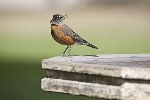
Background imageMandible Collection: American Robin (Turdus migratorius) juvenile, with deformed upper mandible to beak

American Robin (Turdus migratorius) juvenile, with deformed upper mandible to beak, perched on birdbath in garden, North Dakota, U.S.A. September

Background imageMandible Collection: Wood Ant (Formica sp. ) adult workers, fighting, with severed head still grasping with mandibles

Wood Ant (Formica sp. ) adult workers, fighting, with severed head still grasping with mandibles, near Minerve
Wood Ant (Formica sp.) adult workers, fighting, with severed head still grasping with mandibles, near Minerve, Herault, Languedoc-Roussillon, France, may

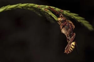
Background imageMandible Collection: Nomad Bee (Nomada lathburiana) adult female, sleeping, with jaws locked onto stem, Leicestershire

Nomad Bee (Nomada lathburiana) adult female, sleeping, with jaws locked onto stem, Leicestershire, England, april

Background imageMandible Collection: Mayfly nymph head, SEM

Mayfly nymph head, SEM
Mayfly nymph head, scanning electron micrograph (SEM). Mayflies (order Ephemeroptera) are the oldest surviving winged insects

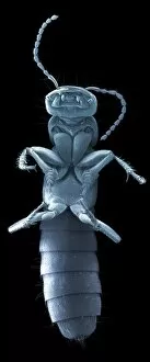
Background imageMandible Collection: Earwig head, SEM

Earwig head, SEM
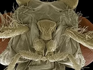
Earwig. Coloured scanning electron micrograph (SEM) of the head of an unidentified earwig (order Dermaptera). The bases of the long, slender antennae (purple, upper frame)

Background imageMandible Collection: Caddisfly head, SEM

Caddisfly head, SEM
Caddisfly head. Coloured scanning electron micrograph (SEM) of the head of a caddisfly (order Trichoptera). At centre are the flys mouthparts

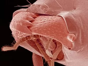
Background imageMandible Collection: Scorpion fly head, SEM

Scorpion fly head, SEM
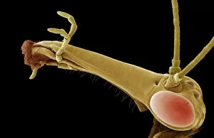
Scorpion fly head. Coloured scanning electron micrograph (SEM) of the head of a scorpion fly (Panorpa sp.). It has a long beak-like extension on its head that has the jaws at the end (left)

Background imageMandible Collection: Hedgehog flea, SEM

Hedgehog flea, SEM
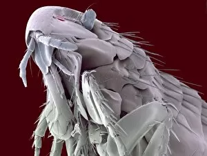
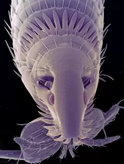
Hedgehog flea (Archaeopsylla erinacei), coloured scanning electron micrograph (SEM). The two structures protruding from the centre of the fleas head are sensory palps

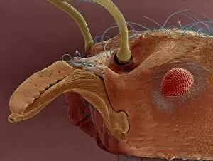
Background imageMandible Collection: Alderfly larva, SEM

Alderfly larva, SEM
Alderfly larva. Coloured scanning electron micrograph (SEM) of the head of an alderfly larva (Sialis lutaria). At right is one of its compound eyes (orange) and at lower centre are its powerful jaws

Background imageMandible Collection: Cat fleas head, SEM

Cat fleas head, SEM

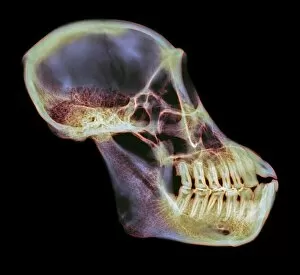
Background imageMandible Collection: Orangutan skull, X-ray

Orangutan skull, X-ray
Orangutan skull. Coloured X-ray of an orangutan (Pongo sp.) skull. The orangutan is native to Malaysia and Indonesia

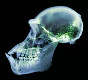
Background imageMandible Collection: Chimpanzee skull, X-ray

Chimpanzee skull, X-ray
Chimpanzee skull. Coloured X-ray of a side view of a chimpanzees (Pan troglodytes) skull. The cranium (upper right) is considerably smaller than in humans

Background imageMandible Collection: Orangutan skull

Orangutan skull. Coloured X-ray of an orangutan (Pongo sp.) skull. The orangutan is native to Malaysia and Indonesia

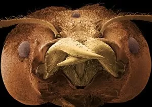
Background imageMandible Collection: Ant mouth, SEM

Ant mouth, SEM
Ant mouthparts. Coloured scanning electron micrograph (SEM) of the mouthparts of an ant (family Formicidae). The powerful mandibles (jaws, centre and upper left) are overlapping

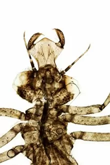
Background imageMandible Collection: Damselfly nymph skin

Damselfly nymph skin. Light micrograph of the head and thorax of a shed skin from a damselfly nymph (Coenagrion sp.). This is the ventral (underside)

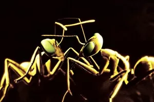
Background imageMandible Collection: Ants fighting

Ants fighting (family Formicidae), illustration. Ants live in large colonies of sometimes millions of individuals. Soldier ants are typically worker ants that have large heads

Background imageMandible Collection: Soldier ant jaws, SEM

Soldier ant jaws, SEM
Soldier ant jaws. Coloured scanning electron micrograph (SEM) of the head of a leaf-cutter ant (Atta sexdens). This is a soldier ant

Background imageMandible Collection: Leaf cutter ant, SEM

Leaf cutter ant, SEM
Leaf cutter ants head. Coloured scanning electron micrograph (SEM) of the head of a leaf cutter ant. This insect lives in colonies and uses its powerful mandibles to remove pieces of leaves

Background imageMandible Collection: Bee mouthparts, light micrograph

Bee mouthparts, light micrograph. The two darker brown upper appendages are the mandibles (jaws), which are used for handling objects and for communicating with other bees

Background imageMandible Collection: Red wood ant, SEM

Red wood ant, SEM
Red wood ant. Coloured scanning electron micrograph (SEM) of the head of a red ant (Formica rufa). Its long sensory antennae are seen protruding from its head at lower centre and lower left

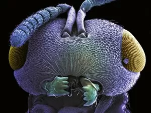
Background imageMandible Collection: Wasp head, SEM

Wasp head, SEM
Wasp head. Coloured scanning electron micrograph (SEM) of a wasps head (order Hymenoptera). Its large compound eyes (yellow) are seen on either side of its head

Background imageMandible Collection: Moth fly, SEM

Moth fly, SEM
Moth fly. Coloured scanning electron micrograph (SEM) of a moth fly (Psychoda sp.). The two compound eyes (pink) dominate the head

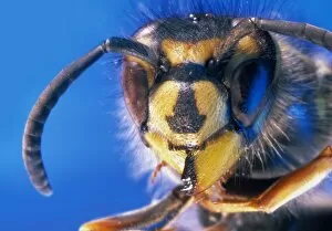
Background imageMandible Collection: Wasp

Wasp. Macrophotograph of the head of a wasp (Vespula sp.), showing its large, compound eyes (black) and segmented sensory antennae

Background imageMandible Collection: Devils coach horse beetle, SEM

Devils coach horse beetle, SEM
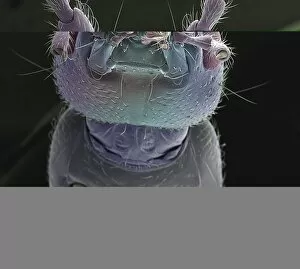
Devils coach horse. Coloured scanning electron micrograph (SEM) of the underside of a devils coach horse beetle (Staphylinus olens)

Background imageMandible Collection: Damselfly nymph

Damselfly nymph (Coenagrion sp.). Head and thorax of a damselfly at the nymph stage. The thorax contains three pairs of legs and a pair of undeveloped wings

Background imageMandible Collection: Red-shouldered ham beetle head, SEM

Red-shouldered ham beetle head, SEM
Red-shouldered ham beetle (Necrobia ruficollis) head, coloured scanning electron micrograph (SEM). Forensic entomologists study this beetle because it feeds on dead bodies

Background imageMandible Collection: Fungus weevil head, SEM

Fungus weevil head, SEM
Weevil head. Coloured scanning electron micrograph (SEM) of the head of an endangered fungus weevil (Notioxenus sp.). The mouthparts are at lower right

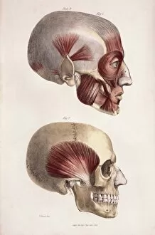
Background imageMandible Collection: Head muscles

Head muscles
Head musculature. Historical artwork of the facial and other head muscles (red) on a human skull. The temporal muscle is shown in the lower frame

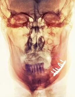
Background imageMandible Collection: Pinned broken jaw, X-ray

Pinned broken jaw, X-ray
Pinned broken jaw. Coloured X-ray of the skull (front view) of a 65 year old man, showing a pinned fracture of the lower jawbone (mandible)

Background imageMandible Collection: Lower jaw of a python

Lower jaw of a python. Coloured scanning electron micrograph (SEM) of the mouth of a python, showing the backward-pointing teeth used for gripping prey

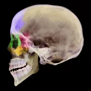
Background imageMandible Collection: Paranasal sinuses, X-ray

Paranasal sinuses, X-ray
Paranasal sinuses. Coloured X-ray of a sagittal section through a human skull. The skull has been sliced in half down the centre and the regions of the paranasal sinuses coloured


For sale as Licensed Images

Choose your image, Select your licence and Download the media

The mandible, also known as the jawbone, is a fascinating and crucial component of our skull anatomy. Leonardo da Vinci's detailed sketches in his "Skull Anatomy" showcase the intricacies of this bone. A panoramic dental X-ray allows us to examine the mandible's structure and its relationship with teeth. Looking back in time, the Paranthropus boisei (Zinjanthropus) cranium (OH5) provides valuable insights into our evolutionary history. The study of human skulls through X-rays reveals not only the mandible but also other vital structures within our head. Nature never fails to amaze us; even tiny creatures like the red-barbed ant can be observed under a scanning electron microscope (SEM), revealing their unique mandibles adapted for various tasks. Meanwhile, a cross-section diagram illustrates how our mouth and jaw work together harmoniously. Insects are no exception when it comes to showcasing intriguing mandibular adaptations. Witnessing a red flour beetle in flight or examining an artwork depicting a Black Death rat flea reminds us of nature's diversity and complexity. However, sometimes unfortunate incidents occur that affect this essential bone. Fractured jawbones captured by X-rays remind us of both medical challenges and advancements in treating such injuries. Zooming further into nature's wonders, we encounter spiny spiders under SEMs - their menacing-looking yet functional jaws ready for capturing prey. Ultimately, whether studying ancient fossils or observing modern-day organisms, understanding the mandible plays an integral role in comprehending head and neck anatomy as depicted beautifully through artworks dedicated to this subject matter.