
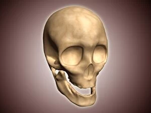
Mandible Collection (page 3)
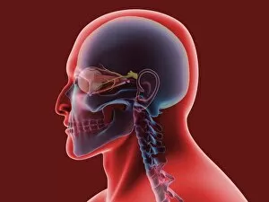
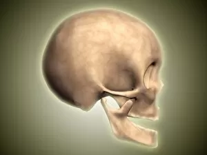
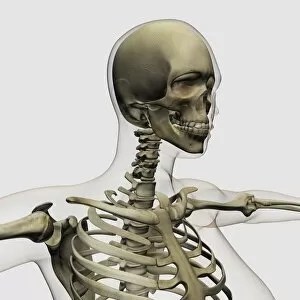
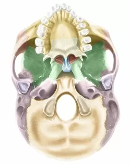
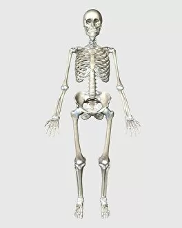
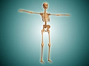
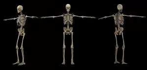
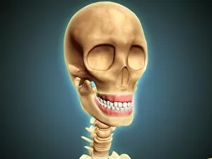
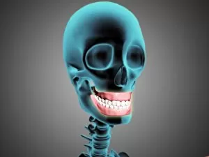
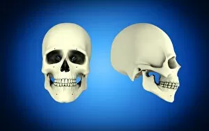
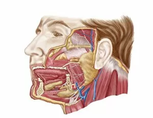
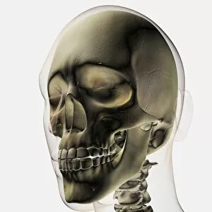
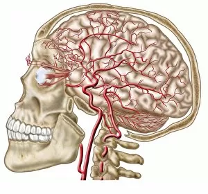
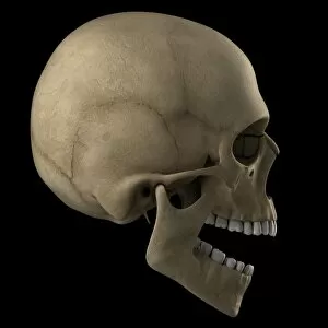
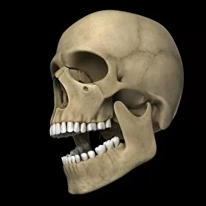
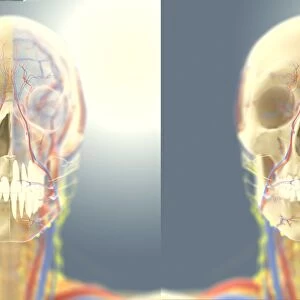
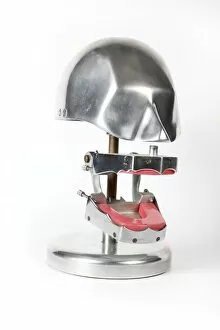
The mandible, also known as the jawbone, is a fascinating and crucial component of our skull anatomy


For sale as Licensed Images
Choose your image, Select your licence and Download the media
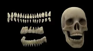
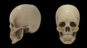
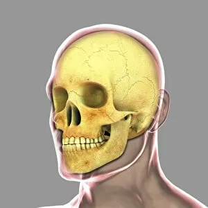
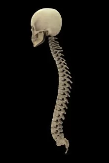
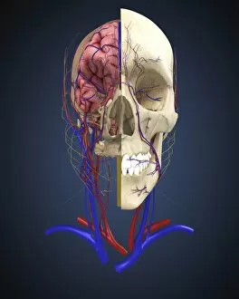
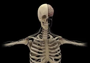
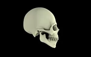
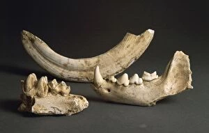
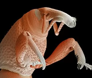
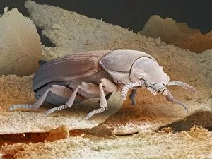
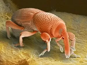
The mandible, also known as the jawbone, is a fascinating and crucial component of our skull anatomy. Leonardo da Vinci's detailed sketches in his "Skull Anatomy" showcase the intricacies of this bone. A panoramic dental X-ray allows us to examine the mandible's structure and its relationship with teeth. Looking back in time, the Paranthropus boisei (Zinjanthropus) cranium (OH5) provides valuable insights into our evolutionary history. The study of human skulls through X-rays reveals not only the mandible but also other vital structures within our head. Nature never fails to amaze us; even tiny creatures like the red-barbed ant can be observed under a scanning electron microscope (SEM), revealing their unique mandibles adapted for various tasks. Meanwhile, a cross-section diagram illustrates how our mouth and jaw work together harmoniously. Insects are no exception when it comes to showcasing intriguing mandibular adaptations. Witnessing a red flour beetle in flight or examining an artwork depicting a Black Death rat flea reminds us of nature's diversity and complexity. However, sometimes unfortunate incidents occur that affect this essential bone. Fractured jawbones captured by X-rays remind us of both medical challenges and advancements in treating such injuries. Zooming further into nature's wonders, we encounter spiny spiders under SEMs - their menacing-looking yet functional jaws ready for capturing prey. Ultimately, whether studying ancient fossils or observing modern-day organisms, understanding the mandible plays an integral role in comprehending head and neck anatomy as depicted beautifully through artworks dedicated to this subject matter.