Neuron Collection (#7)

"Unveiling the Intricacies of Neurons: Exploring the Wonders Within Our Brain" Delving into the depths of our brain

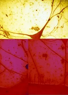
Background imageNeuron Collection: Eye retina C017 / 7784

Eye retina C017 / 7784
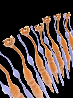
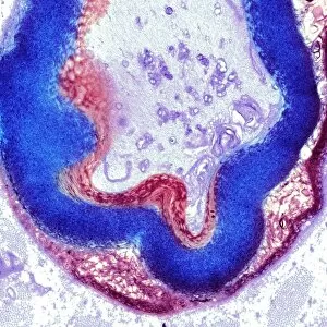
The retina, inside the eye, contains a light-sensitive membranous layer of cells. These are specialized nerve cells: elongated rods (flower like shapes) and cone-tipped cells, that produce vision
Background imageNeuron Collection: Eye retina C017 / 7787

Eye retina C017 / 7787
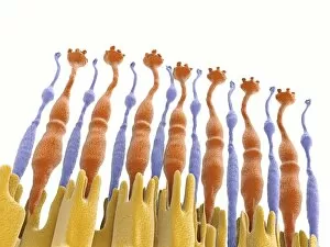
The retina, inside the eye, contains a light-sensitive membranous layer of cells. These are specialized nerve cells: elongated rods (flower like shapes) and cone-tipped cells, that produce vision
Background imageNeuron Collection: Eye retina C017 / 7783

Eye retina C017 / 7783
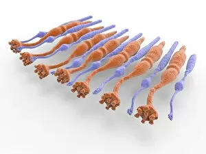
The retina, inside the eye, contains a light-sensitive membranous layer of cells. These are specialized nerve cells: elongated rods (flower like shapes) and cone-tipped cells, that produce vision

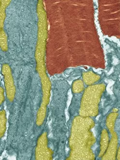
Background imageNeuron Collection: Myelinated nerve, TEM C016 / 5840

Myelinated nerve, TEM C016 / 5840
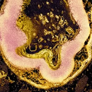
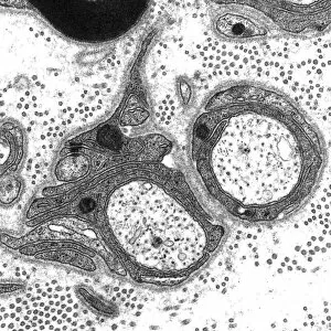
Myelinated nerve. Coloured transmission electron micrograph (TEM) of a section through a myelinated nerve fibre and Schwann cell

Background imageNeuron Collection: Myelinated nerve, TEM C016 / 5839

Myelinated nerve, TEM C016 / 5839
Myelinated nerve. Coloured transmission electron micrograph (TEM) of a section through a myelinated nerve fibre and Schwann cell

Background imageNeuron Collection: Myelinated nerve, TEM C016 / 5838

Myelinated nerve, TEM C016 / 5838
Myelinated nerve. Coloured transmission electron micrograph (TEM) of a section through a myelinated nerve fibre and Schwann cell

Background imageNeuron Collection: Unmyelinated nerve, TEM C016 / 5805

Unmyelinated nerve, TEM C016 / 5805
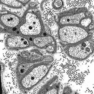
Unmyelinated nerve. Transmission electron micrograph (TEM) of a section through axon (nerve fibre) bundles of unmyelinated nerves

Background imageNeuron Collection: Unmyelinated nerve, TEM C016 / 5804

Unmyelinated nerve, TEM C016 / 5804
Unmyelinated nerve. Transmission electron micrograph (TEM) of a section through axon (nerve fibre) bundles of unmyelinated nerves

Background imageNeuron Collection: Myelinated nerve, TEM C016 / 5448

Myelinated nerve, TEM C016 / 5448
Myelinated nerve. Transmission electron micrograph (TEM) of a section through a myelinated nerve fibre and Schwann cell. Myelin (black)

Background imageNeuron Collection: Myelinated nerve, TEM C016 / 5370

Myelinated nerve, TEM C016 / 5370
Myelinated nerve. Transmission electron micrograph (TEM) of a section through a myelinated nerve fibre and Schwann cell (centre)

Background imageNeuron Collection: Synapses, artwork C014 / 0002

Synapses, artwork C014 / 0002
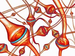
Synapses. Computer artwork of synapses, the junction between nerve cells (orange). Synapses transmit electrical signals from one nerve cell to the next

Background imageNeuron Collection: Synapses, artwork C014 / 0004

Synapses, artwork C014 / 0004
Synapses. Computer artwork of synapses, the junction between nerve cells (blue). Synapses transmit electrical signals from one nerve cell to the next

Background imageNeuron Collection: Synapses, artwork C014 / 0003

Synapses, artwork C014 / 0003
Synapses. Computer artwork of synapses, the junction between nerve cells (blue). Synapses transmit electrical signals from one nerve cell to the next

Background imageNeuron Collection: Nerve cell, conceptual artwork C013 / 9994

Nerve cell, conceptual artwork C013 / 9994
Nerve cell, conceptual computer artwork

Background imageNeuron Collection: Power of the heart, conceptual image C013 / 9982

Power of the heart, conceptual image C013 / 9982
Power of the heart, conceptual image. Computer artwork of a human heart (left) superimposed on an image of an engine (right), representing the physical power needed to pump blood around the body

Background imageNeuron Collection: Heart activity, conceptual artwork C013 / 9981

Heart activity, conceptual artwork C013 / 9981
Heart activity, conceptual computer artwork

Background imageNeuron Collection: Neural network, conceptual image C013 / 9958

Neural network, conceptual image C013 / 9958
Neural network, conceptual image. Computer artwork representing interconnecting nerve cells (neurons)

Background imageNeuron Collection: Neural network, conceptual image C013 / 9957

Neural network, conceptual image C013 / 9957
Neural network, conceptual image. Computer artwork representing interconnecting nerve cells (neurons)

Background imageNeuron Collection: Nerve cell, SEM C013 / 9772

Nerve cell, SEM C013 / 9772
Nerve cell. Coloured scanning electron micrograph (SEM) of a nerve cell (neuron). Neurons are responsible for passing information around the central nervous system (CNS)

Background imageNeuron Collection: Cerebral cortex nerve cells C013 / 9767

Cerebral cortex nerve cells C013 / 9767
Cerebral cortex nerve cells. Light micrograph of a section through neurones (nerve cells, black) in the cerebral cortex of a human brain

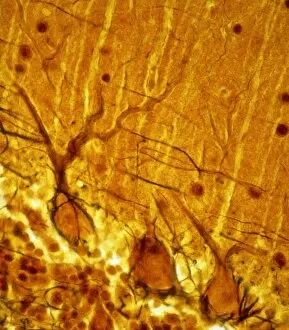
Background imageNeuron Collection: Purkinje nerve cell C013 / 9763

Purkinje nerve cell C013 / 9763
Purkinje nerve cell. Light micrograph of a purkinje nerve cell (orange, centre) from the cerebellum of the brain. The cell comprises a flask-shaped cell body from which numerous processes (dendrites)

Background imageNeuron Collection: Purkinje nerve cells C013 / 9745

Purkinje nerve cells C013 / 9745
Purkinje nerve cells. Light micrograph of three purkinje nerve cells (across bottom) from the cerebellum of the brain. The cells comprise a flask-shaped cell body from which numerous processes

Background imageNeuron Collection: Myelinated nerves, SEM C013 / 7142

Myelinated nerves, SEM C013 / 7142
Myelinated nerves. Coloured scanning electron micrograph (SEM) of a section through the sciatic nerve, showing the myelinated nerve fibres (axons)

Background imageNeuron Collection: Myelinated nerves, SEM C013 / 7141

Myelinated nerves, SEM C013 / 7141
Myelinated nerves. Coloured scanning electron micrograph (SEM) of a section through the sciatic nerve, showing the myelinated nerve fibres (axons)

Background imageNeuron Collection: Myelinated nerves, SEM C013 / 7138

Myelinated nerves, SEM C013 / 7138
Myelinated nerves. Coloured scanning electron micrograph (SEM) of a section through a myelinated nerve fibre (axon, beige, centre) from the sciatic nerve

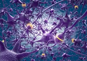
Background imageNeuron Collection: Neural network, artwork C013 / 4636

Neural network, artwork C013 / 4636
Neural network. Computer artwork of nerve cells (neurons) connected by processes (filaments), known as dendrites and axons, to form a neural network

Background imageNeuron Collection: Retinal rod cell, TEM C013 / 4805

Retinal rod cell, TEM C013 / 4805
Retinal rod cell. Transmission electron micrograph (TEM) of a section through a rod cell from the retina of an eye, showing the inner segment (bottom) filled with mitochondria (green)

Background imageNeuron Collection: Retinal rod cell, TEM C013 / 4804

Retinal rod cell, TEM C013 / 4804
Retinal rod cell. Transmission electron micrograph (TEM) of a section through a rod cell from the retina of an eye, showing the inner segment (bottom) filled with mitochondria (green)

Background imageNeuron Collection: Macrophage engulfing a nerve cell, TEM C013 / 4803

Macrophage engulfing a nerve cell, TEM C013 / 4803
Macrophage engulfing a nerve cell. Transmission electron micrograph (TEM) of a section through a macrophage white blood cell (blue) that has engulfed a nerve cell (neuron, centre)

Background imageNeuron Collection: Nerve cell, TEM C013 / 4797

Nerve cell, TEM C013 / 4797
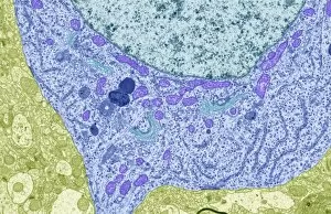
Nerve cell. Transmission electron micrograph (TEM) of a section through a neuron (nerve cell), showing characteristic Nissl body (dark blue lines), numerous golgi apparatus (curved green lines)

Background imageNeuron Collection: Nerve cell, TEM C013 / 4796

Nerve cell, TEM C013 / 4796
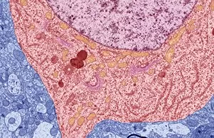
Nerve cell. Transmission electron micrograph (TEM) of a section through a neuron (nerve cell), showing characteristic Nissl body (dark red lines), numerous golgi apparatus (curved pink lines)

Background imageNeuron Collection: Domestic Dog, Golden Retriever, adult, on pavement with disabled owner confined to wheelchair

Domestic Dog, Golden Retriever, adult, on pavement with disabled owner confined to wheelchair, England

Background imageNeuron Collection: Neuromuscular synapse, light micrograph

Neuromuscular synapse, light micrograph
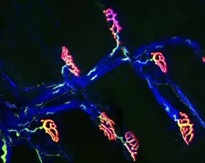
Neuromuscular junction. Fluorescent confocal light micrograph of the junction between a nerve cell and a muscle (not seen). The axon of the nerve cell (neuron) has been tagged with a blue dye

Background imageNeuron Collection: Brain nerve cells

Brain nerve cells
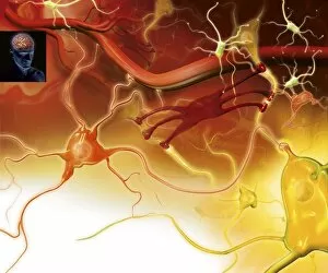
Nerve cells in the brain. Artwork showing the different types of nerve cells in the grey matter of the brain. Neurons (yellow, for example at lower right) relay nerve signals around the brain

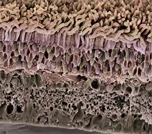
Background imageNeuron Collection: Retina, SEM

Retina, SEM
Retina. Coloured scanning electron micrograph (SEM) of a section through a freeze-fractured retina, showing the light-sensitive rods and cones and their associated neurones

Background imageNeuron Collection: Oligodendrocyte and microglia brain cells

Oligodendrocyte and microglia brain cells
Oligodendrocyte and microglia action in the brain. At top, the axon (output process, orange) of a neuron (nerve cell) is seen

Background imageNeuron Collection: Brain cortex tissue, light micrograph

Brain cortex tissue, light micrograph
Brain cortex tissue. Light micrograph of a section through tissue from the cortex of the brain. The cortex, or grey matter, is the outer layer of the brain

Background imageNeuron Collection: Astrocyte / neuron synapse in the brain

Astrocyte / neuron synapse in the brain
Astrocyte/neuron synapse in the brain. Artwork of a nerve cell (neuron, yellow) synapse in the brain with an associated regulatory astrocyte cell (red)

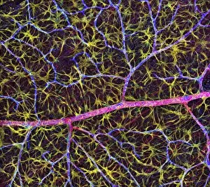
Background imageNeuron Collection: Retina blood vessel and nerve cells

Retina blood vessel and nerve cells
Retina cells. Fluorescent light micrograph of cells in the retina, the light-sensitive membrane that lines the back of the eyeball

Background imageNeuron Collection: Vomiting, artwork

Vomiting, artwork
Vomiting. Computer artwork of synapses, or junctions, (cones) between nerve cells and the exterior wall of the stomach. During the vomit reflex, nerve impulses are sent from the brain to the stomach

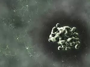
Background imageNeuron Collection: Prion protein plaque, computer artwork

Prion protein plaque, computer artwork
Prion protein plaque (right) surrounded by nerve cells (seen mainly on the left), computer artwork. Prions are infectious agents that cause a group of fatal neurodegenerative diseases

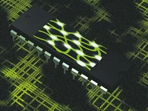
Background imageNeuron Collection: Neural chip

Neural chip. Conceptual computer artwork of a neural network (centre) on a microchip. This could represent hardware which mimics such networks, or contains actual nerve cells

Background imageNeuron Collection: Neuronal network

Neuronal network. Conceptual computer artwork of a neural network. This could represent the interior of a human brain or a biological computer memory which mimics such networks

Background imageNeuron Collection: Optic nerve, light micrograph

Optic nerve, light micrograph
Optic nerve. Coloured light micrograph of a section through the optic nerve. The optic nerve (bright pink) collects impulses from the light sensitive cells of the retina and relays them to the brain

Background imageNeuron Collection: Nerve cells, light micrograph

Nerve cells, light micrograph
Nerve cells, or neurons, light micrograph. Neurons are responsible for passing information around the central nervous system (CNS) and from the CNS to the rest of the body

Background imageNeuron Collection: Purkinje nerve cell, light micrograph

Purkinje nerve cell, light micrograph
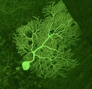
Purkinje nerve cell. Confocal light micrograph of a purkinje cell (stained with green fluorescent protein), a type of neuron (nerve cell)

Background imageNeuron Collection: Creating new neural pathways, artwork

Creating new neural pathways, artwork
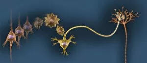
Creating new neural pathways. Artwork showing the process involved in the formation of new nerve cells (neurogenesis) and neural pathways

Background imageNeuron Collection: Spinal nerve ganglion, light micrograph

Spinal nerve ganglion, light micrograph
Spinal nerve ganglion. Light micrograph of a cross-section through a spinal nerve ganglion. This is a node of nerve cells located just outside the spinal cord at the point where it is joined by


For sale as Licensed Images

Choose your image, Select your licence and Download the media

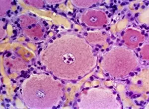
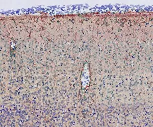
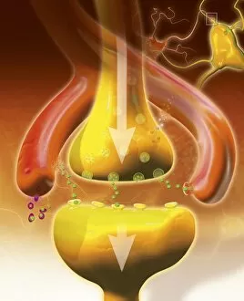
"Unveiling the Intricacies of Neurons: Exploring the Wonders Within Our Brain" Delving into the depths of our brain, we encounter a histological diagram of a mammalian retina. This intricate network showcases the complexity and beauty of neurons that enable us to perceive light and color. Moving further, we explore cerebellum tissue through a light micrograph. The mesmerizing patterns reveal nerve and glial cells working in harmony, orchestrating our body's movements with precision. Zooming in closer, we witness a synapse nerve junction captured by TEM. This microscopic marvel highlights how information is transmitted between neurons, forming connections crucial for our thoughts and actions. Shifting gears to SEM imagery, we are introduced to an awe-inspiring nerve cell. Its intricate structure resembles an elaborate work of art—a testament to nature's ingenuity in crafting these building blocks of intelligence. Tracing back history, we stumble upon Santiago Ramon y Cajal's 1894 drawing depicting cell types within the mammalian cerebellum. His meticulous observations laid foundations for understanding neural networks that govern our motor skills. Venturing deeper into brain tissue, we discover hippocampus tissue—an essential region responsible for memory formation and spatial navigation. Here lies another realm where neurons weave together memories that shape who we are. Intriguingly unique are Purkinje nerve cells found within the cerebellum—majestic giants among their peers. Their distinctive appearance signifies their vital role in coordinating movement and maintaining balance. As if peering through a microscope lens once again, another nerve cell captures our attention—the epitome of elegance amidst complexity; it reminds us how intricately woven life truly is at its core. Diving into glial stem cell culture under bright illumination reveals their remarkable regenerative potential—a beacon of hope for treating neurological disorders as they hold promises yet untapped. Examining brain tissue blood supply uncovers an indispensable lifeline, nourishing neurons with oxygen and nutrients.