Neuron Collection (#6)

"Unveiling the Intricacies of Neurons: Exploring the Wonders Within Our Brain" Delving into the depths of our brain

Background imageNeuron Collection: Acupuncture, artwork

Acupuncture, artwork
Acupuncture. Computer artwork showing an acupuncture needle inserted into a patients skin and disrupting a nerve signal (red arrows)

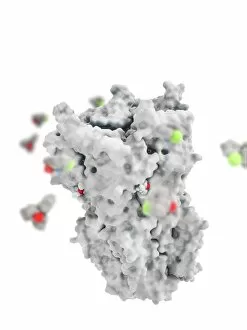
Background imageNeuron Collection: Ketamine drug binding to ion channel

Ketamine drug binding to ion channel, molecular model. Several molecules of the drug ketamine binding to a pentameric ligand-gated ion channel (pLGIC)

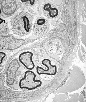
Background imageNeuron Collection: Myelinated nerve tissue, TEM

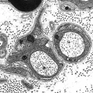
Myelinated nerve tissue, TEM
Myelinated nerve tissue. Transmission electron micrograph (TEM) of a section through myelinated nerve fibres (axons). Each axon is coated with many layers of myelin

Background imageNeuron Collection: Olfactory bulb anatomy, artwork

Olfactory bulb anatomy, artwork
Olfactory bulb anatomy. Artwork of a human nose, showing the anatomy of the olfactory bulb, the organ of smell. This lies above the anterior aspect of the nasal cavity on the ethmoid bone

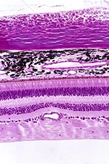
Background imageNeuron Collection: Retina of the eye, light micrograph C016 / 0528

Retina of the eye, light micrograph C016 / 0528
Retina of the eye. Light micrograph of a section through the retina from a human eye. From top down: nerve fibres of the optic nerve and a blood vessel; several layers of neurons (nerve cells)

Background imageNeuron Collection: Nerve cell, illustration C018 / 0740

Nerve cell, illustration C018 / 0740
Nerve cell, or neuron, illustration. Neurons are responsible for passing information around the central nervous system (CNS) and from the CNS to the rest of the body

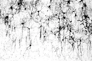
Background imageNeuron Collection: Cerebral cortex nerve cells C018 / 0062

Cerebral cortex nerve cells C018 / 0062
Cerebral cortex nerve cells. Light micrograph of a section through neurones (nerve cells, black) in the cerebral cortex of a human brain

Background imageNeuron Collection: Nervous system cells, artwork C017 / 3423

Nervous system cells, artwork C017 / 3423
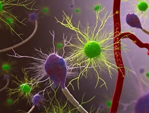
Nervous system cells. Computer artwork showing neurons (nerve cells, purple), astrocytes (green) and a blood vessel (along right)

Background imageNeuron Collection: Nerve synapse, artwork C017 / 3426

Nerve synapse, artwork C017 / 3426
Nerve synapse. Computer artwork of of a junction, or synapse, between two nerve cells (neurons). As the electrical signal reaches the presynaptic end of a neuron it triggers the release of

Background imageNeuron Collection: Cerebrum, light micrograph C014 / 1399

Cerebrum, light micrograph C014 / 1399
Cerebrum. Light micrograph of a section through tissue from the cerebrum, showing neurons (nerve cells, purple). The cerebrum is the largest part of the brain

Background imageNeuron Collection: Cerebrum, light micrograph C014 / 1400

Cerebrum, light micrograph C014 / 1400
Cerebrum. Light micrograph of a section through tissue from the cerebrum, showing neurons (nerve cells, purple). The cerebrum is the largest part of the brain

Background imageNeuron Collection: Rod and cone cells of the eye, SEM C014 / 4865

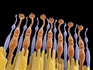
Rod and cone cells of the eye, SEM C014 / 4865
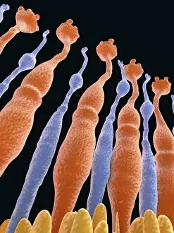
Rod and cone cells of the eye. Coloured scanning electron micrograph (SEM) of rod and cone cells in the retina of a mammalian eye

Background imageNeuron Collection: Induced nerve stem cells, micrograph

Induced nerve stem cells, micrograph
Induced nerve stem cells. Fluorescence light micrograph of neural (nerve) stem cells that have been created (induced) from human adult skin fibroblast cells by gene manipulation

Background imageNeuron Collection: Stem cell-derived nerve cells

Stem cell-derived nerve cells. Fluorescence light micrograph of neural (nerve) stem cells that have been derived from human embryonic stem cells (HESC)

Background imageNeuron Collection: Brain cells, artwork

Brain cells, artwork
Brain cells. Computer artwork representing neurons (nerve cells) in the brain

Background imageNeuron Collection: Brain nerve cells, TEM C014 / 0356

Brain nerve cells, TEM C014 / 0356
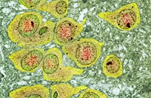
Brain nerve cells. Transmission electron micrograph (TEM) of a section through brain tissue from the cerebral cortex, showing numerous neurons (nerve cells) surrounded by axons and dendrites

Background imageNeuron Collection: Purkinje nerve cell, TEM C014 / 0582

Purkinje nerve cell, TEM C014 / 0582
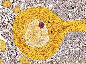
Purkinje nerve cell. Transmission electron micrograph (TEM) of a purkinje nerve cell (bright yellow, centre) from the cerebellum of the brain

Background imageNeuron Collection: Nerve cells, artwork C018 / 2887

Nerve cells, artwork C018 / 2887
Nerve cells, or neurons, computer artwork. Neurons are responsible for passing information around the central nervous system (CNS) and from the CNS to the rest of the body

Background imageNeuron Collection: Eye retina C017 / 7794

Eye retina C017 / 7794
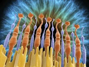
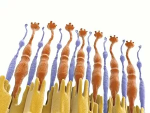
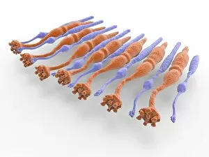
The retina, inside the eye, contains a light-sensitive membranous layer of cells. These are specialized nerve cells: elongated rods (flower like shapes) and cone-tipped cells, that produce vision

Background imageNeuron Collection: Eye retina C017 / 7793

Eye retina C017 / 7793
The retina, inside the eye, contains a light-sensitive membranous layer of cells. These are specialized nerve cells: elongated rods (flower like shapes) and cone-tipped cells, that produce vision

Background imageNeuron Collection: Eye retina C017 / 7792

Eye retina C017 / 7792
The retina, inside the eye, contains a light-sensitive membranous layer of cells. These are specialized nerve cells: elongated rods (flower like shapes) and cone-tipped cells, that produce vision

Background imageNeuron Collection: Eye retina and iris C017 / 7789

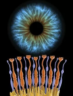
Eye retina and iris C017 / 7789
The retina, inside the eye, contains a light-sensitive membranous layer of cells. These are specialized nerve cells: elongated rods (flower like shapes) and cone-tipped cells, that produce vision

Background imageNeuron Collection: Eye retina C017 / 7790

Eye retina C017 / 7790
The retina, inside the eye, contains a light-sensitive membranous layer of cells. These are specialized nerve cells: elongated rods (flower like shapes) and cone-tipped cells, that produce vision

Background imageNeuron Collection: Eye retina and iris C017 / 7788

Eye retina and iris C017 / 7788
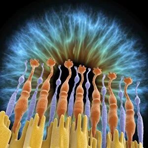
The retina, inside the eye, contains a light-sensitive membranous layer of cells. These are specialized nerve cells: elongated rods (flower like shapes) and cone-tipped cells, that produce vision

Background imageNeuron Collection: Eye retina and iris C017 / 7785

Eye retina and iris C017 / 7785
The retina, inside the eye, contains a light-sensitive membranous layer of cells. These are specialized nerve cells: elongated rods (flower like shapes) and cone-tipped cells, that produce vision

Background imageNeuron Collection: Eye retina C017 / 7786

Eye retina C017 / 7786
The retina, inside the eye, contains a light-sensitive membranous layer of cells. These are specialized nerve cells: elongated rods (flower like shapes) and cone-tipped cells, that produce vision

Background imageNeuron Collection: Eye retina C017 / 7784

Eye retina C017 / 7784
The retina, inside the eye, contains a light-sensitive membranous layer of cells. These are specialized nerve cells: elongated rods (flower like shapes) and cone-tipped cells, that produce vision

Background imageNeuron Collection: Eye retina C017 / 7787

Eye retina C017 / 7787
The retina, inside the eye, contains a light-sensitive membranous layer of cells. These are specialized nerve cells: elongated rods (flower like shapes) and cone-tipped cells, that produce vision

Background imageNeuron Collection: Eye retina C017 / 7783

Eye retina C017 / 7783
The retina, inside the eye, contains a light-sensitive membranous layer of cells. These are specialized nerve cells: elongated rods (flower like shapes) and cone-tipped cells, that produce vision

Background imageNeuron Collection: Myelinated nerve, TEM C016 / 5840

Myelinated nerve, TEM C016 / 5840
Myelinated nerve. Coloured transmission electron micrograph (TEM) of a section through a myelinated nerve fibre and Schwann cell

Background imageNeuron Collection: Myelinated nerve, TEM C016 / 5839

Myelinated nerve, TEM C016 / 5839
Myelinated nerve. Coloured transmission electron micrograph (TEM) of a section through a myelinated nerve fibre and Schwann cell

Background imageNeuron Collection: Myelinated nerve, TEM C016 / 5838

Myelinated nerve, TEM C016 / 5838
Myelinated nerve. Coloured transmission electron micrograph (TEM) of a section through a myelinated nerve fibre and Schwann cell

Background imageNeuron Collection: Unmyelinated nerve, TEM C016 / 5805

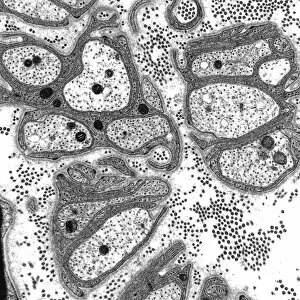
Unmyelinated nerve, TEM C016 / 5805
Unmyelinated nerve. Transmission electron micrograph (TEM) of a section through axon (nerve fibre) bundles of unmyelinated nerves

Background imageNeuron Collection: Unmyelinated nerve, TEM C016 / 5804

Unmyelinated nerve, TEM C016 / 5804
Unmyelinated nerve. Transmission electron micrograph (TEM) of a section through axon (nerve fibre) bundles of unmyelinated nerves

Background imageNeuron Collection: Myelinated nerve, TEM C016 / 5448

Myelinated nerve, TEM C016 / 5448
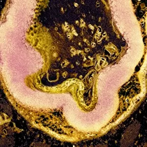
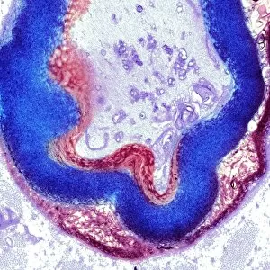
Myelinated nerve. Transmission electron micrograph (TEM) of a section through a myelinated nerve fibre and Schwann cell. Myelin (black)

Background imageNeuron Collection: Myelinated nerve, TEM C016 / 5370

Myelinated nerve, TEM C016 / 5370
Myelinated nerve. Transmission electron micrograph (TEM) of a section through a myelinated nerve fibre and Schwann cell (centre)

Background imageNeuron Collection: Synapses, artwork C014 / 0002

Synapses, artwork C014 / 0002
Synapses. Computer artwork of synapses, the junction between nerve cells (orange). Synapses transmit electrical signals from one nerve cell to the next

Background imageNeuron Collection: Synapses, artwork C014 / 0004

Synapses, artwork C014 / 0004
Synapses. Computer artwork of synapses, the junction between nerve cells (blue). Synapses transmit electrical signals from one nerve cell to the next

Background imageNeuron Collection: Synapses, artwork C014 / 0003

Synapses, artwork C014 / 0003
Synapses. Computer artwork of synapses, the junction between nerve cells (blue). Synapses transmit electrical signals from one nerve cell to the next

Background imageNeuron Collection: Nerve cell, conceptual artwork C013 / 9994

Nerve cell, conceptual artwork C013 / 9994
Nerve cell, conceptual computer artwork

Background imageNeuron Collection: Power of the heart, conceptual image C013 / 9982

Power of the heart, conceptual image C013 / 9982
Power of the heart, conceptual image. Computer artwork of a human heart (left) superimposed on an image of an engine (right), representing the physical power needed to pump blood around the body

Background imageNeuron Collection: Heart activity, conceptual artwork C013 / 9981

Heart activity, conceptual artwork C013 / 9981
Heart activity, conceptual computer artwork

Background imageNeuron Collection: Neural network, conceptual image C013 / 9958

Neural network, conceptual image C013 / 9958
Neural network, conceptual image. Computer artwork representing interconnecting nerve cells (neurons)

Background imageNeuron Collection: Neural network, conceptual image C013 / 9957

Neural network, conceptual image C013 / 9957
Neural network, conceptual image. Computer artwork representing interconnecting nerve cells (neurons)

Background imageNeuron Collection: Nerve cell, SEM C013 / 9772

Nerve cell, SEM C013 / 9772
Nerve cell. Coloured scanning electron micrograph (SEM) of a nerve cell (neuron). Neurons are responsible for passing information around the central nervous system (CNS)

Background imageNeuron Collection: Cerebral cortex nerve cells C013 / 9767

Cerebral cortex nerve cells C013 / 9767
Cerebral cortex nerve cells. Light micrograph of a section through neurones (nerve cells, black) in the cerebral cortex of a human brain

Background imageNeuron Collection: Purkinje nerve cell C013 / 9763

Purkinje nerve cell C013 / 9763
Purkinje nerve cell. Light micrograph of a purkinje nerve cell (orange, centre) from the cerebellum of the brain. The cell comprises a flask-shaped cell body from which numerous processes (dendrites)

Background imageNeuron Collection: Purkinje nerve cells C013 / 9745

Purkinje nerve cells C013 / 9745
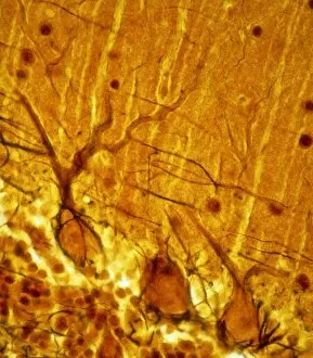
Purkinje nerve cells. Light micrograph of three purkinje nerve cells (across bottom) from the cerebellum of the brain. The cells comprise a flask-shaped cell body from which numerous processes


For sale as Licensed Images

Choose your image, Select your licence and Download the media

"Unveiling the Intricacies of Neurons: Exploring the Wonders Within Our Brain" Delving into the depths of our brain, we encounter a histological diagram of a mammalian retina. This intricate network showcases the complexity and beauty of neurons that enable us to perceive light and color. Moving further, we explore cerebellum tissue through a light micrograph. The mesmerizing patterns reveal nerve and glial cells working in harmony, orchestrating our body's movements with precision. Zooming in closer, we witness a synapse nerve junction captured by TEM. This microscopic marvel highlights how information is transmitted between neurons, forming connections crucial for our thoughts and actions. Shifting gears to SEM imagery, we are introduced to an awe-inspiring nerve cell. Its intricate structure resembles an elaborate work of art—a testament to nature's ingenuity in crafting these building blocks of intelligence. Tracing back history, we stumble upon Santiago Ramon y Cajal's 1894 drawing depicting cell types within the mammalian cerebellum. His meticulous observations laid foundations for understanding neural networks that govern our motor skills. Venturing deeper into brain tissue, we discover hippocampus tissue—an essential region responsible for memory formation and spatial navigation. Here lies another realm where neurons weave together memories that shape who we are. Intriguingly unique are Purkinje nerve cells found within the cerebellum—majestic giants among their peers. Their distinctive appearance signifies their vital role in coordinating movement and maintaining balance. As if peering through a microscope lens once again, another nerve cell captures our attention—the epitome of elegance amidst complexity; it reminds us how intricately woven life truly is at its core. Diving into glial stem cell culture under bright illumination reveals their remarkable regenerative potential—a beacon of hope for treating neurological disorders as they hold promises yet untapped. Examining brain tissue blood supply uncovers an indispensable lifeline, nourishing neurons with oxygen and nutrients.