
Optic Disc Collection
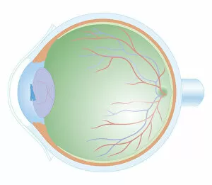
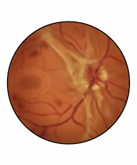
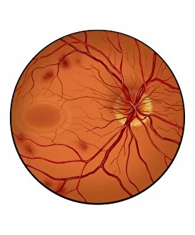
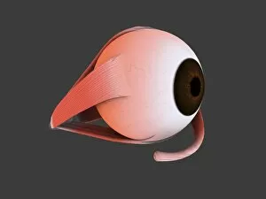
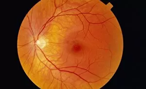
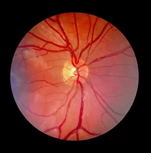
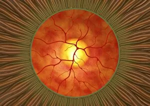
The optic disc, also known as the blind spot, is a crucial structure located at the back of the eye


For sale as Licensed Images
Choose your image, Select your licence and Download the media
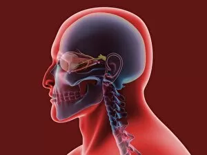
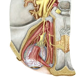
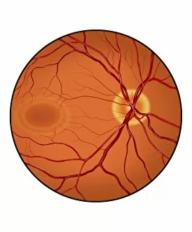
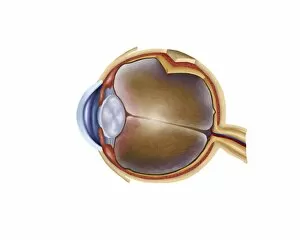
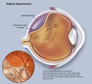
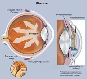
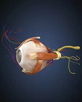
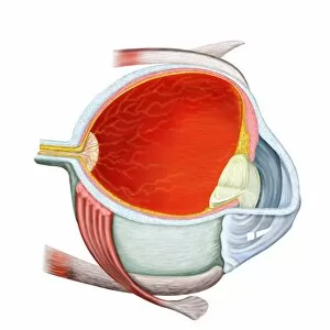
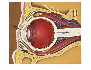
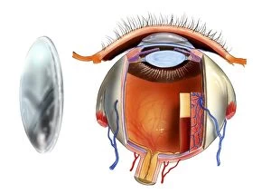
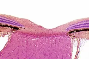
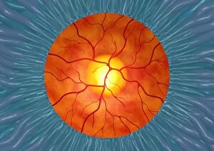
The optic disc, also known as the blind spot, is a crucial structure located at the back of the eye. It serves as an entry point for the optic nerve and plays a vital role in our vision. In this captivating image, we observe a cross-section biomedical illustration of the human eye's anatomy. The intricate details showcase various components such as the retina and its blood vessels. Through scanning electron microscopy (SEM), we get a closer look at these delicate structures that contribute to our visual perception. A conceptual image presents us with an awe-inspiring view of both the human eye and skull, highlighting how intricately connected they are. This representation emphasizes how essential it is to protect our eyes and maintain their health. Another conceptual image portrays macular degeneration affecting the retina—a condition that can lead to vision loss over time if left untreated. This powerful visual reminder urges us to prioritize regular eye check-ups and take necessary precautions for maintaining good ocular health. An orbital cut reveals fascinating details about nerves within our eyes—specifically, showcasing abducent nerve with ciliary ganglion and oculomotor nerve connections. These nerves play significant roles in controlling movement and focus within our eyes. Through multiple conceptual images depicting different angles of human eye anatomy, we gain insight into its complexity. From understanding retinal functions to appreciating its overall structure, these visuals remind us of just how remarkable our sense of sight truly is. Lastly, we encounter a normal retina—an exquisite display demonstrating what healthy ocular tissue looks like under microscopic examination. Appreciating this vibrant portrayal encourages us to value proper care for our eyesight while acknowledging their incredible capabilities.