Organelle Collection (#2)

Organelles: The Tiny Powerhouses Within Our Cells The world of cells is a fascinating one, filled with intricate structures known as organelles

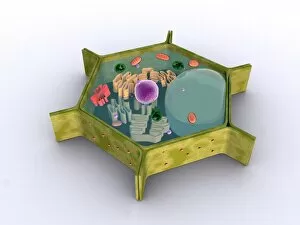
Background imageOrganelle Collection: Conceptual image of a plant cell and its components

Conceptual image of a plant cell and its components

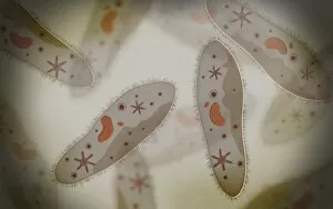
Background imageOrganelle Collection: Microscopic view of paramecium

Microscopic view of paramecium

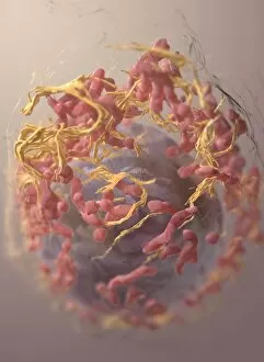
Background imageOrganelle Collection: 3D structure of melanoma cell

3D structure of melanoma cell
3D structure of a melanoma cell derived by ion abrasion scanning electron microscopy

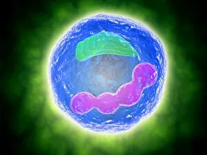
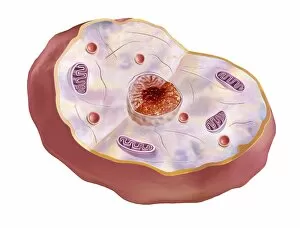
Background imageOrganelle Collection: Conceptual image of human cell

Conceptual image of human cell

Background imageOrganelle Collection: Human cell anatomy

Human cell anatomy

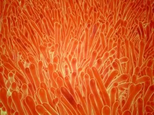
Background imageOrganelle Collection: Microscopic view of intestinal villi inside the small intestine

Microscopic view of intestinal villi inside the small intestine
Microscopic view of intestinal villi which can be found inside of the small intestine

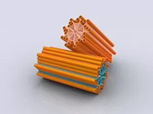
Background imageOrganelle Collection: Conceptual image of centrioles

Conceptual image of centrioles

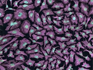
Background imageOrganelle Collection: Multiphoton fluorescence image of HeLa cells

Multiphoton fluorescence image of HeLa cells with cytoskeletal microtubules (magenta) and DNA (cyan)

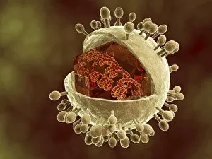
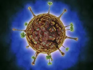
Background imageOrganelle Collection: Conceptual image of human cytomegalovirus

Conceptual image of human cytomegalovirus. Cytomegalovirus is a genus of the viral family Herpesviridae

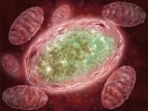
Background imageOrganelle Collection: Microscopic view of Mitochondria

Microscopic view of Mitochondria. Mitochondria are the cells power producers. They convert energy into forms that are usable by the cell. Located in the cytoplasmcells activities

Background imageOrganelle Collection: Conceptual image of cytoskeleton

Conceptual image of cytoskeleton. The cytoskeleton is a cellular scaffolding or skeleton contained within a cells cytoplasm, and is present in all cells
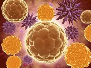
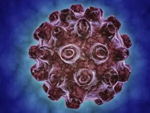
Background imageOrganelle Collection: Microscopic view of samllpox

Microscopic view of samllpox

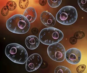
Background imageOrganelle Collection: Microscopic view of human cell

Microscopic view of human cell

Background imageOrganelle Collection: The interior of an eukaryotic cell

The interior of an eukaryotic cell. This is the most common generic type of cell and its present in all mammals. In the center is the nucleus (the perforated sphere)

Background imageOrganelle Collection: Conceptual image of mast cells

Conceptual image of mast cells

Background imageOrganelle Collection: Conceptual image of Encephalitis

Conceptual image of Encephalitis. Encephalitis can cause flu-like symptoms, such as a fever or severe headaches. It can also cause confused thinking, seizures, or problems with senses

Background imageOrganelle Collection: Microscopic view of a mast cell

Microscopic view of a mast cell. Mast cells are found resident in tissues throughout the body, particularly in association with structures such as blood vessels and nerves

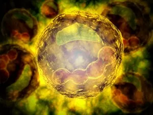
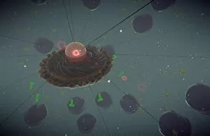
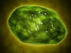
Background imageOrganelle Collection: Microscopic view of Paramecium bursaria

Microscopic view of Paramecium bursaria, a species of ciliate protozoan that has a mutualistic symbiotic relationship with green algae called Zoochlorella

Background imageOrganelle Collection: Microscopic view of protozoa

Microscopic view of protozoa. Leishmaniasis and dysentery are examples of diseases involving protozoa, often transmitted to humans by the bite of a parasite-carrying insect

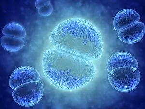
Background imageOrganelle Collection: Conceptual image of mitosis

Conceptual image of mitosis. Mitosis is the process in the cell cycle by which a cell duplicates into two genetically identical sets of chromosomes

Background imageOrganelle Collection: Microscopic view of coronavirus

Microscopic view of coronavirus

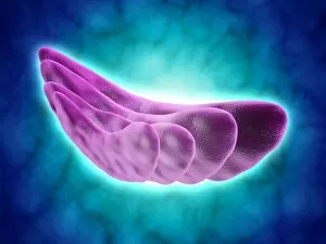
Background imageOrganelle Collection: Conceptual image of the Golgi apparatus

Conceptual image of the Golgi apparatus. The Golgi apparatus is an organelle found in most eukaryotic cells

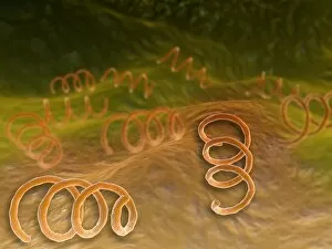
Background imageOrganelle Collection: Microscopic view of Syphillis

Microscopic view of Syphillis. Syphilis is a sexually transmitted infection caused by the spirochete bacterium, Treponema pallidum

Background imageOrganelle Collection: Microscopic view of Rickettsia

Microscopic view of Rickettsia. Rickettsia is a gram-negative bacteria carried by parasites such as ticks, fleas and lice

Background imageOrganelle Collection: Conceptual image of the coronavirus

Conceptual image of the coronavirus. Coronaviruses primarily infect the upper respiratory and gastrointestinal tract

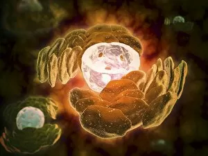
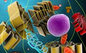
Background imageOrganelle Collection: Conceptual image of endoplasmic reticulum around a cell nucleus

Conceptual image of endoplasmic reticulum around a cell nucleus. Endoplasmic reticulum is an organelle that forms a continuous membrane system of flattened sacs within the cytoplasm of a eukaryotic

Background imageOrganelle Collection: Microscopic view of herpes virus

Microscopic view of herpes virus

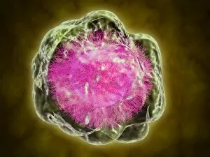
Background imageOrganelle Collection: Conceptual image of cell nucleus

Conceptual image of cell nucleus. The cell nucleus acts like the brain of the cell. It helps control eating, movement, and reproduction

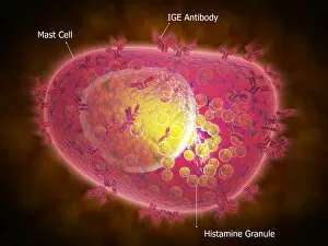
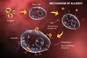
Background imageOrganelle Collection: Mast cell releasing histamine due to allergic reaction

Mast cell releasing histamine due to allergic reaction

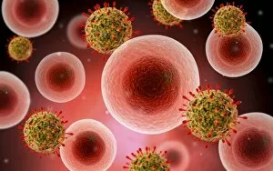
Background imageOrganelle Collection: Microscopic view of cell and virus

Microscopic view of cell and virus

Background imageOrganelle Collection: Microscopic view of centrioles within a human cell

Microscopic view of centrioles within a human cell
Microscopic view of barrel shaped structure of centrioles within a human cell. A centriole is a cylinder shaped cell structure found in most eukaryotic cells

Background imageOrganelle Collection: Microscopic view of Legionella pneumophila

Microscopic view of Legionella pneumophila. Legionella pneumophila is a thin, aerobic, pleomorphic, flagellated, non-spore forming, gram-negative bacterium of the genus Legionella

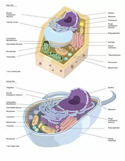
Background imageOrganelle Collection: Comparative illustration of plant and animal cell anatomy (with labels)

Comparative illustration of plant and animal cell anatomy (with labels)

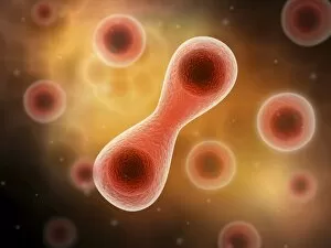
Background imageOrganelle Collection: Microscopic view of cell division

Microscopic view of cell division

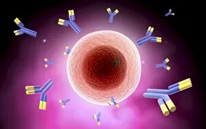
Background imageOrganelle Collection: Microscopic view of cell and antibody

Microscopic view of cell and antibody

Background imageOrganelle Collection: Microscopic view of animal cell nucleus

Microscopic view of animal cell nucleus

Background imageOrganelle Collection: Conceptual image of flagellate bacterium

Conceptual image of flagellate bacterium

Background imageOrganelle Collection: Conceptual image of a ubiquitous virus

Conceptual image of a ubiquitous virus. A ubiquitous virus is contagious in early childhood through the respiratory tract

Background imageOrganelle Collection: Microscopic view of Henipavirus

Microscopic view of Henipavirus. Henipavirus is a established group of paramyxoviruses comprising the Hendra virus, Nipah virus and Cedar virus

Background imageOrganelle Collection: Conceptual image of lysosome

Conceptual image of lysosome. Lysosomes are cellular organelles that contain acid hydrolase enzymes that break down waste materials and cellular debris

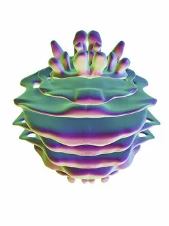
Background imageOrganelle Collection: Chloroplast, artwork

Chloroplast, artwork
Chloroplast, computer artwork

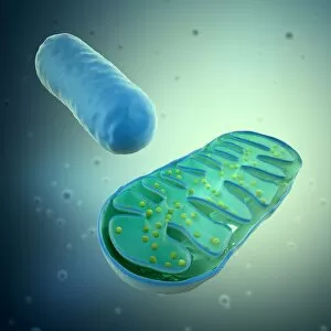
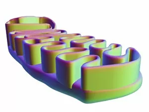
Background imageOrganelle Collection: Mitochondria, artwork

Mitochondria, artwork
Mitochondria, computer artwork

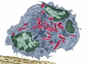
Background imageOrganelle Collection: Eosinophil white blood cell, TEM C014 / 1438

Eosinophil white blood cell, TEM C014 / 1438
Eosinophil white blood cell. Transmission electron micrograph (TEM) of a section through an eosinophil. Eosinophils, like all white blood cells, are part of the bodys immune system

Background imageOrganelle Collection: Nucleus and endoplasmic reticulum F006 / 9196

Nucleus and endoplasmic reticulum F006 / 9196
Computer artwork showing part of a human or eukaryotic cell. In the middle the nucleus which has a membrane with nuclear pores. Inside the nucleus is the DNA

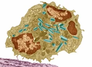
Background imageOrganelle Collection: Eosinophil white blood cell, TEM C014 / 1437

Eosinophil white blood cell, TEM C014 / 1437
Eosinophil white blood cell. Transmission electron micrograph (TEM) of a section through an eosinophil. Eosinophils, like all white blood cells, are part of the bodys immune system

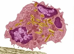
Background imageOrganelle Collection: Eosinophil white blood cell, TEM C014 / 1439

Eosinophil white blood cell, TEM C014 / 1439
Eosinophil white blood cell. Transmission electron micrograph (TEM) of a section through an eosinophil. Eosinophils, like all white blood cells, are part of the bodys immune system

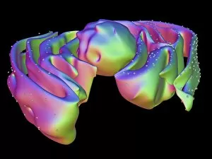
Background imageOrganelle Collection: Golgi apparatus, artwork F006 / 9211

Golgi apparatus, artwork F006 / 9211
Computer artwork of the Golgi apparatus of the human cell. This organelle functions as a central delivery system for the cell

Background imageOrganelle Collection: Mitochondrial structure, artwork F006 / 9207

Mitochondrial structure, artwork F006 / 9207
Mitochondrial structure. Computer artwork of a mitochondrion, showing a loop of mitochondrial DNA (deoxyribonucleic acid, mtDNA), the organelles genetic material


For sale as Licensed Images

Choose your image, Select your licence and Download the media

Organelles: The Tiny Powerhouses Within Our Cells The world of cells is a fascinating one, filled with intricate structures known as organelles. These tiny entities play crucial roles in maintaining the functionality and survival of our cells. From budding yeast to HeLa cells, each cell type possesses its unique set of organelles that contribute to their specialized functions. Under the lens of a light microscope, the HeLa cells reveal mesmerizing patterns resembling abstract artwork. Their delicate structures are visible, showcasing the complexity within these microscopic powerhouses. Meanwhile, another image captures nerve cells in all their glory - long and slender extensions reaching out like branches from a tree. Zooming in further using transmission electron microscopy (TEM), we get an up-close look at some specific organelles. The rough endoplasmic reticulum appears as a network of interconnected membranes studded with ribosomes responsible for protein synthesis. Mitochondria steal the spotlight next; these bean-shaped powerhouses generate energy for cellular activities through respiration. But it's not just animal cells that possess remarkable organelles; plant cells have their own unique features too. Chloroplasts, depicted beautifully through artwork, capture sunlight and convert it into energy via photosynthesis—a process vital for sustaining life on Earth. Another TEM image reveals Purkinje nerve cells—large neurons found in our brain's cerebellum—showcasing their intricate branching structure responsible for coordinating movement and balance. Finally, scanning electron microscopy (SEM) unveils the Golgi apparatus—an organelle involved in processing proteins and packaging them into vesicles for transportation throughout the cell or secretion outside it. These glimpses into different types of organelles highlight their diverse forms and functions within our cellular world. They remind us that even though they may be invisible to the naked eye, these minuscule entities hold immense importance in keeping our bodies functioning harmoniously at every level.