Organelle Collection (#3)

Organelles: The Tiny Powerhouses Within Our Cells The world of cells is a fascinating one, filled with intricate structures known as organelles

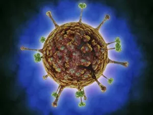
Background imageOrganelle Collection: Microscopic view of herpes virus

Microscopic view of herpes virus

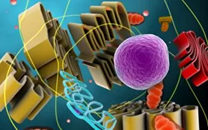
Background imageOrganelle Collection: Conceptual image of cell nucleus

Conceptual image of cell nucleus. The cell nucleus acts like the brain of the cell. It helps control eating, movement, and reproduction

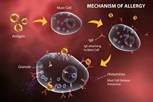
Background imageOrganelle Collection: Mast cell releasing histamine due to allergic reaction

Mast cell releasing histamine due to allergic reaction

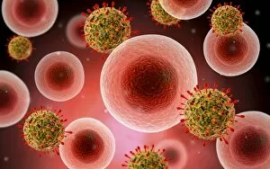
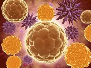
Background imageOrganelle Collection: Microscopic view of cell and virus

Microscopic view of cell and virus

Background imageOrganelle Collection: Microscopic view of centrioles within a human cell

Microscopic view of centrioles within a human cell
Microscopic view of barrel shaped structure of centrioles within a human cell. A centriole is a cylinder shaped cell structure found in most eukaryotic cells

Background imageOrganelle Collection: Microscopic view of Legionella pneumophila

Microscopic view of Legionella pneumophila. Legionella pneumophila is a thin, aerobic, pleomorphic, flagellated, non-spore forming, gram-negative bacterium of the genus Legionella

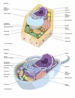
Background imageOrganelle Collection: Comparative illustration of plant and animal cell anatomy (with labels)

Comparative illustration of plant and animal cell anatomy (with labels)

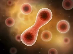
Background imageOrganelle Collection: Microscopic view of cell division

Microscopic view of cell division

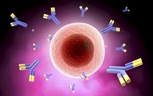
Background imageOrganelle Collection: Microscopic view of cell and antibody

Microscopic view of cell and antibody

Background imageOrganelle Collection: Microscopic view of animal cell nucleus

Microscopic view of animal cell nucleus

Background imageOrganelle Collection: Conceptual image of flagellate bacterium

Conceptual image of flagellate bacterium

Background imageOrganelle Collection: Conceptual image of a ubiquitous virus

Conceptual image of a ubiquitous virus. A ubiquitous virus is contagious in early childhood through the respiratory tract

Background imageOrganelle Collection: Microscopic view of Henipavirus

Microscopic view of Henipavirus. Henipavirus is a established group of paramyxoviruses comprising the Hendra virus, Nipah virus and Cedar virus

Background imageOrganelle Collection: Conceptual image of lysosome

Conceptual image of lysosome. Lysosomes are cellular organelles that contain acid hydrolase enzymes that break down waste materials and cellular debris

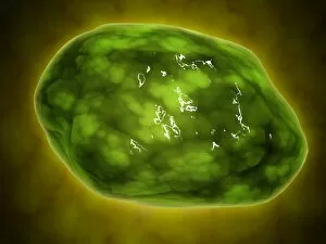
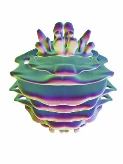
Background imageOrganelle Collection: Chloroplast, artwork

Chloroplast, artwork
Chloroplast, computer artwork

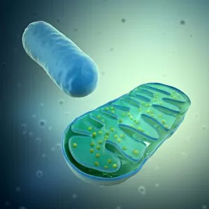
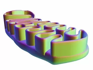
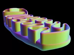
Background imageOrganelle Collection: Mitochondria, artwork

Mitochondria, artwork
Mitochondria, computer artwork

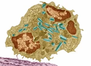
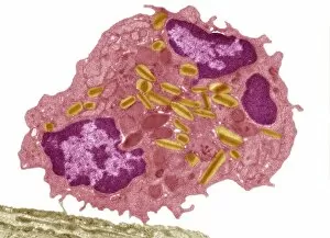
Background imageOrganelle Collection: Eosinophil white blood cell, TEM C014 / 1438

Eosinophil white blood cell, TEM C014 / 1438
Eosinophil white blood cell. Transmission electron micrograph (TEM) of a section through an eosinophil. Eosinophils, like all white blood cells, are part of the bodys immune system

Background imageOrganelle Collection: Nucleus and endoplasmic reticulum F006 / 9196

Nucleus and endoplasmic reticulum F006 / 9196
Computer artwork showing part of a human or eukaryotic cell. In the middle the nucleus which has a membrane with nuclear pores. Inside the nucleus is the DNA

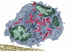
Background imageOrganelle Collection: Eosinophil white blood cell, TEM C014 / 1437

Eosinophil white blood cell, TEM C014 / 1437
Eosinophil white blood cell. Transmission electron micrograph (TEM) of a section through an eosinophil. Eosinophils, like all white blood cells, are part of the bodys immune system

Background imageOrganelle Collection: Eosinophil white blood cell, TEM C014 / 1439

Eosinophil white blood cell, TEM C014 / 1439
Eosinophil white blood cell. Transmission electron micrograph (TEM) of a section through an eosinophil. Eosinophils, like all white blood cells, are part of the bodys immune system

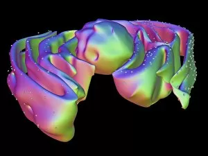
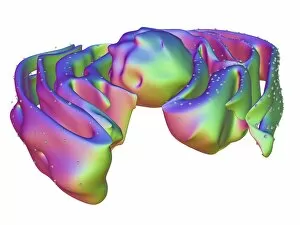
Background imageOrganelle Collection: Golgi apparatus, artwork F006 / 9211

Golgi apparatus, artwork F006 / 9211
Computer artwork of the Golgi apparatus of the human cell. This organelle functions as a central delivery system for the cell

Background imageOrganelle Collection: Mitochondrial structure, artwork F006 / 9207

Mitochondrial structure, artwork F006 / 9207
Mitochondrial structure. Computer artwork of a mitochondrion, showing a loop of mitochondrial DNA (deoxyribonucleic acid, mtDNA), the organelles genetic material

Background imageOrganelle Collection: Nucleus and endoplasmic reticulum F006 / 9201

Nucleus and endoplasmic reticulum F006 / 9201
Computer artwork showing part of a human or eukaryotic cell. In the middle the nucleus which has a membrane with nuclear pores. Inside the nucleus is the DNA

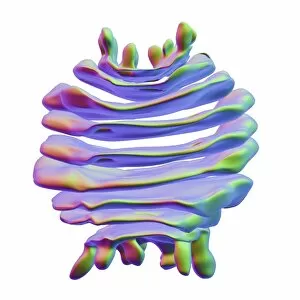
Background imageOrganelle Collection: Golgi apparatus, artwork F006 / 9199

Golgi apparatus, artwork F006 / 9199
Computer artwork of the Golgi apparatus of the human cell. This organelle functions as a central delivery system for the cell

Background imageOrganelle Collection: Mitochondrial structure, artwork F006 / 9198

Mitochondrial structure, artwork F006 / 9198
Mitochondrial structure. Computer artwork of a mitochondrion, showing a loop of mitochondrial DNA (deoxyribonucleic acid, mtDNA), the organelles genetic material

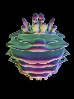
Background imageOrganelle Collection: Golgi apparatus, artwork F006 / 9189

Golgi apparatus, artwork F006 / 9189
Computer artwork of the Golgi apparatus of the human cell. This organelle functions as a central delivery system for the cell

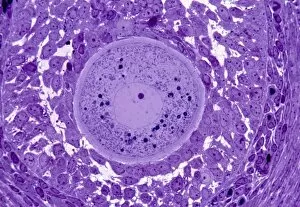
Background imageOrganelle Collection: Oocyte, light micrograph

Oocyte, light micrograph
Oocyte. Light micrograph of a section through an oocyte within a early antral follicle in an ovary. Oocytes are immature ova, or egg cells

Background imageOrganelle Collection: Vault ribonucleoprotein molecule

Vault ribonucleoprotein molecule
Vault cytoplasmic ribonucleoprotein, molecular model. This molecule is made up from proteins and RNA (ribonucleic acid) and has a 39-fold symmetry
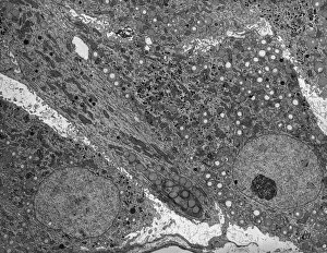
Background imageOrganelle Collection: Corpus luteum, TEM

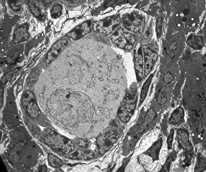
Corpus luteum, TEM
Corpus luteum. Transmission electron micrograph (TEM) of a section through the corpus luteum of an ovary, showing several luteal cells

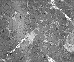
Background imageOrganelle Collection: Pancreatic exocrine cells, TEM

Pancreatic exocrine cells, TEM
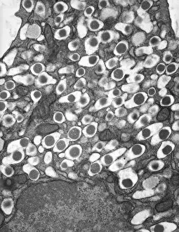
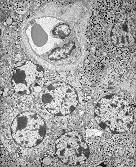
Pancreatic exocrine cells. Transmission electron micrograph (TEM) of a section through exocrine cells in the pancreas, showing numerous zymogen granules (circles), rough endoplasmic reticulum (ER)

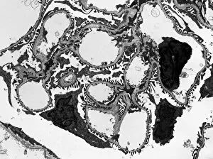
Background imageOrganelle Collection: Glomerulus, TEM

Glomerulus, TEM
Glomerulus. Transmission electron micrograph (TEM) of a section through a nephron in a human kidney, showing a cluster of capillaries within a renal corpuscle, also known as a glomerulus

Background imageOrganelle Collection: Gastric gland, TEM

Gastric gland, TEM
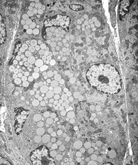
Gastric gland. Transmission electron micrograph (TEM) of a section through the deep region of a gastric (fundus) gland, showing several enzyme-secreting chief cells with many secretory granules

Background imageOrganelle Collection: Islet of Langerhans beta cell, TEM

Islet of Langerhans beta cell, TEM
Islet of Langerhans beta cell. Transmission electron micrograph (TEM) of a section through a beta cell in an islet of Langerhans in the pancreas
Background imageOrganelle Collection: Pancreatic acinar cell, TEM

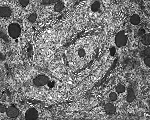
Pancreatic acinar cell, TEM
Pancreatic acinar cell. Transmission electron micrograph (TEM) of a section through an enzyme-secreting acinar cell in the human pancreas, showing the nucleus (dark purple, centre)
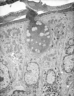
Background imageOrganelle Collection: Basophil white blood cell, TEM

Basophil white blood cell, TEM
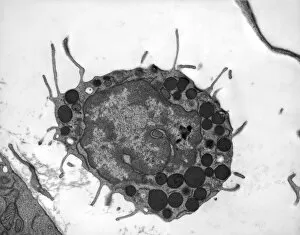
Basophil white blood cell. Transmission electron micrograph (TEM) of section through a circulating basophil cell. Basophils are the smallest and least common of the white blood cells

Background imageOrganelle Collection: Myenteric nerve plexus, TEM

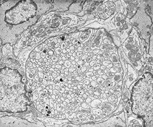
Myenteric nerve plexus, TEM
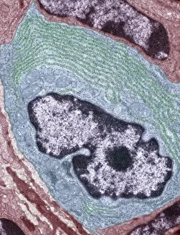
Myenteric nerve plexus. Transmission electron micrograph (TEM) of a section through a bundle of unmyelinated nerve fibres in the myenteric nerve plexus of the intestinal tract

Background imageOrganelle Collection: Golgi membranes, TEM

Golgi membranes, TEM
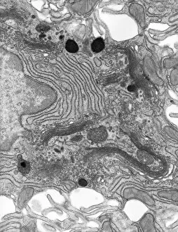
Golgi membranes. Transmission electron micrograph (TEM) of a section through a cell, showing the membranes (dark lines) of the Golgi apparatus

Background imageOrganelle Collection: Goblet cell, TEM

Goblet cell, TEM
Goblet cell. Transmission electron micrograph (TEM) of a section through a goblet cell in the lining (epithelium, top) of the small intestine

Background imageOrganelle Collection: Smooth muscle of gut wall, TEM

Smooth muscle of gut wall, TEM
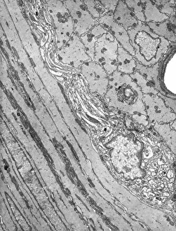
Smooth muscle of gut wall. Transmission electron micrograph (TEM) of a section through the outermost wall of the intestine

Background imageOrganelle Collection: Lymphocyte white blood cell, TEM

Lymphocyte white blood cell, TEM
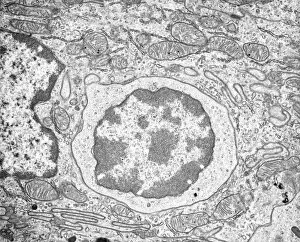
Lymphocyte white blood cell. Transmission electron micrograph (TEM) of a section through lymphocyte, showing the nucleus (large, round, centre), endoplasmic reticulum (ER)

Background imageOrganelle Collection: Unmyelinated nerve, TEM

Unmyelinated nerve, TEM
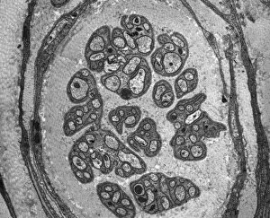
Unmyelinated nerve. Transmission electron micrograph (TEM) of a section through axon (nerve fibre) bundles of unmyelinated nerves

Background imageOrganelle Collection: Steroid secreting cell, TEM

Steroid secreting cell, TEM
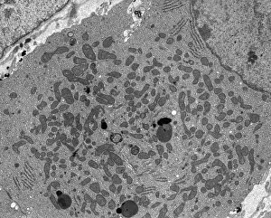
Steroid secreting cell. Transmission electron micrograph (TEM) of a section through a cell involved with steroid synthesis and secretion (steroidogenesis)

Background imageOrganelle Collection: Enterocyte, TEM

Enterocyte, TEM
Enterocyte. Transmission electron micrograph (TEM) of a section through the cytoplasm and part of the nucleus of an enterocyte cell located in a crypt of Lieberkuhn of the small intestine

Background imageOrganelle Collection: Islet of Langerhans, TEM

Islet of Langerhans, TEM
Islet of Langerhans. Transmission electron micrograph (TEM) of a section through cells clustered in an islet of Langerhans in the pancreas

Background imageOrganelle Collection: Capillary, TEM

Capillary, TEM
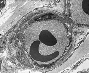
Capillary. Transmission electron micrograph (TEM) of a section through a capillary, showing two red blood cells (erythrocytes, black) in its interior

Background imageOrganelle Collection: Kidney mitochondria, TEM

Kidney mitochondria, TEM
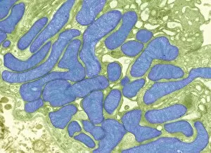
Kidney mitochondria. Transmission electron micrograph (TEM) of a section through a kidney tubule, showing numerous mitochondria (blue)

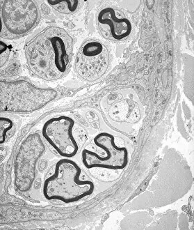
Background imageOrganelle Collection: Myelinated nerve tissue, TEM

Myelinated nerve tissue, TEM
Myelinated nerve tissue. Transmission electron micrograph (TEM) of a section through myelinated nerve fibres (axons). Each axon is coated with many layers of myelin

Background imageOrganelle Collection: Ovarian follicle, TEM

Ovarian follicle, TEM
Ovarian follicle. Transmission electron micrograph (TEM) of a section through an ovary, showing a primary follicle. Primary follicles contain a central oocyte (female germ cell)


For sale as Licensed Images

Choose your image, Select your licence and Download the media

Organelles: The Tiny Powerhouses Within Our Cells The world of cells is a fascinating one, filled with intricate structures known as organelles. These tiny entities play crucial roles in maintaining the functionality and survival of our cells. From budding yeast to HeLa cells, each cell type possesses its unique set of organelles that contribute to their specialized functions. Under the lens of a light microscope, the HeLa cells reveal mesmerizing patterns resembling abstract artwork. Their delicate structures are visible, showcasing the complexity within these microscopic powerhouses. Meanwhile, another image captures nerve cells in all their glory - long and slender extensions reaching out like branches from a tree. Zooming in further using transmission electron microscopy (TEM), we get an up-close look at some specific organelles. The rough endoplasmic reticulum appears as a network of interconnected membranes studded with ribosomes responsible for protein synthesis. Mitochondria steal the spotlight next; these bean-shaped powerhouses generate energy for cellular activities through respiration. But it's not just animal cells that possess remarkable organelles; plant cells have their own unique features too. Chloroplasts, depicted beautifully through artwork, capture sunlight and convert it into energy via photosynthesis—a process vital for sustaining life on Earth. Another TEM image reveals Purkinje nerve cells—large neurons found in our brain's cerebellum—showcasing their intricate branching structure responsible for coordinating movement and balance. Finally, scanning electron microscopy (SEM) unveils the Golgi apparatus—an organelle involved in processing proteins and packaging them into vesicles for transportation throughout the cell or secretion outside it. These glimpses into different types of organelles highlight their diverse forms and functions within our cellular world. They remind us that even though they may be invisible to the naked eye, these minuscule entities hold immense importance in keeping our bodies functioning harmoniously at every level.