Organelle Collection (#8)

Organelles: The Tiny Powerhouses Within Our Cells The world of cells is a fascinating one, filled with intricate structures known as organelles

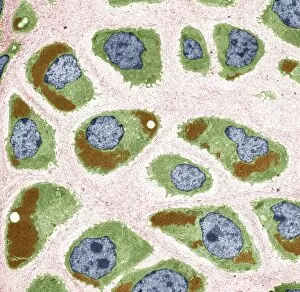
Background imageOrganelle Collection: Cell nucleus, TEM

Cell nucleus, TEM
Cell nucleus. Coloured transmission electron micrograph (TEM) of a section through a cell, showing the nucleus (large, spherical), and mitochondria (green)

Background imageOrganelle Collection: Chloroplasts, light micrograph

Chloroplasts, light micrograph. Chloroplasts are one of the features that distinguish a plant cell from an animal cell. They contain chlorophyll

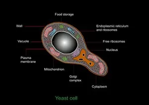
Background imageOrganelle Collection: Yeast cell, artwork

Yeast cell, artwork
Yeast cell. Computer artwork showing the structure of a yeast cell

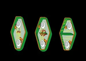
Background imageOrganelle Collection: Cytokinesis, artwork

Cytokinesis, artwork
Cytokinesis. Artwork showing the stage of cell division that involves the splitting of the cell cytoplasm between two daughter cells

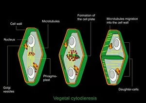
Background imageOrganelle Collection: Cytokinesis, diagram

Cytokinesis, diagram
Cytokinesis. Diagram showing the stage of cell division that involves the splitting of the cell cytoplasm between two daughter cells

Background imageOrganelle Collection: Cilium and flagellum structure, artwork

Cilium and flagellum structure, artwork. Cilia and flagella (collectively known as undulipodia) are hair-like protrusions from a cell membrane

Background imageOrganelle Collection: Chloroplast structures, artwork

Chloroplast structures, artwork
Chloroplast structures. Cutaway artwork showing the internal structure of a chloroplast, the organelle in plant cells responsible for photosynthesis

Background imageOrganelle Collection: Eukaryotic flagellum structure, artwork

Eukaryotic flagellum structure, artwork
Eukaryotic flagellum structure. Cutaway artwork showing the internal structure of the flagellum in eukaryotic cells. Flagella are tail-like projections used for cell locomotion

Background imageOrganelle Collection: Kidney cells, light micrograph

Kidney cells, light micrograph
Kidney cells. Quantum dot fluorescence micrograph of a section through kidney tissue showing its cells

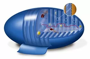
Background imageOrganelle Collection: Mitochondrion, artwork

Mitochondrion, artwork
Mitochondrion. Cutaway artwork showing the internal structure of a mitochondrion. This structure, found in eukaryotic cells, is the site of energy production

Background imageOrganelle Collection: Animal cell processes, artwork

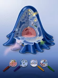
Animal cell processes, artwork
Animal cell processes. Cutaway artwork showing the structures inside an animal cell and four different processes that take place inside it or on its membrane (all marked by magnifying glasses)

Background imageOrganelle Collection: Animal cell structure, artwork

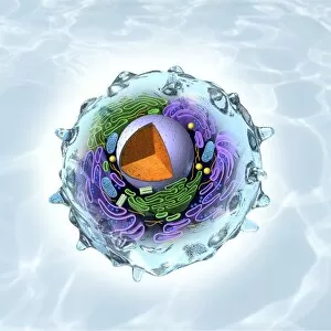
Animal cell structure, artwork
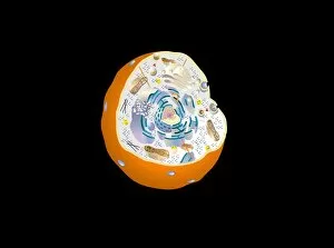
Animal cell structure. Artwork showing the internal structure of an animal cell. Inside the cell, the cellular structures (organelles) include the Golgi apparatus (green)

Background imageOrganelle Collection: Golgi apparatus, artwork

Golgi apparatus, artwork. This structure is an organelle found within eukaryotic cells. It receives proteins and lipids that are synthesised elsewhere on the endoplasmic reticulum

Background imageOrganelle Collection: Cytoskeleton, TEM

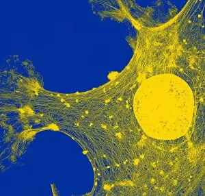
Cytoskeleton, TEM
Cytoskeleton. Coloured transmission electron micrograph (TEM) of the cytoskeleton of a human skin cell. The cell nucleus is at centre right

Background imageOrganelle Collection: Cytoskeleton, SEM

Cytoskeleton, SEM
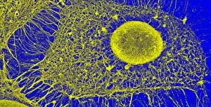
Cytoskeleton. Coloured scanning electron micrograph (SEM) of the cytoskeleton of a human skin cell. The cell nucleus is oval. The rest of the cells contents have been biochemically extracted

Background imageOrganelle Collection: Keratinocyte skin cells, light micrograph

Keratinocyte skin cells, light micrograph
Keratinocyte skin cells. Fluorescent light micrograph of the cytoskeleton of human keratinocyte skin cells. Cell nuclei are oval. The rest of the cells contents have been biochemically extracted

Background imageOrganelle Collection: Volvent nematocyst, artwork

Volvent nematocyst, artwork
Volvent nematocyst Artwork of a nematocyst, or cnidocyst, before (left) and after (right) discharge. Nematocysts are usually venomous threads that are found in Cnidaria, which includes jellyfish

Background imageOrganelle Collection: Milk-producing cell, artwork

Milk-producing cell, artwork
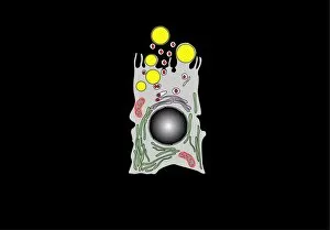
Milk-producing cell. Artwork of the anatomical structure of a lacteal (milk-producing) cell. Normal cell organelles include the nucleus (black, centre), the golgi apparatus (white)

Background imageOrganelle Collection: Milk-producing cell, diagram

Milk-producing cell, diagram
Milk-producing cell. Diagram of the anatomical structure of a lacteal (milk-producing) cell. Normal cell organelles include the nucleus (black, centre), the golgi apparatus (white)

Background imageOrganelle Collection: Animal cell anatomy, artwork

Animal cell anatomy, artwork
Animal cell anatomy. Artwork showing the internal and external anatomy of an animal cell

Background imageOrganelle Collection: mRNA leaving the nucleus, artwork

mRNA leaving the nucleus, artwork. mRNA (messenger ribonucleic acid, orange) is the intermediary molecule between DNA (deoxyribonucleic acid) and its protein product

Background imageOrganelle Collection: Animal cell, artwork

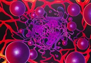
Animal cell, artwork
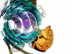
Animal cell. Computer artwork of an expanded animal cell. At centre left is the nucleus (purple sphere), which contains the cells genetic information in the form of DNA (deoxyribonucleic acid)

Background imageOrganelle Collection: Heart muscle fibres, SEM

Heart muscle fibres, SEM
Heart muscle fibres. Coloured scanning electron micrograph (SEM) of cardiac muscle fibrils (pink) from a healthy heart. The muscle fibrils, or myofibrils

Background imageOrganelle Collection: Cartilage cells, TEM

Cartilage cells, TEM
Cartilage cells. Coloured transmission electron micrograph (TEM) of a section through chondrocytes from nasal hyaline cartilage

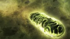
Background imageOrganelle Collection: Mitochondrial energy, conceptual image

Mitochondrial energy, conceptual image. Computer artwork of a glowing mitochondrion, representing the energy produced by these cell organelles

Background imageOrganelle Collection: Nucleolus, SEM

Nucleolus, SEM
Nucleolus, coloured scanning electron micrograph (SEM). The nucleolus is responsible for producing components of ribosomes, the cells protein-manufacturing organelles

Background imageOrganelle Collection: Plant cell, SEM

Plant cell, SEM
Plant cell. Coloured scanning electron micrograph (SEM) of a section through a plant cell, revealing its internal structure. The cell is encased in a cellulose, hemicellulose and pectin cell wall

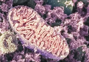
Background imageOrganelle Collection: Mitochondrion, SEM

Mitochondrion, SEM
Mitochondrion. Coloured scanning electron micrograph (SEM) of a mitochondrion in a nerve cell. Mitochondria are a type of organelle found in the cytoplasm of eukaryotic cells

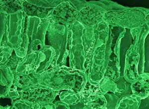
Background imageOrganelle Collection: Leaf section, SEM

Leaf section, SEM
Leaf section. Coloured scanning electron micrograph (SEM) of a section through a fractured leaf. At top is a single layer of cells that forms the epidermis of the leaf

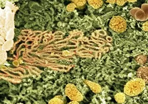
Background imageOrganelle Collection: Cell organelles, SEM

Cell organelles, SEM
Cell organelles. Coloured scanning electron micrograph (SEM) of a section through a cell from a kidney proximal tubule. The cell nucleus (partially seen at top left)

Background imageOrganelle Collection: Smooth endoplasmic reticulum, SEM

Smooth endoplasmic reticulum, SEM
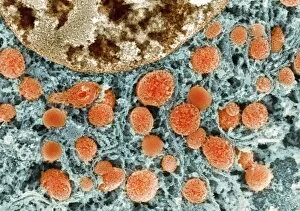
Endoplasmic reticulum. Coloured scanning electron micrograph (SEM) of smooth endoplasmic reticulum (SER) (orange, centre left) in the cytoplasm of a kidney cell

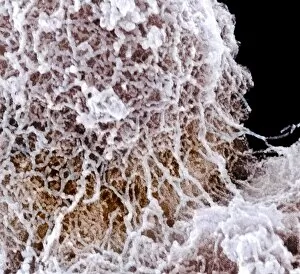
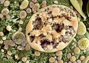
Background imageOrganelle Collection: Cell nucleus, SEM

Cell nucleus, SEM
Cell nucleus. Coloured scanning electron micrograph (SEM) of a section through the nucleus (centre right) of a kidney cell


For sale as Licensed Images

Choose your image, Select your licence and Download the media

Organelles: The Tiny Powerhouses Within Our Cells The world of cells is a fascinating one, filled with intricate structures known as organelles. These tiny entities play crucial roles in maintaining the functionality and survival of our cells. From budding yeast to HeLa cells, each cell type possesses its unique set of organelles that contribute to their specialized functions. Under the lens of a light microscope, the HeLa cells reveal mesmerizing patterns resembling abstract artwork. Their delicate structures are visible, showcasing the complexity within these microscopic powerhouses. Meanwhile, another image captures nerve cells in all their glory - long and slender extensions reaching out like branches from a tree. Zooming in further using transmission electron microscopy (TEM), we get an up-close look at some specific organelles. The rough endoplasmic reticulum appears as a network of interconnected membranes studded with ribosomes responsible for protein synthesis. Mitochondria steal the spotlight next; these bean-shaped powerhouses generate energy for cellular activities through respiration. But it's not just animal cells that possess remarkable organelles; plant cells have their own unique features too. Chloroplasts, depicted beautifully through artwork, capture sunlight and convert it into energy via photosynthesis—a process vital for sustaining life on Earth. Another TEM image reveals Purkinje nerve cells—large neurons found in our brain's cerebellum—showcasing their intricate branching structure responsible for coordinating movement and balance. Finally, scanning electron microscopy (SEM) unveils the Golgi apparatus—an organelle involved in processing proteins and packaging them into vesicles for transportation throughout the cell or secretion outside it. These glimpses into different types of organelles highlight their diverse forms and functions within our cellular world. They remind us that even though they may be invisible to the naked eye, these minuscule entities hold immense importance in keeping our bodies functioning harmoniously at every level.