Phloem Collection (#3)

Phloem, the intricate network of vascular bundles responsible for transporting nutrients and sugars throughout plants, is a fascinating subject to explore

Background imagePhloem Collection: Horse-tail stem, light micrograph

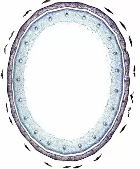
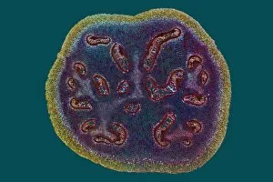
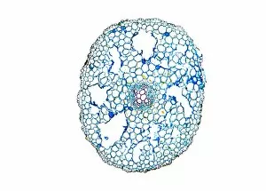
Horse-tail stem, light micrograph
Rice grass stem. Light micrograph of a section through a rice grass stem (Oryza sativa). Vascular bundles (small oval structures) can be seen containing xylem (larger openings)

Background imagePhloem Collection: Plant root development, artwork

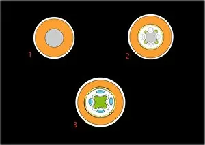
Plant root development, artwork
Plant root development. Artwork showing the first three stages in the development of a plant root. The first of these three stages (upper left)

Background imagePhloem Collection: Plant root development, diagram

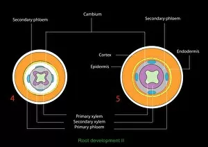
Plant root development, diagram
Plant root development. Diagram showing the the fourth (left) and fifth (right) stages in a series showing the development of a plant root

Background imagePhloem Collection: Grape vine stem, light micrograph

Grape vine stem, light micrograph
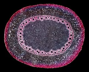
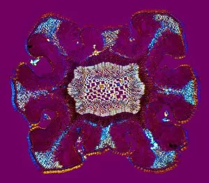
Grape vine stem. Polarised light micrograph of a cross-section through a young stem from the grape vine (Vitis vinifera). Most of the stem is a large central pith made up of parenchyma tissue (blue)

Background imagePhloem Collection: Tree growth rings, light micrograph

Tree growth rings, light micrograph
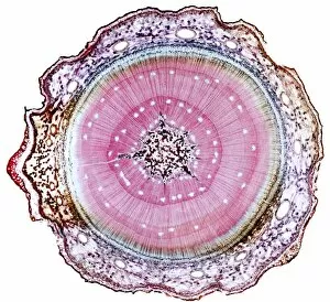
Tree growth rings. Polarised light micrograph of a cross-section through a woody stem, showing a portion of an annual ring of xylem tissue with periods of seasonal growth

Background imagePhloem Collection: Virginia creeper stem, light micrograph

Virginia creeper stem, light micrograph
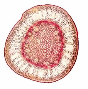
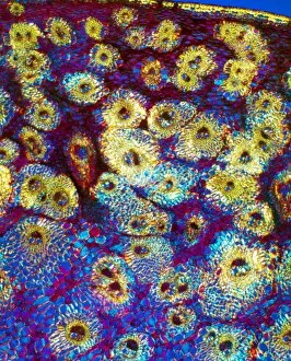
Virginia creeper stem. Polarised light micrograph of a cross-section through a stem from a Virginia creeper (Parthenocissus tricuspidatus)

Background imagePhloem Collection: Celery stalk, light micrograph

Celery stalk, light micrograph
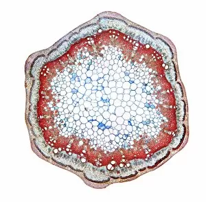
Celery stalk. Light micrograph of a cross-section through a stalk from a celery plant (Apium graveolens). Red eosin dye has been used to stain the xylem tissues

Background imagePhloem Collection: Tree-of-heaven stem, light micrograph

Tree-of-heaven stem, light micrograph
Tree-of-heaven stem. Polarised light micrograph of a cross-section through the stem of the tree-of-heaven (Ailanthus glandulosa). Below the outer layers (red) is a ring of vascular bundles

Background imagePhloem Collection: Aralia leaf stalk, light micrograph

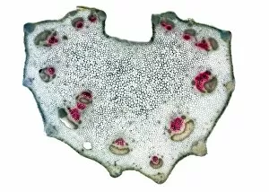
Aralia leaf stalk, light micrograph
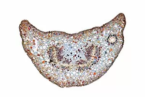
Aralia leaf stalk. Light micrograph of a cross-section through a leaf stalk (petiole) from an Araliatree. The outer layer directly below the epidermis is a supporting collenchyma ring (red)

Background imagePhloem Collection: Bracken fern rhizome, light micrograph

Bracken fern rhizome, light micrograph
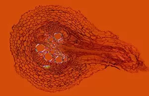
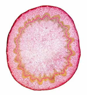
Bracken fern rhizome. Polarised light micrograph of a cross-section through a rhizome from a bracken fern (Pteris aquilinum). The outer layer (yellow-green) consists of supporting fibres

Background imagePhloem Collection: Corn (Zea mays) root, light micrograph

Corn (Zea mays) root, light micrograph
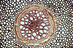
Maize root. Polarised light micrograph of a section through the root of a maize plant (Zea mays) showing a vascular cylinder

Background imagePhloem Collection: Fern rhizome, light micrograph

Fern rhizome, light micrograph. Transverse section through the center of a rhizome from the Killarney fern (Trichomanes speciosum)

Background imagePhloem Collection: Clubmoss stem, light micrograph

Clubmoss stem, light micrograph
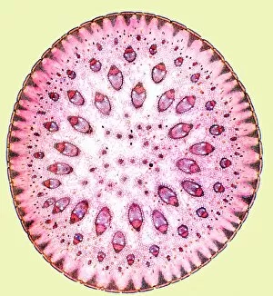
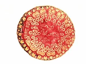
Clubmoss stem, polarised light micrograph. Transverse section through the stem of the clubmoss Lycopodium clavatum. This is the central portion of the stem consisting of the inner cortex (red)

Background imagePhloem Collection: Butchers broom stem, light micrograph

Butchers broom stem, light micrograph
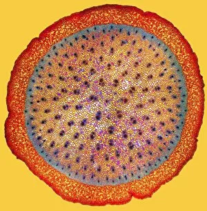
Butchers broom stem. Polarised light micrograph of a section through a butchers broom stem (Ruscus aculeatis) showing many vascular bundles (dark clusters) within the plant cortex (orange)

Background imagePhloem Collection: Umbrella grass stem, light micrograph

Umbrella grass stem, light micrograph
Umbrella grass stem. Polarised light micrograph of a section through a umbrella grass stem (Cyperus alternifolius). Vascular bundles (brown) can be seen containing xylem (larger openings)

Background imagePhloem Collection: Flax plant stem, light micrograph

Flax plant stem, light micrograph
Flax plant stem. Light micrograph of a transverse section through a stem of the flax plant (Linum usitatissimum). The layers from outer to inner (some very thin) are the epidermis (bottom)

Background imagePhloem Collection: Purple moor grass stem, light micrograph

Purple moor grass stem, light micrograph
Purple moor grass stem. Polarised light micrograph of a section through a purple moor grass stem (Molinia caerula). Vascular bundles (round orange structures)

Background imagePhloem Collection: Bamboo stem, light micrograph

Bamboo stem, light micrograph
Bamboo stem. Polarised light micrograph of a section through a bamboo stem (Bambusa vulgaris). Vascular bundles can be seen (yellow/green) containing xylem (larger openings)

Background imagePhloem Collection: Maize stem, light micrograph

Maize stem, light micrograph
Maize stem. Polarised light micrograph of a section through a maize plant stem (Zea mays). A vascular bundles (brown) can be seen containing xylem (larger openings)

Background imagePhloem Collection: Spiny rush stem, light micrograph

Spiny rush stem, light micrograph
Spiny rush stem. Polarised light micrograph of a section through a spiny rush stem (Juncus actus). Vascular bundles can be seen (purple/red) containing xylem (larger openings)

Background imagePhloem Collection: Wheat stem, light micrograph

Wheat stem, light micrograph
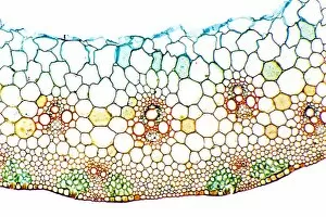
Wheat stem. Light micrograph of a section through the stem of a wheat grass (Triticum aestivium). The circular structures (orange and green) are vascular bundles

Background imagePhloem Collection: Sugar cane stem, light micrograph

Sugar cane stem, light micrograph
Sugar cane stem. Polarised light micrograph of a section through a sugar cane stem (Saccharum officinareum). Vascular bundles (round blue/purple structures)

Background imagePhloem Collection: Reed canary grass stem, light micrograph

Reed canary grass stem, light micrograph
Reed canary grass stem. Light micrograph of a section through a reed canary grass stem (Phalaris arundinacea). Vascular bundles (oval yellow and red structures)

Background imagePhloem Collection: Cotton plant root, light micrograph

Cotton plant root, light micrograph
Cotton plant root. Light micrograph of a transverse section through a root of the cotton plant (Gossypium hirsutum). The layers from outer to centre (some very thin)

Background imagePhloem Collection: Dragon tree stem, light micrograph

Dragon tree stem, light micrograph
Dragon tree stem. Light micrograph of a section through a dragon tree stem (Dracaena draco). Vascular bundles (round dark structures) can be seen containing xylem (larger openings)

Background imagePhloem Collection: Peanut plant stem, light micrograph

Peanut plant stem, light micrograph
Peanut plant stem. Light micrograph of a transverse section through a stem of the peanut plant (Arachis hypogaea). Below the stems outer layer (epidermis) is a cortex of parenchyma cells (blue)

Background imagePhloem Collection: Sarsaparilla plant stem, light micrograph

Sarsaparilla plant stem, light micrograph
Maize root. Polarised light micrograph of a section through the root of a sarsaparilla plant (Smilax regelii) showing a vascular cylinder

Background imagePhloem Collection: Stinging nettle stem, light micrograph

Stinging nettle stem, light micrograph
Stinging nettle stem. Polarised light micrograph of a transverse section through a stem of the stinging nettle plant (Urtica dioica)

Background imagePhloem Collection: Tea leaf, light micrograph

Tea leaf, light micrograph
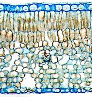
Tea leaf. Light micrograph of a cross-section through a tea (Camellia sinensis) leaf. The upper and lower epidermis on the surfaces of the leaf are blue

Background imagePhloem Collection: Kidney bean stem, light micrograph

Kidney bean stem, light micrograph
Kidney bean stem. Light micrograph of a section through the stem of a kidney bean (Phaseolus vulgaris) plant. The outer layer is the cuticle (brown), with a cortex of parenchyma (yellow) beneath it

Background imagePhloem Collection: Broad bean root, light micrograph

Broad bean root, light micrograph
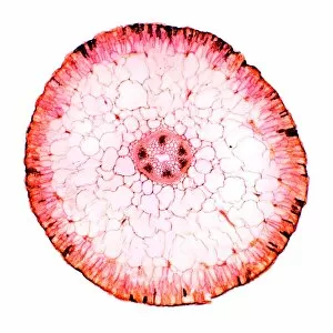
Broad bean root. Light micrograph of a transverse section through the young root of a broad bean (Vicia faba) plant. The outer epidermis is covered in root hairs

Background imagePhloem Collection: Kidney bean root, light micrograph

Kidney bean root, light micrograph
Kidney bean root. Light micrograph of a section through the root of a kidney bean (Phaseolus vulgaris) plant. At centre is the pith (blue)

Background imagePhloem Collection: Common broom stem, light micrograph

Common broom stem, light micrograph
Common broom stem. Light micrograph of a transverse section through the stem of a common broom (Salicornia europaea) plant

Background imagePhloem Collection: Phloem plant cells, light micrograph

Phloem plant cells, light micrograph
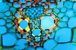
Phloem plant cells. Light micrograph of a longitudinal section through phloem tissue in a Cucurbita plant. Most of this tissue is large phloem tubes (stained blue)

Background imagePhloem Collection: Oleander stem, light micrograph

Oleander stem, light micrograph
Oleander stem. Light micrograph of a cross-section through the stem of an oleander (Nerium oleander) tree. The outer layer is a thick cuticle (yellow) and then cork cambium (blue-green)

Background imagePhloem Collection: Young pine tree stem, light micrograph

Young pine tree stem, light micrograph
Young pine tree stem. Light micrograph of a transverse section through a two and a half-year-old stem of a pine tree (Pinus sp.)

Background imagePhloem Collection: Black burdock stem, light micrograph

Black burdock stem, light micrograph
Black burdock stem. Polarised light micrograph of a transverse section through the stem of a black burdock (Arctium lappa) plant

Background imagePhloem Collection: Goosefoot root, light micrograph

Goosefoot root, light micrograph
Goosefoot root. Light micrograph of a section through the root of a goosefoot (Cheopodium sp.) plant. Xylem cells, which transport water around the plant are red

Background imagePhloem Collection: Dyers greenweed stem, light micrograph

Dyers greenweed stem, light micrograph
Dyers greenweed stem. Polarised light micrograph of a transverse section through the stem of a dyers greenweed (Genista tinctoria) plant

Background imagePhloem Collection: Yew tree root, light micrograph

Yew tree root, light micrograph
Yew tree root. Light micrograph of a transverse section through a root of a yew tree, (Taxus baccata). Most of the outer cortex (dark blue)

Background imagePhloem Collection: Marsh samphire stem, light micrograph

Marsh samphire stem, light micrograph
Marsh samphire stem. Light micrograph of a transverse section through the stem of a marsh samphire, (Salicornia europaea) plant. This is a succulent, xerophytic, halophyte plant

Background imagePhloem Collection: Ginkgo leaf stalk, light micrograph

Ginkgo leaf stalk, light micrograph
Ginkgo leaf stalk. Light micrograph of a transverse section through the leaf stalk (petiole) of a ginkgo tree (Ginkgo biloba)

Background imagePhloem Collection: Begonia stem, light mictograph

Begonia stem, light mictograph
Begoinia stem. Light micrograph of a transverse section through the stem of a begonia (Begonia sp.) plant. At centre is a large area of pith, which contains starch grains (orange) a store of energy

Background imagePhloem Collection: Blackcurrant plant stem, light micrograph

Blackcurrant plant stem, light micrograph
Blackcurrant plant stem. Light micrograph of a cross-section through the stem of a blackcurrant (Ribes nigrum) plant. The outer cortex (blue and pale brown) is made of cork cambium tissue

Background imagePhloem Collection: Sunflower root, light micrograph

Sunflower root, light micrograph
Sunflower root. Polarised light micrograph of a transverse section through the root of a sunflower (Helianthus annuus) plant

Background imagePhloem Collection: Spikenard stem, light micrograph

Spikenard stem, light micrograph
Spikenard stem. Light micrograph of a cross-section through the stem of a Spikenard (Aralia sp.) tree. The dark red cells just beneath the surface bark are collenchyma cells

Background imagePhloem Collection: Buttercup root, light micrograph

Buttercup root, light micrograph
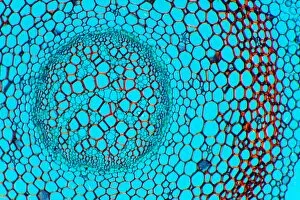
Buttercup root. Light micrograph of a transverse section through the root of a buttercup (Ranunculus sp.) plant. At centre is the vascular bundle, comprising xylem (red) and phloem (blue) tissues

Background imagePhloem Collection: Rose stem, light micrograph

Rose stem, light micrograph
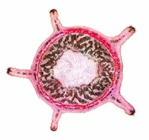
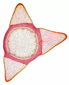
Rose stem. Light micrograph of a cross-section through the stem of a rose (Rosa sp.). The three triangular protrusions are thorns, which protect the plant from being eaten by animals


For sale as Licensed Images

Choose your image, Select your licence and Download the media

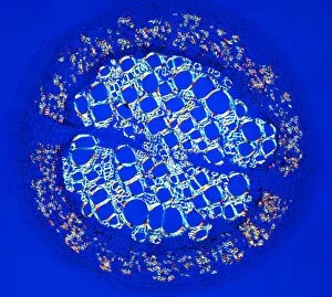
Phloem, the intricate network of vascular bundles responsible for transporting nutrients and sugars throughout plants, is a fascinating subject to explore. Through scanning electron microscopy (SEM) and light micrographs, we can delve into the hidden world in various plant species. In a lime tree stem, a light micrograph reveals the delicate structure cells intertwined within the vascular bundle. These specialized cells form sieve tubes that efficiently transport organic compounds from leaves to other parts of the plant. Similarly, in a maize root captured under a light microscope, we witness the intricate web-like arrangement tissue. This vital system ensures essential nutrients are distributed from roots to shoots for growth and development. The water lily leaf showcases another mesmerizing view through a light micrograph. Here, we observe elongated phloem fibers running parallel to each other like veins on this aquatic beauty's surface. Switching gears to SEM imaging, we encounter an enchanting sight on a silver birch twig. The high-resolution image captures xylem vessels alongside phloem cells with astonishing detail—each playing its crucial role in maintaining fluid balance and nutrient transport within this majestic tree. A closer look at water fern rhizome under a light microscope uncovers an interconnected network tissues supporting its underground growth. These structures ensure efficient distribution of resources necessary for survival in challenging environments. Examining pine tree stems through light microscopy allows us to appreciate how intricately woven strands contribute to their robustness and longevity—a testament to nature's engineering marvels. Delving into tomato leaf anatomy via light micrographs reveals densely packed clusters of sieve tube elements forming part of its extensive phloem network—an integral component enabling fruit production and overall plant health. Pondweed stems provide yet another captivating glimpse into the world beneath our feet when observed using advanced microscopic techniques. Phloem fibers intertwine with xylem vessels, showcasing the plant's ability to adapt and thrive in aquatic environments.