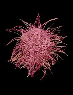
Protozoan Collection (#2)

"Exploring the Microscopic World: Unveiling the Diversity Life" In this captivating image series

Background imageProtozoan Collection: Cross section biomedical illustration on grid of Worm, Fungi, Protozoa

Cross section biomedical illustration on grid of Worm, Fungi, Protozoa, and Bacteria infection and infestation

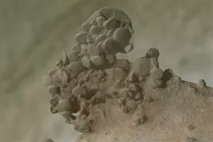
Background imageProtozoan Collection: Egypt, Eastern Saharan Libyan Desert, Qattara Depression, Protozoan nummulites

Egypt, Eastern Saharan Libyan Desert, Qattara Depression, Protozoan nummulites
Egypt. Eastern Saharan Libyan Desert. Qattara Depression. Protozoan nummulites

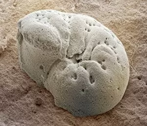
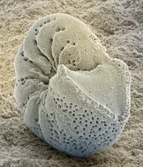
Background imageProtozoan Collection: Foraminiferan fossil, SEM

Foraminiferan fossil, SEM
Foraminiferan fossil. Coloured scanning electron micrograph (SEM) of the fossilised shell (test) of a foraminiferan. Foraminifera are single-celled marine protozoa that construct

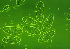
Background imageProtozoan Collection: LM of the protozoan Paramecium sp

LM of the protozoan Paramecium sp
Light micrograph of the ciliate protozoan, Paramecium sp. They are one of the most highly- evolved of all unicellular animals, and are plentiful in fresh water ponds

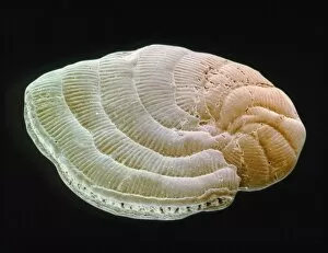
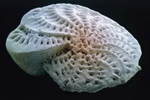
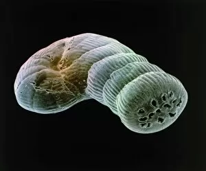
Background imageProtozoan Collection: Coloured SEM of foraminiferan, Peneroplis sp

Coloured SEM of foraminiferan, Peneroplis sp
Foraminiferan shell. Coloured scanning electron micrograph (SEM) of the test (shell) of the Mediterranean foraminiferan, Peneroplis proteus

Background imageProtozoan Collection: SEM of Paramecium

SEM of Paramecium
Paramecium. Coloured scanning electron micrograph (SEM) of the single-celled protozoan, Paramecium caudatum. This ciliate animal has cilia on its cell membrane surface enabling it to swim with an

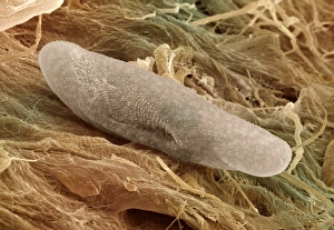
Background imageProtozoan Collection: Chilodonella ciliate protozoan, SEM

Chilodonella ciliate protozoan, SEM
Chilodonella ciliate protozoa, coloured scanning electron micrograph (SEM). These tiny single- celled organisms are found in fresh water

Background imageProtozoan Collection: LM of of a single Paramecium sp

LM of of a single Paramecium sp
Light micrograph of the ciliate protozoan Paramecium sp. The internal structure of the organism is particularly clear. The large dark area toward the top of the frame is the meganucleus

Background imageProtozoan Collection: Coloured SEM of Podocyrtis mitra, a radiolarian

Coloured SEM of Podocyrtis mitra, a radiolarian
Radiolarian. Coloured scanning electron micrograph (SEM) of the test (skeleton) of a Podocyrtis mitra, a type of radiolarian protozoan

Background imageProtozoan Collection: LM of paramecium sp

LM of paramecium sp
Light micrograph in phase contrast illumination of the ciliate protozoan, Paramecium sp. The internal structure of Paramecium is visible here with many food and water vacuoles seen as bright spots

Background imageProtozoan Collection: Protozoan sexual reproduction, SEM Z105 / 0240

Protozoan sexual reproduction, SEM Z105 / 0240
Protozoan sexual reproduction. Coloured scanning electron micrograph (SEM) of two Paramecium caudatum protozoa reproducing sexually by a method called conjugation

Background imageProtozoan Collection: Coloured SEM of foraminiferan, Elphidium sp

Coloured SEM of foraminiferan, Elphidium sp
Foraminiferan shell. Coloured scanning electron micrograph (SEM) of the test (shell) of the British formaminiferan, Elphidium crispum

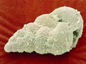
Background imageProtozoan Collection: Coloured SEM of Foraminifera Textularia sp. fossil

Coloured SEM of Foraminifera Textularia sp. fossil
Fossil foraminifera. Coloured scanning electron micrograph (SEM) of the fossil shell of the single-celled protozoan Textularia sp. belonging to the order Foraminifera

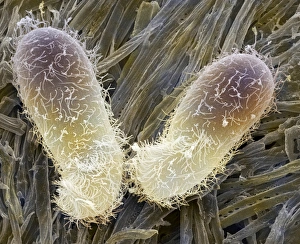
Background imageProtozoan Collection: Paramecium protozoa, SEM Z105 / 0239

Paramecium protozoa, SEM Z105 / 0239
Paramecium protozoa. Coloured scanning electron micrograph (SEM) of two Paramecium caudatum protozoa on filaments (green) of Oscillatoria sp. blue-green algae. P

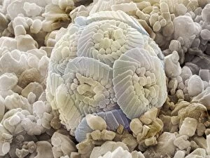
Background imageProtozoan Collection: Calcareous phytoplankton fossil, SEM Z100 / 0212

Calcareous phytoplankton fossil, SEM Z100 / 0212
Calcareous phytoplankton fossil. Coloured scanning electron micrograph (SEM) of a fossil of the skeleton (coccosphere) of a coccolithotrope, a small marine algal organism

Background imageProtozoan Collection: Chilodonella ciliate protozoa, LM

Chilodonella ciliate protozoa, LM
Chilodonella protozoa. Light micrograph of several Chilodonella sp. ciliate protozoa. Chilodonella is a single-celled animal that is found in freshwater

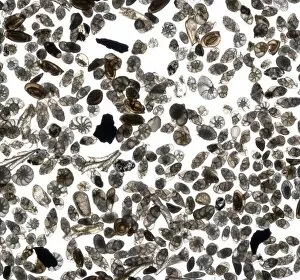
Background imageProtozoan Collection: Fossilised foraminiferans, SEM

Fossilised foraminiferans, SEM
Fossilised foraminiferans. Coloured scanning electron micrograph (SEM) of the fossilised shells of assorted foraminiferan protozoa (order Foraminifera)

Background imageProtozoan Collection: Amoebae, SEM

Amoebae, SEM
Amoebae. Coloured scanning electron micrograph (SEM) of two large protozoan amoebae (Amoeba proteus). A. proteus is commonly found on the bottom of pools and puddles

Background imageProtozoan Collection: Coloured SEM of foraminiferan, Spirolina sp

Coloured SEM of foraminiferan, Spirolina sp
Foraminiferan shell. Coloured scanning electron micrograph (SEM) of the test (shell) of the Mediterranean foraminiferan, Spirolina areitinus

Background imageProtozoan Collection: Foraminiferan tests

Foraminiferan tests (shells), macrophotograph. Species seen here include Bulimina gibba, Elphidium crispum, E. articulatum, Lagena sulcata, and Miliolinella circularis

Background imageProtozoan Collection: Foraminiferan fossil, SEM Z110 / 0239

Foraminiferan fossil, SEM Z110 / 0239
Foraminiferan fossil. Coloured scanning electron micrograph (SEM) of the fossilised shell (test) of a foraminiferan. Foraminifera are single-celled marine protozoa that construct

Background imageProtozoan Collection: Giardia protozoan, artwork F005 / 0579

Giardia protozoan, artwork F005 / 0579
Giardia protozoan, computer artwork

Background imageProtozoan Collection: Radiolarian skeleton, artwork F005 / 0194

Radiolarian skeleton, artwork F005 / 0194
Radiolarian skeleton. Computer artwork of the skeleton of a radiolarian. Radiolaria are single-celled protozoans that are found in marine plankton

Background imageProtozoan Collection: Radiolarian skeleton, artwork F005 / 0190

Radiolarian skeleton, artwork F005 / 0190
Radiolarian skeleton. Computer artwork of the skeleton of a radiolarian. Radiolaria are single-celled protozoans that are found in marine plankton

Background imageProtozoan Collection: Giardia protozoan, artwork F005 / 0580

Giardia protozoan, artwork F005 / 0580
Giardia protozoan, computer artwork

Background imageProtozoan Collection: Giardia protozoan, artwork F005 / 0578

Giardia protozoan, artwork F005 / 0578
Giardia protozoan, computer artwork

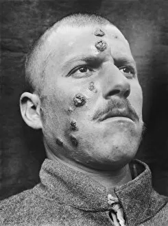
Background imageProtozoan Collection: Cutaneous leishmaniasis, 1917

Cutaneous leishmaniasis, 1917
Cutaneous leishmaniasis. Lesions on the face of a man in Palestine in 1917 in a case of cutaneous leishmaniasis. The local name for this condition at the time was Jericho buttons

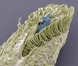
Background imageProtozoan Collection: Holosticha ciliate protozoan, SEM

Holosticha ciliate protozoan, SEM
Holosticha ciliate protozoan. Coloured scanning electron micrograph (SEM) of a Holosticha sp. ciliate protozoan ingesting diatoms (blue). Holosticha is a tiny single-celled aquatic organism

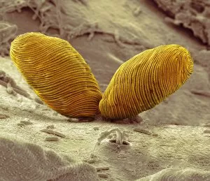
Background imageProtozoan Collection: Euplotes protozoa, SEM C016 / 9040

Euplotes protozoa, SEM C016 / 9040
Euplotes protozoa. Coloured scanning electron micrograph (SEM) showing the upperside (bottom) and underside (top) of two Euplotes sp. hypotrich ciliate protozoa

Background imageProtozoan Collection: Climacostomum protozoan, SEM C016 / 9063

Climacostomum protozoan, SEM C016 / 9063
Climacostomum protozoan. Coloured scanning electron micrograph (SEM) of a Climacostomum sp. unicellular ciliate protozoan, showing the cells large oral apparatus (round, centre)

Background imageProtozoan Collection: Euglena flagellate protozoa, SEM C016 / 9103

Euglena flagellate protozoa, SEM C016 / 9103
Euglena flagellate protozoa. Coloured scanning electron micrograph (SEM) of two Euglena sp. flagellate protozoa. Euglena sp

Background imageProtozoan Collection: Climacostomum protozoan, SEM C016 / 9121

Climacostomum protozoan, SEM C016 / 9121
Climacostomum protozoan. Coloured scanning electron micrograph (SEM) of a Climacostomum sp. unicellular ciliate protozoan, showing the cells large oral apparatus (round, centre)

Background imageProtozoan Collection: Climacostomum protozoan, SEM C016 / 9061

Climacostomum protozoan, SEM C016 / 9061
Climacostomum protozoan. Coloured scanning electron micrograph (SEM) of a Climacostomum sp. unicellular ciliate protozoan, showing the cells large oral apparatus (round, upper right)

Background imageProtozoan Collection: Climacostomum protozoan, SEM C016 / 9120

Climacostomum protozoan, SEM C016 / 9120
Climacostomum protozoan. Coloured scanning electron micrograph (SEM) of a Climacostomum sp. unicellular ciliate protozoan, showing the cells large oral apparatus (round, centre)

Background imageProtozoan Collection: Climacostomum protozoan, SEM C016 / 9065

Climacostomum protozoan, SEM C016 / 9065
Climacostomum protozoan. Coloured scanning electron micrograph (SEM) of a Climacostomum sp. unicellular ciliate protozoan, showing the cells large oral apparatus (round, upper right)

Background imageProtozoan Collection: Euglena flagellate protozoa, SEM C016 / 9104

Euglena flagellate protozoa, SEM C016 / 9104
Euglena flagellate protozoa. Coloured scanning electron micrograph (SEM) of two Euglena sp. flagellate protozoa. Euglena sp

Background imageProtozoan Collection: Climacostomum protozoan, SEM C016 / 9064

Climacostomum protozoan, SEM C016 / 9064
Climacostomum protozoan. Coloured scanning electron micrograph (SEM) of a Climacostomum sp. unicellular ciliate protozoan, showing the cells large oral apparatus (round, centre)

Background imageProtozoan Collection: Climacostomum protozoan, SEM C016 / 9122

Climacostomum protozoan, SEM C016 / 9122
Climacostomum protozoan. Coloured scanning electron micrograph (SEM) of a Climacostomum sp. unicellular ciliate protozoan, showing the cells large oral apparatus (round, centre)

Background imageProtozoan Collection: Hartmannella vermiformis protozoa cysts C016 / 9402

Hartmannella vermiformis protozoa cysts C016 / 9402
Hartmannella vermiformis protozoa cysts. Transmission electron micrograph (TEM) of a section through cysts (round) containing Hartmannella vermiformis protozoa. H

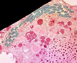
Background imageProtozoan Collection: Legionella infected protozoan, TEM C016 / 9412

Legionella infected protozoan, TEM C016 / 9412
Legionella infected protozoan. Transmission electron micrograph (TEM) of a section through a single-celled Tetrahymena pyriformis protozoan, infected with Legionella pneumophila bacteria (yellow)

Background imageProtozoan Collection: Heliozoan, SEM C016 / 9024

Heliozoan, SEM C016 / 9024
Heliozoan. Coloured scanning electron micrograph (SEM) of a heliozoan freshwater protozoan. This single-celled organism has many projections, known as axopods, radiating from its cell body

Background imageProtozoan Collection: Euplotes protozoa, SEM C016 / 9041

Euplotes protozoa, SEM C016 / 9041
Euplotes protozoa. Coloured scanning electron micrograph (SEM) showing the upperside (bottom) and underside (top) of two Euplotes sp. hypotrich ciliate protozoa

Background imageProtozoan Collection: Climacostomum protozoan, SEM C016 / 9062

Climacostomum protozoan, SEM C016 / 9062
Climacostomum protozoan. Coloured scanning electron micrograph (SEM) of a Climacostomum sp. unicellular ciliate protozoan, showing the cells large oral apparatus (round, upper right)

Background imageProtozoan Collection: Heliozoan, SEM C016 / 9023

Heliozoan, SEM C016 / 9023
Heliozoan. Coloured scanning electron micrograph (SEM) of a heliozoan freshwater protozoan. This single-celled organism has many projections, known as axopods, radiating from its cell body

Background imageProtozoan Collection: Giardia protozoan, SEM C016 / 9398

Giardia protozoan, SEM C016 / 9398
Giardia protozoan. Coloured scanning electron micrograph (SEM) of a Giardia sp. trophozoite protozoan (yellow) attached to the lining of a human intestine

Background imageProtozoan Collection: Ciliate protozoan ingesting algae

Ciliate protozoan ingesting algae. Darkfield illuminated light micrograph of a ciliate protozoan ingesting green algae (green)

Background imageProtozoan Collection: Tetrahymena protozoan, SEM

Tetrahymena protozoan, SEM
Tetrahymena protozoan. Coloured scanning electron micrograph (SEM) of a Tetrahymena sp. ciliate protozoan. Tetrahymena are free-living freshwater unicellular organisms found in ponds

Background imageProtozoan Collection: Amoeboid protozoa, SEM

Amoeboid protozoa, SEM
Amoeboid protozoa. Differential interference contrast (DIC) micrograph of four Thecamoeba sp. amoebozoa. Amoebozoa are a major group of amoeboid protozoa, motile single-celled organisms


For sale as Licensed Images

Choose your image, Select your licence and Download the media

"Exploring the Microscopic World: Unveiling the Diversity Life" In this captivating image series, we delve into the fascinating realm of protozoa – a diverse group of single-celled organisms that inhabit our planet. From ancient calcareous phytoplankton fossils to intricate SEM images, prepare to be amazed by their incredible forms and functions. Starting with a glimpse into the past, we encounter an exquisite fossilized specimen of calcareous phytoplankton. Preserved in time, it offers us a window into Earth's history and evolution. Moving forward, our attention is captured by an artistic representation of Trypanosome protozoan – a notorious parasite responsible for causing sleeping sickness. Its mesmerizing artwork reminds us of both its beauty and danger lurking within nature. Shifting gears towards human health concerns, we come across Plasmodium sp. , the malarial parasite that has plagued humanity for centuries. This microscopic culprit serves as a stark reminder of the ongoing battle against malaria worldwide. Protozoa are known for their unique feeding strategies; some scavenge particles and microorganisms like bacteria while others absorb nutrients from their surroundings. Their adaptability is showcased through stunning SEM images featuring diatoms - intricately patterned unicellular algae - and Acrosphaera radiolarian - delicate marine organisms with intricate skeletal structures. The exploration continues as we encounter another menacing parasite responsible for mouse malaria. Through high-resolution SEM imagery, its complex morphology comes to life before our eyes. Diving deeper into this hidden world reveals Oxytricha ciliate protozoan – an organism characterized by its hair-like projections called cilia. The detailed SEM image showcases its extraordinary structure in vivid detail. Finally, we marvel at the intricate design of Foraminiferan tests (shells) captured using scanning electron microscopy (SEM). These tiny shells serve as protective homes for these remarkable creatures living in aquatic environments.