Protozoan Collection (#8)

"Exploring the Microscopic World: Unveiling the Diversity Life" In this captivating image series

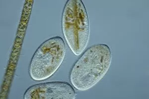
Background imageProtozoan Collection: Frontonia protozoa, light micrograph

Frontonia protozoa, light micrograph
Frontonia protozoa. Light micrograph of a group of Frontonia sp. ciliate protozoa

Background imageProtozoan Collection: Euglena flagellate protozoan, SEM

Euglena flagellate protozoan, SEM
Euglena flagellate protozoan. Coloured scanning electron micrograph (SEM) of a Euglena sp. flagellate protozoan present in a urine sample taken from someone with a urinary tract infection (UTI)

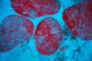
Background imageProtozoan Collection: Frontonia protozoan, light micrograph

Frontonia protozoan, light micrograph
Frontonia protozoan. Light micrograph of a Frontonia sp. ciliate protozoan

Background imageProtozoan Collection: Foraminiferan, light micrograph

Foraminiferan, light micrograph. Foraminifera are marine single-celled protozoa that construct and inhabit shells composed of several chambers

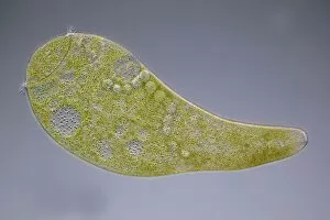
Background imageProtozoan Collection: Stentor coeruleus protozoan, micrograph

Stentor coeruleus protozoan, micrograph
Stentor coeruleus protozoan. Light micrograph of Stentor coeruleus amongst green algae. This large single-celled protist measures 0.5 to 2 millimetres when fully extended

Background imageProtozoan Collection: Vorticella protozoan, SEM

Vorticella protozoan, SEM
Vorticella protozoan. Coloured scanning electron micrograph (SEM) of a Vorticella sp. ciliate protozoan. This single-celled organism consists of a bell-shaped head

Background imageProtozoan Collection: Paramecium protozoan, light micrograph

Paramecium protozoan, light micrograph
Paramecium protozoan. Differential interference contrast micrograph of a Paramecium sp. protozoan. This ciliate protozoan inhabits freshwater, where it feeds mainly on bacteria

Background imageProtozoan Collection: Epistylis protozoan colony on algae

Epistylis protozoan colony on algae. Light micrograph of a colony of Epistylis sp. protozoa (centre right) attached to a green algae filament (lower left)

Background imageProtozoan Collection: Proteus amoeba, light micrograph

Proteus amoeba, light micrograph
Proteus amoeba. Differential interference contrast micrograph of an Amoeba proteus protozoan. This freshwater single-celled organism feeds on bacteria and smaller protozoa

Background imageProtozoan Collection: Echinocactus pentacanthus cactus, artwork

Echinocactus pentacanthus cactus, artwork
Proteus amoeba. Differential interference contrast micrograph of an Amoeba proteus protozoan. This freshwater single-celled organism feeds on bacteria and smaller protozoa

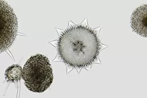
Background imageProtozoan Collection: Radiolarian, light micrograph

Radiolarian, light micrograph
Radiolarian, differential interference contrast micrograph. Radiolaria are single-celled protozoans that are found in marine plankton

Background imageProtozoan Collection: Stentor ciliate protozoan, micrograph

Stentor ciliate protozoan, micrograph
Stentor ciliate protozoan. Light micrograph of a Stentor polymorphus ciliate protozoan. This ciliate protozoan inhabits freshwater, where it feeds mainly on bacteria

Background imageProtozoan Collection: Radiolaria, light micrograph

Radiolaria, light micrograph
Radiolaria. Light micrograph of various radiolaria. Radiolaria are single-celled protozoans that are found in marine plankton

Background imageProtozoan Collection: Stentor ciliate protozoa light micrograph

Stentor ciliate protozoa light micrograph

Background imageProtozoan Collection: Giant clam mantle

Giant clam mantle. Close-up of the mantle of a giant clam (Tridacna Tridacna gigas). The bright colours in the mantle come from symbiotic zooxanthellae (protozoa) in the tissue

Background imageProtozoan Collection: Macrophage attacking a foreign body, SEM

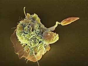
Macrophage attacking a foreign body, SEM
Macrophage attacking a foreign body. Coloured scanning electron micrograph (SEM) of a macrophage white blood cell (round, left) engulfing a Leishmania mexicana protozoan parasite (long, right)

Background imageProtozoan Collection: Paramecium protozoan, artwork

Paramecium protozoan, artwork
Paramecium protozoan. Computer artwork of a Paramecium sp. ciliate protozoan. Its single cell is covered in numerous cilia (white lines), hair-like structures that it beats for propulsion

Background imageProtozoan Collection: Vorticella protozoa, artwork

Vorticella protozoa, artwork. This ciliate protozoan (three present here) consists of a bell-shaped head (bottom) and a stalk that is anchored to a substrate (top)

Background imageProtozoan Collection: Loxophyllum ciliate, light micrograph

Loxophyllum ciliate, light micrograph
Loxophyllum ciliate. Light micrograph of a Loxophyllum helus ciliate protozoan. This tiny single-celled organism is found in marine environments

Background imageProtozoan Collection: Protozoan tentacle, TEM

Protozoan tentacle, TEM
Protozoan tentacle. Coloured transmission electron micrograph (TEM) of a cross-section through a tentacle of a Dendrocometes sp. protozoan showing the microtubule (purple circles) structure

Background imageProtozoan Collection: Protozoa, historical artwork

Protozoa, historical artwork
Protozoa. 1839 artwork by the naturalist Christian Gottfried Ehrenberg of Protozoa as seen under his microscope

Background imageProtozoan Collection: Trichodina parasite, SEM

Trichodina parasite, SEM
Trichodina parasite. Coloured scanning electron micrograph (SEM) of a Trichodina sp. protist on a freshwater minnow. This ciliate parasite lives on the skin

Background imageProtozoan Collection: Malaria infection, artwork

Malaria infection, artwork
Malaria infection. Computer artwork of a macrophage white blood cell (right) extending pseudopodia towards a red blood cell (left) infected with the Plasmodium fulciparum malaria parasite

Background imageProtozoan Collection: Dinoflagellate, SEM

Dinoflagellate, SEM
Dinoflagellate. Coloured scanning electron micrograph (SEM) of the dinoflagellate Prorocentrum balticum. Dinoflagellates are unicellular (single-celled) protozoans

Background imageProtozoan Collection: Euglena flagellate protozoans, SEM

Euglena flagellate protozoans, SEM
Euglena flagellate protozoans. Scanning electron micrograph (SEM) of Euglena sp. flagellate protozoans. Euglena sp. is a freshwater unicellular (single-celled)
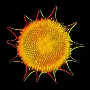
Background imageProtozoan Collection: Radiolarian, SEM

Radiolarian, SEM
Radiolarian. Coloured scanning electron micrograph (SEM) of the shell of a radiolarian. Radiolaria are single-celled protozoans that are found in marine plankton

Background imageProtozoan Collection: Actinosphaerium protozoan

Actinosphaerium protozoan. Differential interference contrast micrograph of an Actinoshaerium sp. heliozoa protozoan. This single-celled freshwater organism has many pseudopodia radiating outwards

Background imageProtozoan Collection: Euglena protozoan, light micrograph

Euglena protozoan, light micrograph
Euglena protozoan. Differential interference contrast micrograph of the flagellate protozoan Euglena spirogyra. This freshwater single-celled organism can either obtain energy from sunlight

Background imageProtozoan Collection: Shelled amoeba, light micrograph

Shelled amoeba, light micrograph
Shelled amoeba. Differential interference contrast micrograph of the testate (shelled) amoeba Arcella sp.. The circular shell is formed form the polysaccharide chitin

Background imageProtozoan Collection: Euglena protozoa, light micrograph

Euglena protozoa, light micrograph
Euglena protozoa. Differential interference contrast micrograph of the flagellate protozoa Euglena acus. These freshwater single-celled organisms can either obtain energy from sunlight

Background imageProtozoan Collection: Vorticella protozoa, light micrograph

Vorticella protozoa, light micrograph
Vorticella protozoa. Differential interference contrast micrograph of a colony of Vorticella sp.. This ciliate protozoan consists of a bell-shaped head and a stalk that is anchored to the substrate

Background imageProtozoan Collection: Radiolarian skeleton, artwork

Radiolarian skeleton, artwork
Radiolarian skeleton. Computer artwork of the skeleton (test) of a radiolarian. Radiolaria are single-celled protozoans that are found in marine plankton

Background imageProtozoan Collection: Marine protozoa shells, SEM

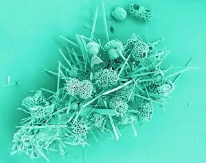
Marine protozoa shells, SEM
Marine protozoa shells, coloured scanning electron micrograph (SEM). The shells (tests) seen here include those of foraminifera, as well as coral spicules, radiolarian tests and cocccoliths

Background imageProtozoan Collection: Ciliate protozoan, SEM

Ciliate protozoan, SEM
Ciliate protozoan. Coloured scanning electron micrograph (SEM) of a Paramecium cilliate protozoan. Protozoa are a type of unicellular animal

Background imageProtozoan Collection: Ciliate protozoan membranelles, SEM

Ciliate protozoan membranelles, SEM
Ciliate protozoan membranelles, coloured scanning electron micrograph (SEM). Protozoa are a type of unicellular animal. The ciliate protozoa are so named because of the hair-like cilia which are used

Background imageProtozoan Collection: Kerona protozoan, light micrograph

Kerona protozoan, light micrograph
Kerona protozoan. Differential interference contrast micrograph of the ciliate protozoan Kerona polyporum. This protozoan is parasitic on freshwater hydras.Magnification

Background imageProtozoan Collection: Radiolarian shell, light micrograph

Radiolarian shell, light micrograph
Radiolarian. Light micrograph of a fossilised radiolarian shell. Radiolaria are single-celled protozoans that are found in marine plankton. They have a silicate skeleton

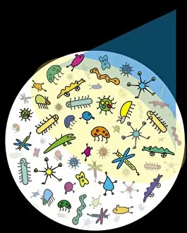
Background imageProtozoan Collection: Microbes

Microbes. Conceptual computer artwork of microbes under a light microscope

Background imageProtozoan Collection: Hexastylus radiolarian, artwork

Hexastylus radiolarian, artwork
Hexastylus radiolarian, historical artwork. Radiolaria are single-celled protozoans that are found in marine plankton. This radiolarian has a spherical silicate skeleton with pores through which

Background imageProtozoan Collection: Protozoan RNA-binding protein complex

Protozoan RNA-binding protein complex
RNA-binding protein complex. Computer model showing a guide RNA-binding protein complex (green and blue), bound to guide RNA (gRNA, yellow and red))

Background imageProtozoan Collection: Paramecium protozoan, SEM

Paramecium protozoan, SEM
Paramecium protozoan, coloured scanning electron micrograph (SEM). Paramecia are a group of unicellular ciliate protozoa. They inhabit fresh water, and feed mainly on bacteria and smaller protozoa


For sale as Licensed Images

Choose your image, Select your licence and Download the media

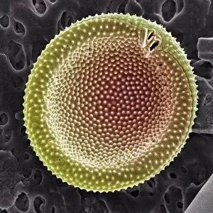
"Exploring the Microscopic World: Unveiling the Diversity Life" In this captivating image series, we delve into the fascinating realm of protozoa – a diverse group of single-celled organisms that inhabit our planet. From ancient calcareous phytoplankton fossils to intricate SEM images, prepare to be amazed by their incredible forms and functions. Starting with a glimpse into the past, we encounter an exquisite fossilized specimen of calcareous phytoplankton. Preserved in time, it offers us a window into Earth's history and evolution. Moving forward, our attention is captured by an artistic representation of Trypanosome protozoan – a notorious parasite responsible for causing sleeping sickness. Its mesmerizing artwork reminds us of both its beauty and danger lurking within nature. Shifting gears towards human health concerns, we come across Plasmodium sp. , the malarial parasite that has plagued humanity for centuries. This microscopic culprit serves as a stark reminder of the ongoing battle against malaria worldwide. Protozoa are known for their unique feeding strategies; some scavenge particles and microorganisms like bacteria while others absorb nutrients from their surroundings. Their adaptability is showcased through stunning SEM images featuring diatoms - intricately patterned unicellular algae - and Acrosphaera radiolarian - delicate marine organisms with intricate skeletal structures. The exploration continues as we encounter another menacing parasite responsible for mouse malaria. Through high-resolution SEM imagery, its complex morphology comes to life before our eyes. Diving deeper into this hidden world reveals Oxytricha ciliate protozoan – an organism characterized by its hair-like projections called cilia. The detailed SEM image showcases its extraordinary structure in vivid detail. Finally, we marvel at the intricate design of Foraminiferan tests (shells) captured using scanning electron microscopy (SEM). These tiny shells serve as protective homes for these remarkable creatures living in aquatic environments.