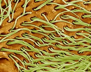
Protozoan Collection (#5)

"Exploring the Microscopic World: Unveiling the Diversity Life" In this captivating image series

Background imageProtozoan Collection: Minakatella longifila, slime mould

Minakatella longifila, slime mould

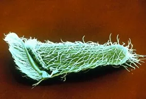
Background imageProtozoan Collection: Metopus protozoan

Metopus protozoan

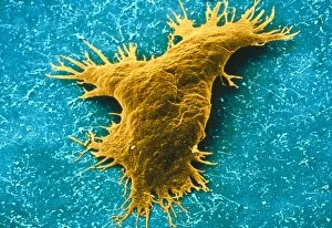
Background imageProtozoan Collection: Thalassomyxa australis protozoan

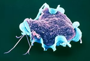
Thalassomyxa australis protozoan
Thalassomyxa protozoan. Coloured scanning electron micrograph (SEM) of a Thalassomyxa australis prot- ozoan (single-celled animal)

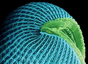
Background imageProtozoan Collection: Lembadion protozoan

Lembadion protozoan. Coloured scanning electron micrograph (SEM) of a Lembadion bullinum protozoan (single-celled animal)

Background imageProtozoan Collection: TEM of Giardia lamblia

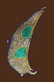
TEM of Giardia lamblia
Coloured Transmission Electron Micrograph of Giardia lamblia (orange), a parasitic flagellate protozoan which causes the disease lambliasis (giardiasis)

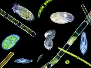
Background imageProtozoan Collection: Ciliate protozoa, light micrograph

Ciliate protozoa, light micrograph
Ciliate protozoa. Light micrograph of a mixed population of freshwater ciliate protozoa and algae. The algae are the green filaments crossing the field

Background imageProtozoan Collection: Radiolarian planktonic protozoan, SEM

Radiolarian planktonic protozoan, SEM
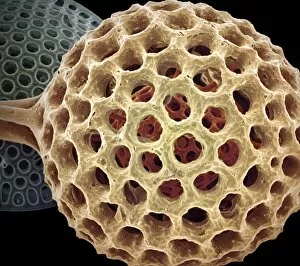
Radiolarian protozoan. Coloured scanning electron micrograph (SEM) of the shell (test) of an unidentified radiolarian protozoan

Background imageProtozoan Collection: Calcareous phytoplankton fossil, artwork

Calcareous phytoplankton fossil, artwork
Calcareous phytoplankton fossil. Artwork of a fossil of the skeleton (coccosphere) of a coccolithotrope, a small marine algal organism

Background imageProtozoan Collection: Leishmania protozoa

Leishmania protozoa
False-colour light micrograph (LM) of a colony of Leishmania tropica. a parasitic flagellate protozoan that causes the disease leishmaniasis in humans

Background imageProtozoan Collection: Ciliate protozoa dividing

Ciliate protozoa dividing
Ciliate protozoa. Light micrograph of unidentified ciliate protozoa (class Ciliata) undergoing asexual reproduction. These unicellular micro-organisms can multiply by either of two methods

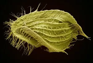
Background imageProtozoan Collection: Euplotes ciliate protozoan, SEM

Euplotes ciliate protozoan, SEM
Euplotes ciliate protozoan. Coloured scanning electron micrograph (SEM) of a ciliate protozoan (Euplotes sp.). This single-celled organism is found in fresh and marine water

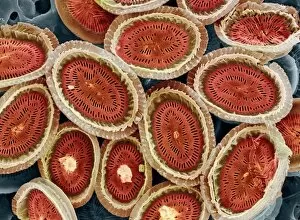
Background imageProtozoan Collection: Plankton cell wall, SEM

Plankton cell wall, SEM
Plankton cell wall. Coloured scanning electron micrograph (SEM) of the geometrical plates making up the mineralised cell wall of a planktonic alga (Coronosphaera mediterranea)

Background imageProtozoan Collection: Spirochona protozoa

Spirochona protozoa

Background imageProtozoan Collection: Euglena protozoan, SEM

Euglena protozoan, SEM
Euglena protozoan (Euglena sp.), coloured scanning electron micrograph (SEM). This protozoan is described as flagellate because it has a flagellum (thin tail-like structure)

Background imageProtozoan Collection: LM of assorted fossil Foraminifera shells

LM of assorted fossil Foraminifera shells
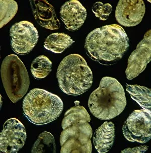
Foraminiferan shells. Light micrograph of assorted shells of fossil species belonging to the order: Foraminifera. Single-celled protozoans once inhabited these shells which are composed of several
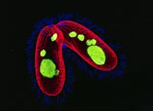
Background imageProtozoan Collection: Protozoan, SEM

Protozoan, SEM
Protozoan. Coloured scanning electron micrograph (SEM) of a protozoan. Protozoa are unicellular eukaryotic organisms, that usually are not photosynthetic

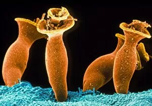
Background imageProtozoan Collection: Ciliate protozoans, SEM

Ciliate protozoans, SEM
Ciliate protozoans. Coloured scanning electron micrograph (SEM) of sessile (anchored) ciliate protozoan (subclass Peritrichia)

Background imageProtozoan Collection: Giardia protozoan, TEM

Giardia protozoan, TEM
Giardia protozoan. Coloured transmission electron micrograph (TEM) of a Giardia sp. protozoan. It is a single-celled organism that has two cell nuclei (green)

Background imageProtozoan Collection: Calcareous phytoplankton plates, SEM

Calcareous phytoplankton plates, SEM
Calcareous phytoplankton plates. Coloured scanning electron micrograph (SEM) of parts of the skeleton (coccosphere) of the coccolithophore Emiliania huxleyi, a small marine algal organism

Background imageProtozoan Collection: Trichomytopsis protozoan

Trichomytopsis protozoan

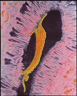
Background imageProtozoan Collection: Coloured SEM of Giardia lamblia in human intestine

Coloured SEM of Giardia lamblia in human intestine
Giardia lamblia. Coloured Scanning Electron Micro- graph (SEM) of the parasite Giardia lamblia in the human small intestine

Background imageProtozoan Collection: Ciliate protozoa, SEM

Ciliate protozoa, SEM
Ciliate protozoa. Coloured scanning electron micrograph (SEM) of cilliate protozoa (unicellular animals). The ciliates are so named because of the hair-like cilia surrounding their bodies which are

Background imageProtozoan Collection: LM of a colony of Volvox

LM of a colony of Volvox
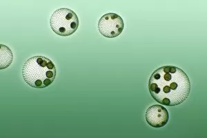
Light micrograph of a colony of Volvox containing eight asexual daughter colonies within. Volvox is an organism that lies between the plant & animal kingdom

Background imageProtozoan Collection: Naegleria fowleri protozoa, TEM

Naegleria fowleri protozoa, TEM
Naegleria fowleri protozoan. Coloured transmission electron micrograph (TEM) of a section through a Naegleria fowleri protozoan

Background imageProtozoan Collection: Flagellate protozoan, SEM

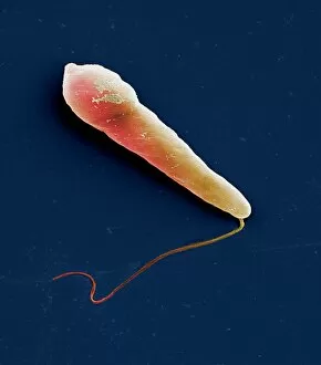
Flagellate protozoan, SEM
Flagellate protozoan, coloured scanning electron micrograph (SEM). This is a protozoan that has a flagellum (thin tail-like structure, lower left)

Background imageProtozoan Collection: Parasitic protozoan, TEM

Parasitic protozoan, TEM
Parasitic protozoan. Coloured transmission electron micrograph (TEM) of a trypanosome protozoan (Trypanosoma sp.). The nucleus (centre), cell membrane (outline)

Background imageProtozoan Collection: Enterocytozoon sp. parasites, TEM

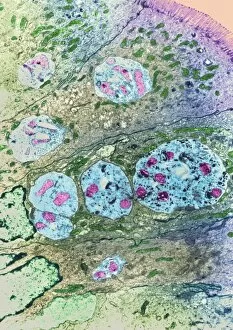
Enterocytozoon sp. parasites, TEM

Background imageProtozoan Collection: Isospora sp. parasites, TEM

Isospora sp. parasites, TEM
Isospora sp. parasites. Coloured transmission electron micrograph (TEM) of Isospora sp. parasites (yellow) in a sectioned cell

Background imageProtozoan Collection: Lychnocanium radiolarian, SEM

Lychnocanium radiolarian, SEM
Lychnocanium radiolarian. Coloured scanning electron micrograph (SEM) of the shell of a Lychnocanium sp. radiolarian. Radiolaria are single-celled protozoans that are found in marine plankton

Background imageProtozoan Collection: Alga-covered protozoan, SEM

Alga-covered protozoan, SEM
Alga-covered protozoan, coloured scanning electron micrograph (SEM). Algae (blue) are seen on the body of the protozoan, and cilia (green) are used to waft the algae to the protozoans feeding area

Background imageProtozoan Collection: TEM of Leishmania sp

TEM of Leishmania sp
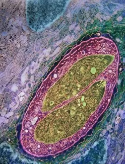
False-colour transmission electron micrograph (TEM) of Leishmania sp. a parasitic flagellate protozoan that causes the disease leishmaniasis in humans

Background imageProtozoan Collection: Naegleria fowleri protozoan, TEM

Naegleria fowleri protozoan, TEM
Naegleria fowleri protozoan. Coloured transmission electron micrograph (TEM) of a section through a Naegleria fowleri protozoan

Background imageProtozoan Collection: Paramecium protozoa, SEM

Paramecium protozoa, SEM
Paramecium protozoa (Paramecium sp.), coloured scanning electron micrograph (SEM). These protozoa (two seen) are described as ciliate because many cilia (hair-like structures) cover them

Background imageProtozoan Collection: Hartmannella amoeba and bacterium

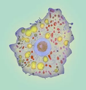
Hartmannella amoeba and bacterium. Computer artwork of a Hartmannella vermiformis amoeba (green) engulfing a Legionella pneumophila bacterium (orange) with a pseudopodium

Background imageProtozoan Collection: Protozoan cilia, SEM

Protozoan cilia, SEM
Protozoan cilia. Coloured scanning electron micrograph (SEM) of cilia (microscopic hairs) from a ciliate protozoan. Ciliate protozoans are single-celled aquatic organisms

Background imageProtozoan Collection: LM of Paramecium bursaris

LM of Paramecium bursaris
Light micrograph of Paramecium bursaris, a member of the species of green paramecia. The green colouration is due to the presence of unicellulate green algae which form organic substances & produce

Background imageProtozoan Collection: Breslauides ciliate protozoan, SEM

Breslauides ciliate protozoan, SEM
Breslauides ciliate protozoan. Coloured scanning electron micrograph (SEM) of a ciliate protozoan (Breslauides sp.). This tiny single-celled organism is found in water

Background imageProtozoan Collection: Tetrahymena protozoa

Tetrahymena protozoa. Immunofluorescent light micrograph of two Tetrahymena thermophila protozoa (single-celled animals). Nuclei are green, cell walls red and cilia (hairs) blue. T

Background imageProtozoan Collection: Amphisphaerina radiolarian, SEM

Amphisphaerina radiolarian, SEM
Amphisphaerina radiolarian. Coloured scanning electron micrograph (SEM) of the shell of a Amphisphaerina sp. radiolarian. Radiolaria are single-celled protozoans that are found in marine plankton

Background imageProtozoan Collection: Acrosphaera radiolarian, SEM

Acrosphaera radiolarian, SEM
Acrosphaera radiolarian. Coloured scanning electron micrograph (SEM) of the shell of a Acrosphaera sp. radiolarian. Radiolaria are single-celled protozoans that are found in marine plankton

Background imageProtozoan Collection: Dendrocometes protozoan

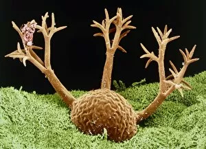
Dendrocometes protozoan. Coloured scanning electron micrograph (SEM) of a Dendrocometes paradoxus protozoan (single-celled animal)

Background imageProtozoan Collection: Green algae colonies

Green algae colonies. Light micrograph of green algae colonies (Volvox sp.). These colonies are made up of approximately 500 individual flagellate cells, arranged in a glycoprotein filled sphere

Background imageProtozoan Collection: Amoeba, SEM

Amoeba, SEM
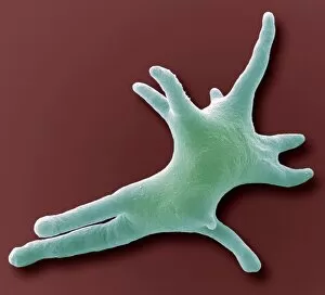
Amoeba. Coloured scanning electron micrograph (SEM) of an Amoeba proteus protozoan. This is a freshwater single-celled organism that feeds on bacteria and smaller protozoa

Background imageProtozoan Collection: LM of assorted Foraminifera shells

LM of assorted Foraminifera shells
Foraminiferan shells. Light micrograph of assorted species of shells belonging to the order Foramini- fera. Single-celled protozoans inhabit these shells which are composed of several chambers

Background imageProtozoan Collection: Protozoan cilia, SEM

Protozoan cilia, SEM
Protozoan cilia. Coloured scanning electron micrograph (SEM) of cilia (microscopic hairs) from a ciliate protozoan. Ciliate protozoans are single-celled aquatic organisms

Background imageProtozoan Collection: Blepharisma ciliate protozoan, SEM

Blepharisma ciliate protozoan, SEM
Blepharisma ciliate protozoan. Coloured scanning electron micrograph (SEM) of a ciliate protozoan (Blepharisma sp.). This tiny single- celled organism is found in freshwater


For sale as Licensed Images

Choose your image, Select your licence and Download the media

"Exploring the Microscopic World: Unveiling the Diversity Life" In this captivating image series, we delve into the fascinating realm of protozoa – a diverse group of single-celled organisms that inhabit our planet. From ancient calcareous phytoplankton fossils to intricate SEM images, prepare to be amazed by their incredible forms and functions. Starting with a glimpse into the past, we encounter an exquisite fossilized specimen of calcareous phytoplankton. Preserved in time, it offers us a window into Earth's history and evolution. Moving forward, our attention is captured by an artistic representation of Trypanosome protozoan – a notorious parasite responsible for causing sleeping sickness. Its mesmerizing artwork reminds us of both its beauty and danger lurking within nature. Shifting gears towards human health concerns, we come across Plasmodium sp. , the malarial parasite that has plagued humanity for centuries. This microscopic culprit serves as a stark reminder of the ongoing battle against malaria worldwide. Protozoa are known for their unique feeding strategies; some scavenge particles and microorganisms like bacteria while others absorb nutrients from their surroundings. Their adaptability is showcased through stunning SEM images featuring diatoms - intricately patterned unicellular algae - and Acrosphaera radiolarian - delicate marine organisms with intricate skeletal structures. The exploration continues as we encounter another menacing parasite responsible for mouse malaria. Through high-resolution SEM imagery, its complex morphology comes to life before our eyes. Diving deeper into this hidden world reveals Oxytricha ciliate protozoan – an organism characterized by its hair-like projections called cilia. The detailed SEM image showcases its extraordinary structure in vivid detail. Finally, we marvel at the intricate design of Foraminiferan tests (shells) captured using scanning electron microscopy (SEM). These tiny shells serve as protective homes for these remarkable creatures living in aquatic environments.