Red Blood Cell Collection (#9)

Red blood cells, also known as erythrocytes, are the most abundant type of blood cell in our bodies

Background imageRed Blood Cell Collection: Coloured SEM of a blood clot due to an injury

Coloured SEM of a blood clot due to an injury

Background imageRed Blood Cell Collection: Computer artwork of a blood clot

Computer artwork of a blood clot
Blood clot. Computer artwork of a blood clot, showing disc-like red blood cells and fibrin. Fibrin (yellow fibres) is a long insoluble protein produced from the blood protein fibrinogen

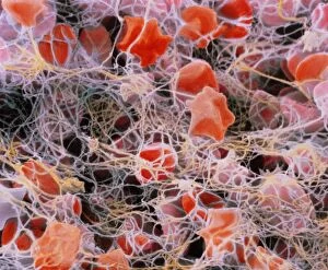
Background imageRed Blood Cell Collection: Coloured SEM of red blood cells forming a clot

Coloured SEM of red blood cells forming a clot
Blood clot. Coloured Scanning Electron Micrograph (SEM) showing the formation of a blood clot. Red blood cells (erythrocytes)

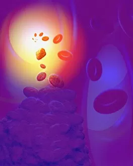
Background imageRed Blood Cell Collection: Illustration of a dissolving blood clot (thrombus)

Illustration of a dissolving blood clot (thrombus)
Dissolving blood clot. Illustration of a human blood clot (thrombus) in the process of dissolving. At upper centre is the blood clot

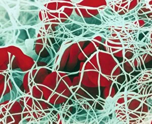
Background imageRed Blood Cell Collection: Blood clot

Blood clot
False-colour scanning electron micrograph (SEM) of a blood clot. Red blood cells have been trapped by a web of thin yellow-white strands of fibrin

Background imageRed Blood Cell Collection: False-colour SEM of a human blood clot

False-colour SEM of a human blood clot
False-colour scanning electron micrograph (SEM) of a human blood clot (thrombus), showing strands of fibrin around red blood cells

Background imageRed Blood Cell Collection: Red blood cell crenation, SEM

Red blood cell crenation, SEM
Red blood cell crenation. Coloured scanning electron micrograph (SEM) of two red blood cells (erythrocytes). The cell on the bottom is normal

Background imageRed Blood Cell Collection: Blood cells, computer artwork

Blood cells, computer artwork
Blood cells. Computer artwork of red blood cells, white blood, white blood cells (blue) and platelets (yellow) travelling through the lumen of the blood vessel

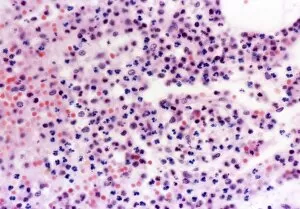
Background imageRed Blood Cell Collection: Human blood cells, light micrograph

Human blood cells, light micrograph
Human blood cells. Light micrograph of blood cells at a site of inflammation, showing red blood cells (erythrocytes, red) surrounded by many granulocytes (blue)

Background imageRed Blood Cell Collection: Red blood cell

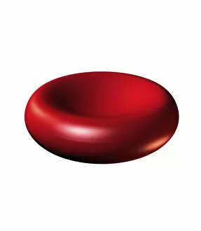
Red blood cell. Computer artwork of a human red blood cell (erythrocyte). Red blood cells are biconcave, giving them a large surface area for gas exchange, and highly elastic

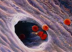
Background imageRed Blood Cell Collection: Red blood cells in blood vessel

Red blood cells in blood vessel
Red blood cells. Coloured scanning electron micro- graph (SEM) of red blood cells in a human blood vessel. The red blood cells (erythrocytes, at top) are filling the lumen of the blood vessel

Background imageRed Blood Cell Collection: Red blood cells, SEM

Red blood cells, SEM
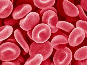
Red blood cells. Coloured scanning electron micrograph (SEM) of red blood cells (erythrocytes). Red blood cells are biconcave, disc-shaped cells that transport oxygen from the lungs to body cells

Background imageRed Blood Cell Collection: TEM of erythrocytes passing through capillary wall

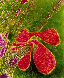
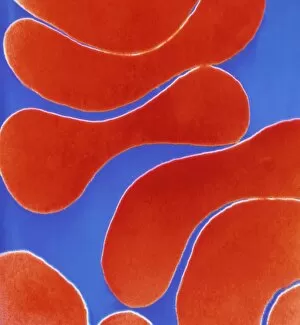
TEM of erythrocytes passing through capillary wall
False-colour transmission electron micrograph showing the flexibility of red blood cells, or erythrocytes. Three of them are seen in a deformed state as they pass through the wall of a small

Background imageRed Blood Cell Collection: LM of human red blood cells stacked like coins

LM of human red blood cells stacked like coins
Light micrograph of human red blood cells stacked together like coins. This is the way they line up in blood vessels. Magnification: X 450 at 35mm size, X 900 at 6x7cm size

Background imageRed Blood Cell Collection: Coloured SEM of red blood cells on vessel wall

Coloured SEM of red blood cells on vessel wall
Red blood cells. Coloured scanning electron micrograph (SEM) of human red blood cells (erythrocytes) on the wall of a blood vessel. Red blood cells carry oxygen and carbon dioxide around the body

Background imageRed Blood Cell Collection: Coloured SEM of red blood cells (erythrocytes)

Coloured SEM of red blood cells (erythrocytes)
Red blood cells. Coloured scanning electron micrograph of red blood cells (erythrocytes). These biconcave discs contain the red pigment haemoglobin with which they transport

Background imageRed Blood Cell Collection: Colour TEM of red blood cells, rouleau formation

Colour TEM of red blood cells, rouleau formation
Red blood cells. Coloured Transmission Electron Micrograph (TEM) of sectioned human red blood cells (erythrocytes) in a rouleau formation

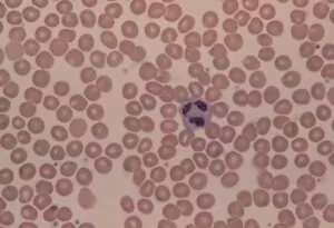
Background imageRed Blood Cell Collection: LM of human blood smear showing red & white cells

LM of human blood smear showing red & white cells
Healthy blood smear. Light micrograph of a smear of human blood consisting of a field of red blood cells (erythrocytes). Near centre is a single white blood cell (leucocyte) with a purple nucleus

Background imageRed Blood Cell Collection: False-colour SEM of a group of red blood cells

False-colour SEM of a group of red blood cells
Red blood cells (erythrocytes). False-colour scanning electron micrograph of a group of red blood cells on the endocardium, the innermost layer lining the heart

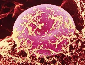
Background imageRed Blood Cell Collection: Coloured SEM of red blood cell covered in fibrin

Coloured SEM of red blood cell covered in fibrin
False-colour scanning electron micrograph (SEM) showing a human red blood cell (erythrocyte) with a fine covering of fibrin, the protein involved in clot formation

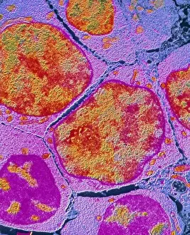
Background imageRed Blood Cell Collection: Colour TEM of an erythroblast cell in bone marrow

Colour TEM of an erythroblast cell in bone marrow

Background imageRed Blood Cell Collection: Coloured SEM of red blood cells in blood vessel

Coloured SEM of red blood cells in blood vessel
Red blood cells. Coloured scanning electron micrograph of a group of red blood cells (erythrocytes). They are travelling through a large vessel and are just entering a small vessel

Background imageRed Blood Cell Collection: Computer graphics of haemoglobin blood substitute

Computer graphics of haemoglobin blood substitute
Computer graphics representation of haemoglobin, the oxygen-carrying molecule of blood, constructed during research for a blood substitute at the U.K

Background imageRed Blood Cell Collection: F / col TEM of erythroblast in bone marrow

F / col TEM of erythroblast in bone marrow
False-colour transmission electron micrograph of erythroblasts, primitive red blood cells, in human bone marrow. Erythropoiesis, the formation of mature red cells

Background imageRed Blood Cell Collection: Haemoglobin molecule

Haemoglobin molecule
Computer graphic representation of the haemoglobin molecule, the oxygen-carrying substance of human blood. The roughly tubular features (in orange and blue) are polypeptide (protein) chains

Background imageRed Blood Cell Collection: SEM of bone marrow

SEM of bone marrow

Background imageRed Blood Cell Collection: Computer graphics of haemoglobin molecule

Computer graphics of haemoglobin molecule
Computer graphic representation of part of the haemoglobin molecule, the oxygen-carrying substance of human blood, showing its four polypeptide (protein) chains

Background imageRed Blood Cell Collection: Illustration showing interior of a blood vessel

Illustration showing interior of a blood vessel
Illustration of the interior of a human blood vessel, showing the various cells that are suspended in blood plasma. The large saucer-like discs are red blood cells (erythrocytes)

Background imageRed Blood Cell Collection: Heart and red blood cells, artwork

Heart and red blood cells, artwork
Heart and red blood cells, computer artwork. The heart is a hollow muscle that pumps blood around the body. Deoxygenated blood from the body is carried into the right side of the heart (left)

Background imageRed Blood Cell Collection: Diagram showing the structure of the haem group

Diagram showing the structure of the haem group
Diagram showing the atomic structure of the haem group of the haemoglobin molecule, the oxygen- transporting protein in red blood cells that transfers oxygen from the lungs to the bodys tissues

Background imageRed Blood Cell Collection: Types of blood cell

Types of blood cell
Blood cells. Artwork (based on a scanning electron micrograph) showing various blood cells escaping from a sectioned small vein (top right)

Background imageRed Blood Cell Collection: ECG and red blood cells

ECG and red blood cells. Computer artwork of an electrocardiogram (ECG) trace and red blood cells (erythrocytes). Red blood cells are biconcave, giving them a large surface area for gas exchange

Background imageRed Blood Cell Collection: Heart and red blood cells

Heart and red blood cells (erythrocytes), computer artwork. The coronary blood vessels are seen surrounding the heart. The heart is a muscular pump that moves blood around the body


For sale as Licensed Images

Choose your image, Select your licence and Download the media

Red blood cells, also known as erythrocytes, are the most abundant type of blood cell in our bodies. These tiny cells play a crucial role in maintaining our overall health and well-being. In athlete physiology they can especially important as they carry oxygen from the lungs to every part of the body. This ensures that muscles receive an adequate supply of oxygen during physical activity, enhancing performance and endurance. Artwork depicting the intricate structure of red blood cells showcases their unique shape - biconcave discs without nuclei. This design allows for flexibility and efficient transport through narrow capillaries. The process of blood coagulation cascade is essential for wound healing and preventing excessive bleeding. Artwork illustrating this complex mechanism highlights how red blood cells interact with platelets and clotting factors to form a stable clot, sealing off damaged vessels. Scanning electron microscopy (SEM) images provide detailed views of various aspects related to red blood cells. One such image displays a close-up view of a blood clot formed by these specialized cells (SEM C016 / 9747). Another SEM image reveals infected red blood cells invaded by mouse malaria parasites (SEM). A diagram showcasing the bloodstream inside a vein demonstrates how red and white blood cells along with platelets flow together within our circulatory system. It emphasizes their collective effort in delivering nutrients, removing waste products, and defending against pathogens. Computer artwork beautifully portrays vibrant red blood cells flowing through arteries and veins, emphasizing their vital role in sustaining life throughout our bodies. Lastly, highlighting the connection between red blood cells and heart reminds us that these remarkable microscopic entities work tirelessly alongside our cardiovascular system to ensure proper circulation throughout every organ and tissue.