Red Blood Cell Collection (#4)

Red blood cells, also known as erythrocytes, are the most abundant type of blood cell in our bodies

Background imageRed Blood Cell Collection: Elliptocytosis, light micrograph

Elliptocytosis, light micrograph
Elliptocytosis. Light micrograph of red blood cells in a case of elliptocytosis. Red blood cells (erythrocytes) carry oxygen and carbon dioxide to and from body tissues

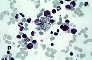
Background imageRed Blood Cell Collection: Cystine in bone marrow, light micrograph

Cystine in bone marrow, light micrograph
Cystine in bone marrow. Light micrograph of crystals of cystine among blood cells in a sample of bone marrow. Cystine is an amino acid that can form crystals in urine

Background imageRed Blood Cell Collection: Bone marrow blood cells, light micrograph

Bone marrow blood cells, light micrograph

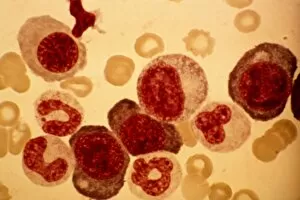
Background imageRed Blood Cell Collection: Pernicious anaemia, light micrograph

Pernicious anaemia, light micrograph
Pernicious anaemia. Light micrograph of megaloblast blood cells from bone marrow in a case of pernicious anaemia, also known as Biermers anaemia

Background imageRed Blood Cell Collection: Blood cell cancer, light micrograph

Blood cell cancer, light micrograph
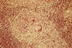
Blood cell cancer. Light micrograph of blood cells from a lymphatic ganglion in a case of a blood cancer of a mixed cell type

Background imageRed Blood Cell Collection: Erythroblast blood cell, light micrograph

Erythroblast blood cell, light micrograph
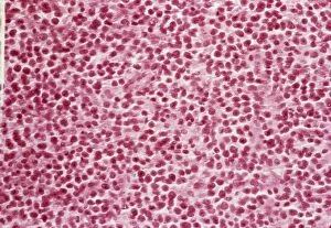
Erythroblast blood cell. Light micrograph of cells from a sample of bone marrow, including a polychromatic erythroblast (upper left). This one has multiple cell nuclei (dark red, four in total)

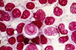
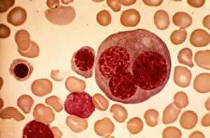
Background imageRed Blood Cell Collection: Acute erythroid leukaemia, micrograph

Acute erythroid leukaemia, micrograph
Acute erythroid leukaemia. Light micrograph of blood cells from bone marrow in a case of acute erythroid leukaemia. The cells include dystrophic (degenerated) erythroblasts (nuclei stained dark red)

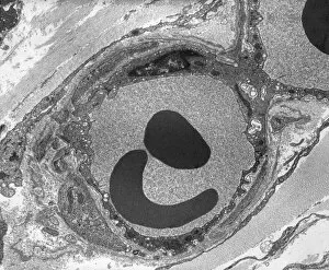
Background imageRed Blood Cell Collection: Capillary, TEM

Capillary, TEM
Capillary. Transmission electron micrograph (TEM) of a section through a capillary, showing two red blood cells (erythrocytes, black) in its interior

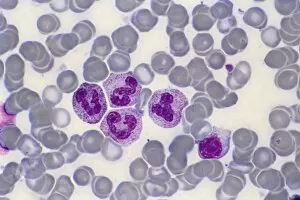
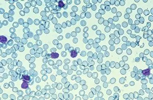
Background imageRed Blood Cell Collection: Blood cells, light micrograph

Blood cells, light micrograph
Blood cells. Light micrograph of red blood cells (erythrocytes, light blue) and white blood cells (leucocytes, nuclei stained purple)

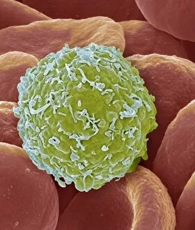
Background imageRed Blood Cell Collection: Leukaemia cell, SEM

Leukaemia cell, SEM
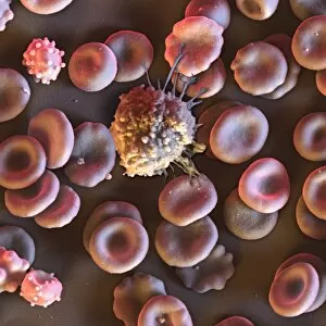
Leukaemia cell. Coloured scanning electron micrograph (SEM) of a leukaemic (cancerous) lymphocyte white blood cell (green), amongst normal red blood cells (erythrocytes, red)

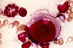
Background imageRed Blood Cell Collection: Reticulosarcoma, light micrograph

Reticulosarcoma, light micrograph
Reticulosarcoma. Light micrograph of reticulocyte blood cells (red, one at upper left) from a lymphatic ganglion in a case of reticulosarcoma

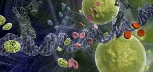
Background imageRed Blood Cell Collection: Drug effect on viruses, conceptual image C016 / 6253

Drug effect on viruses, conceptual image C016 / 6253
Drug effect on viruses, conceptual image. Computer artwork showing a single strand of DNA (deoxyribonucleic acid, spiral, centre), red blood cells (pink), virus particles (virions, green, small)

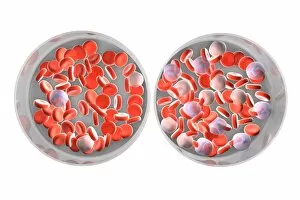
Background imageRed Blood Cell Collection: Normal and leukaemic blood, artwork C016 / 6261

Normal and leukaemic blood, artwork C016 / 6261
Normal and leukaemic blood. Computer artwork comparing the appearance of normal blood (left) and that affected by leukaemia (right)

Background imageRed Blood Cell Collection: Heart tissue, SEM C015 / 9598

Heart tissue, SEM C015 / 9598
Heart tissue. Coloured scanning electron micrograph (SEM) of heart tissue with red blood cells (erythrocytes, red) and connective tissue (orange)

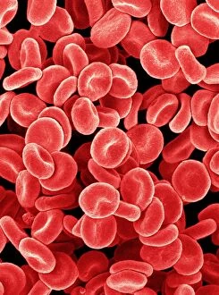
Background imageRed Blood Cell Collection: Red blood cells, SEM C015 / 8789

Red blood cells, SEM C015 / 8789
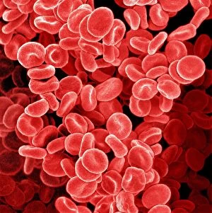
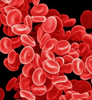
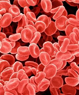
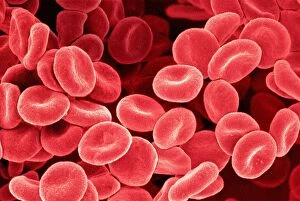
Red blood cells. Coloured scanning electron micrograph (SEM) of human red blood cells (erythrocytes). Red blood cells are biconcave, giving them a large surface area for gas exchange

Background imageRed Blood Cell Collection: Red blood cells, SEM C015 / 8792

Red blood cells, SEM C015 / 8792
Red blood cells. Coloured scanning electron micrograph (SEM) of human red blood cells (erythrocytes). Red blood cells are biconcave, giving them a large surface area for gas exchange

Background imageRed Blood Cell Collection: Red blood cells, SEM C015 / 8794

Red blood cells, SEM C015 / 8794
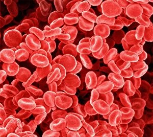
Red blood cells. Coloured scanning electron micrograph (SEM) of human red blood cells (erythrocytes). Red blood cells are biconcave, giving them a large surface area for gas exchange

Background imageRed Blood Cell Collection: Red blood cells, SEM C015 / 8796

Red blood cells, SEM C015 / 8796
Red blood cells. Coloured scanning electron micrograph (SEM) of human red blood cells (erythrocytes). Red blood cells are biconcave, giving them a large surface area for gas exchange

Background imageRed Blood Cell Collection: Red blood cells, SEM C015 / 8790

Red blood cells, SEM C015 / 8790
Red blood cells. Coloured scanning electron micrograph (SEM) of human red blood cells (erythrocytes). Red blood cells are biconcave, giving them a large surface area for gas exchange

Background imageRed Blood Cell Collection: Blood clot, SEM C015 / 9608

Blood clot, SEM C015 / 9608
Blood clot. Coloured scanning electron micrograph (SEM) of a blood clot from the inner wall of the left ventricle of a heart

Background imageRed Blood Cell Collection: Heart tissue, SEM C015 / 9541

Heart tissue, SEM C015 / 9541
Heart tissue. Coloured scanning electron micrograph (SEM) of heart tissue with red blood cells (erythrocytes)
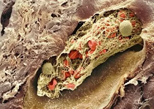
Background imageRed Blood Cell Collection: Reticulosarcoma, light micrograph C015 / 7133

Reticulosarcoma, light micrograph C015 / 7133
Reticulosarcoma. Light micrograph of reticulocyte blood cells (red, one at upper left) from a lymphatic ganglion in a case of reticulosarcoma

Background imageRed Blood Cell Collection: Red blood cells, SEM C015 / 8793

Red blood cells, SEM C015 / 8793
Red blood cells. Coloured scanning electron micrograph (SEM) of human red blood cells (erythrocytes). Red blood cells are biconcave, giving them a large surface area for gas exchange

Background imageRed Blood Cell Collection: Red blood cells, SEM C015 / 8795

Red blood cells, SEM C015 / 8795
Red blood cells. Coloured scanning electron micrograph (SEM) of human red blood cells (erythrocytes). Red blood cells are biconcave, giving them a large surface area for gas exchange

Background imageRed Blood Cell Collection: Red blood cells, SEM C015 / 8787

Red blood cells, SEM C015 / 8787
Red blood cells. Coloured scanning electron micrograph (SEM) of human red blood cells (erythrocytes). Red blood cells are biconcave, giving them a large surface area for gas exchange

Background imageRed Blood Cell Collection: Red blood cells, SEM C015 / 8791

Red blood cells, SEM C015 / 8791
Red blood cells. Coloured scanning electron micrograph (SEM) of human red blood cells (erythrocytes). Red blood cells are biconcave, giving them a large surface area for gas exchange

Background imageRed Blood Cell Collection: Red blood cells, SEM C015 / 8788

Red blood cells, SEM C015 / 8788
Red blood cells. Coloured scanning electron micrograph (SEM) of human red blood cells (erythrocytes). Red blood cells are biconcave, giving them a large surface area for gas exchange

Background imageRed Blood Cell Collection: Blood cells, light micrograph C015 / 7130

Blood cells, light micrograph C015 / 7130
Blood cells. Light micrograph of red blood cells (erythrocytes, red) and white blood cells (leucocytes, nuclei stained pink)

Background imageRed Blood Cell Collection: Blood cells, illustration C018 / 0802

Blood cells, illustration C018 / 0802
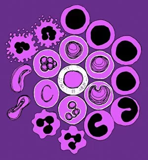
Blood cells. All cellular blood components originate from the same cell, the haematopoietic stem cell. The stem cell differentiates into two types of progenitor cells

Background imageRed Blood Cell Collection: Blood clot, SEM C016 / 9744

Blood clot, SEM C016 / 9744
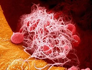
Blood clot, coloured scanning electron micrograph (SEM). Red blood cells (erythrocytes) are trapped within a fibrin protein mesh (beige)

Background imageRed Blood Cell Collection: Blood clot, SEM C014 / 0381

Blood clot, SEM C014 / 0381
Blood clot. Coloured scanning electron micrograph (SEM) of a blood clot (thrombus) in an arteriole (small blood vessel) of a salivary gland

Background imageRed Blood Cell Collection: Blood clot, SEM C014 / 0380

Blood clot, SEM C014 / 0380
Blood clot. Coloured scanning electron micrograph (SEM) of a blood clot (thrombus) in an arteriole (small blood vessel) of a salivary gland

Background imageRed Blood Cell Collection: Arteriole, TEM

Arteriole, TEM
Arteriole. Transmission electron micrograph (TEM) of a section through a small calibre arterial vessel called an arteriole
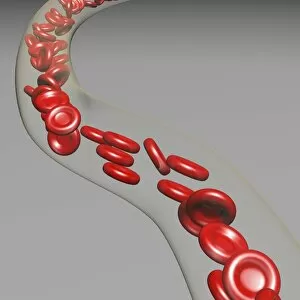
Background imageRed Blood Cell Collection: Red blood cells, artwork C016 / 8542

Red blood cells, artwork C016 / 8542
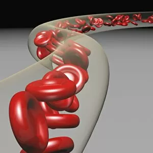
Red blood cells in a blood vessel, computer artwork. Red blood cells are biconcave, giving them a large surface area for gas exchange, and highly elastic

Background imageRed Blood Cell Collection: Red blood cells, artwork C016 / 8547

Red blood cells, artwork C016 / 8547
Red blood cells in a blood vessel, computer artwork. Red blood cells are biconcave, giving them a large surface area for gas exchange, and highly elastic

Background imageRed Blood Cell Collection: Red blood cells, artwork C016 / 8543

Red blood cells, artwork C016 / 8543
Red blood cells in a blood vessel, computer artwork. Red blood cells are biconcave, giving them a large surface area for gas exchange, and highly elastic

Background imageRed Blood Cell Collection: Red blood cells, artwork C016 / 8548

Red blood cells, artwork C016 / 8548
Red blood cells in a blood vessel, computer artwork. Red blood cells are biconcave, giving them a large surface area for gas exchange, and highly elastic

Background imageRed Blood Cell Collection: Red blood cells, artwork C016 / 8546

Red blood cells, artwork C016 / 8546
Red blood cells in a blood vessel, computer artwork. Red blood cells are biconcave, giving them a large surface area for gas exchange, and highly elastic

Background imageRed Blood Cell Collection: Red blood cells, artwork C016 / 8544

Red blood cells, artwork C016 / 8544
Red blood cells in a blood vessel, computer artwork. Red blood cells are biconcave, giving them a large surface area for gas exchange, and highly elastic

Background imageRed Blood Cell Collection: Blood cells, SEM C016 / 8026

Blood cells, SEM C016 / 8026
Blood cells. Coloured scanning electron micrograph (SEM) of a lymphocyte white blood cell (centre) and red blood cells (erythrocytes, circular)

Background imageRed Blood Cell Collection: Red blood cells, SEM C016 / 8027

Red blood cells, SEM C016 / 8027
Red blood cells. Coloured scanning electron micrograph (SEM) of red blood cells (erythrocytes). The main function of red blood cells is to distribute oxygen to body tissues

Background imageRed Blood Cell Collection: Red blood cells, artwork C013 / 5932

Red blood cells, artwork C013 / 5932
Red blood cells (erythrocytes), computer artwork

Background imageRed Blood Cell Collection: Malarial blood cell, SEM C017 / 8307

Malarial blood cell, SEM C017 / 8307
Malarial blood cell. Coloured scanning electron micrograph (SEM) of an erythrocyte (red blood cell) infected with Plasmodium parasites, the cause of malaria

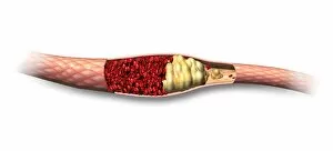
Background imageRed Blood Cell Collection: Atheromatous artery, artwork C015 / 6445

Atheromatous artery, artwork C015 / 6445
Atheromatous artery. Computer artwork showing the interior of an artery affected by atherosclerosis. Atherosclerosis is narrowing of an artery (stenosis) due to fatty deposits (beige)

Background imageRed Blood Cell Collection: Healthy artery, artwork C015 / 6444

Healthy artery, artwork C015 / 6444
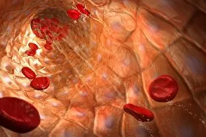
Healthy artery. Computer artwork of the inside of an healthy artery, showing red blood cells (circular) and the epithelial cells (square) lining the vessels wall

Background imageRed Blood Cell Collection: Atheromatous artery, artwork C015 / 6446

Atheromatous artery, artwork C015 / 6446
Atheromatous artery. Cutaway computer artwork showing the interior of an artery affected by atherosclerosis. Atherosclerosis is narrowing of an artery (stenosis) due to fatty deposits (beige)

Background imageRed Blood Cell Collection: Sickle cell anaemia, light micrograph C015 / 6395

Sickle cell anaemia, light micrograph C015 / 6395
Sickle cell anaemia. Light micrograph of a blood smear showing normally rounded red blood cells that have become curved, elongated sickle-shaped cells

Background imageRed Blood Cell Collection: Kidney glomerulus, TEM C016 / 5831

Kidney glomerulus, TEM C016 / 5831
Kidney glomerulus. Transmission electron micrograph (TEM) of a section through a renal glomerulus. At centre left is a capillary with a red blood cell (black) in its lumen


For sale as Licensed Images

Choose your image, Select your licence and Download the media

Red blood cells, also known as erythrocytes, are the most abundant type of blood cell in our bodies. These tiny cells play a crucial role in maintaining our overall health and well-being. In athlete physiology they can especially important as they carry oxygen from the lungs to every part of the body. This ensures that muscles receive an adequate supply of oxygen during physical activity, enhancing performance and endurance. Artwork depicting the intricate structure of red blood cells showcases their unique shape - biconcave discs without nuclei. This design allows for flexibility and efficient transport through narrow capillaries. The process of blood coagulation cascade is essential for wound healing and preventing excessive bleeding. Artwork illustrating this complex mechanism highlights how red blood cells interact with platelets and clotting factors to form a stable clot, sealing off damaged vessels. Scanning electron microscopy (SEM) images provide detailed views of various aspects related to red blood cells. One such image displays a close-up view of a blood clot formed by these specialized cells (SEM C016 / 9747). Another SEM image reveals infected red blood cells invaded by mouse malaria parasites (SEM). A diagram showcasing the bloodstream inside a vein demonstrates how red and white blood cells along with platelets flow together within our circulatory system. It emphasizes their collective effort in delivering nutrients, removing waste products, and defending against pathogens. Computer artwork beautifully portrays vibrant red blood cells flowing through arteries and veins, emphasizing their vital role in sustaining life throughout our bodies. Lastly, highlighting the connection between red blood cells and heart reminds us that these remarkable microscopic entities work tirelessly alongside our cardiovascular system to ensure proper circulation throughout every organ and tissue.