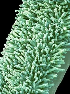
Scanning Electron Microscope Collection (#10)

"Unveiling the Hidden World

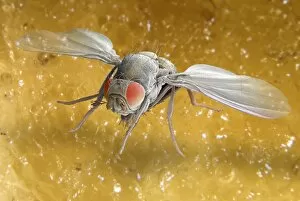
Background imageScanning Electron Microscope Collection: Fruit fly, SEM Z340 / 0662

Fruit fly, SEM Z340 / 0662
Fruit fly. Coloured scanning electron micrograph (SEM) of a fruit fly (Drosophila busckii) on a banana. Its compound eyes (red) are seen and its wings are outstretched

Background imageScanning Electron Microscope Collection: Hedgehog hair, coloured SEM

Hedgehog hair, coloured SEM
Hedgehog hair. Coloured scanning electron micrograph of the hairs from a hedgehog (Erinaceus europaeus). Magnification: x130 when printed 10cm high

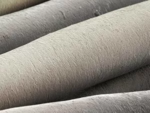
Background imageScanning Electron Microscope Collection: Horse tail hair, coloured SEM

Horse tail hair, coloured SEM
Horse hair. Coloured scanning electron micrograph (SEM) of hair from the tail of a horse (Equus caballus). Magnification: x200 when printed 10cm high

Background imageScanning Electron Microscope Collection: Fruit fly, SEM Z340 / 0663

Fruit fly, SEM Z340 / 0663
Fruit fly. Coloured scanning electron micrograph (SEM) of a fruit fly (Drosophila busckii) on a banana. Its compound eyes (red) are seen and its wings are outstretched

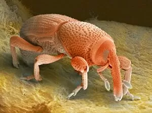
Background imageScanning Electron Microscope Collection: Grain weevil Z330 / 0408

Grain weevil Z330 / 0408
Grain weevil. Coloured scanning electron micrograph of a grain weevil (Sitophilus granarius). This beetle infests and damages stored grain, using the mandibles at the end of its elongated snout

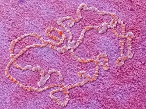
Background imageScanning Electron Microscope Collection: Giant chromosomes, SEM

Giant chromosomes, SEM
Giant chromosomes. Colured scanning electron micrograph (SEM) of giant (polytene) chromosomes from a fruit fly (Drosophila busckii)

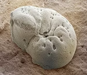
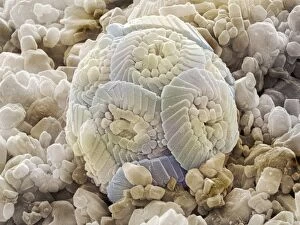
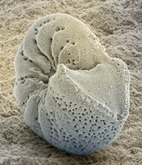
Background imageScanning Electron Microscope Collection: Foraminiferan fossil, SEM

Foraminiferan fossil, SEM
Foraminiferan fossil. Coloured scanning electron micrograph (SEM) of the fossilised shell (test) of a foraminiferan. Foraminifera are single-celled marine protozoa that construct

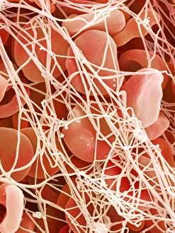
Background imageScanning Electron Microscope Collection: Blood clot, SEM P260 / 0123

Blood clot, SEM P260 / 0123
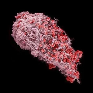
Blood clot. Coloured scanning electron micrograph (SEM) of a blood clot. The red blood cells (erythrocytes) are trapped in filaments of fibrin protein (pink)

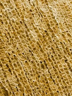
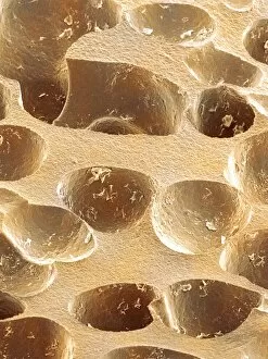
Background imageScanning Electron Microscope Collection: Wine cork, SEM

Wine cork, SEM
Wine cork. Coloured scanning electron micrograph (SEM) of a section through a wine cork. The structure of the cells can be seen. Cork is taken from the bark of the cork oak tree (Quercus suber)

Background imageScanning Electron Microscope Collection: Giant chromosomes, SEM P657 / 0034

Giant chromosomes, SEM P657 / 0034
Giant chromosomes. Coloured scanning electron micrograph (SEM) of giant (polytene) chromosomes from a fruit fly (Drosophila busckii)

Background imageScanning Electron Microscope Collection: Microchip, SEM

Microchip, SEM
Microchip. Coloured scanning electron micrograph (SEM) of a microchip from a chip and pin credit card. The circuit is composed of tracks made by various deposition and etching techniques

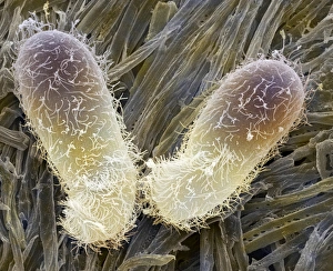
Background imageScanning Electron Microscope Collection: Chilodonella ciliate protozoan, SEM

Chilodonella ciliate protozoan, SEM
Chilodonella ciliate protozoa, coloured scanning electron micrograph (SEM). These tiny single- celled organisms are found in fresh water

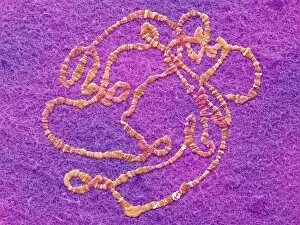
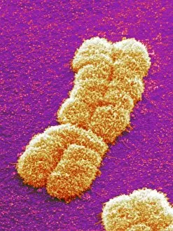
Background imageScanning Electron Microscope Collection: Human chromosome pair, SEM

Human chromosome pair, SEM
Human chromosome. Coloured scanning electron micrograph (SEM) of a human chromosome as a pair of identical copies called chromatids. These form as part of chromosome replication during cell division

Background imageScanning Electron Microscope Collection: Chip connector, SEM

Chip connector, SEM
Chip connector, coloured scanning electron micrograph (SEM). This connecting wire (yellow, top) has been soldered to a terminal pad at the edge of a silicon microchip

Background imageScanning Electron Microscope Collection: Light bulb filament, SEM

Light bulb filament, SEM
Tungsten light bulb filament, coloured scanning electron micrograph (SEM). The heat generated by an electrical current flowing through the filament makes it glow white hot

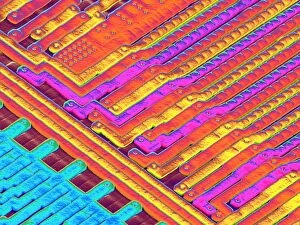
Background imageScanning Electron Microscope Collection: Microchip surface, SEM

Microchip surface, SEM
Microchip surface. Coloured scanning electron micrograph (SEM) of a few of the many tracks that are etched on the surface of a microchip

Background imageScanning Electron Microscope Collection: Calcareous phytoplankton fossil, SEM Z100 / 0212

Calcareous phytoplankton fossil, SEM Z100 / 0212
Calcareous phytoplankton fossil. Coloured scanning electron micrograph (SEM) of a fossil of the skeleton (coccosphere) of a coccolithotrope, a small marine algal organism

Background imageScanning Electron Microscope Collection: Household dust, SEM

Household dust, SEM
Household dust, coloured scanning electron micrograph (SEM). Magnification: x130 when printed at 10 centimetres wide

Background imageScanning Electron Microscope Collection: Amoebae, SEM

Amoebae, SEM
Amoebae. Coloured scanning electron micrograph (SEM) of two large protozoan amoebae (Amoeba proteus). A. proteus is commonly found on the bottom of pools and puddles

Background imageScanning Electron Microscope Collection: Electric blanket heating element, SEM

Electric blanket heating element, SEM
Heating element. Coloured scanning electron micrograph (SEM) of part of the heating element from an electric blanket. A wire (gold)

Background imageScanning Electron Microscope Collection: Synthetic cork, SEM

Synthetic cork, SEM
Synthetic cork. Coloured scanning electron micrograph (SEM) of a section through a plastic wine bottle cork. The air holes within the cork allow it to shrink and expand to fit the bottle neck

Background imageScanning Electron Microscope Collection: Foraminiferan fossil, SEM Z110 / 0239

Foraminiferan fossil, SEM Z110 / 0239
Foraminiferan fossil. Coloured scanning electron micrograph (SEM) of the fossilised shell (test) of a foraminiferan. Foraminifera are single-celled marine protozoa that construct

Background imageScanning Electron Microscope Collection: Electric razor, SEM

Electric razor, SEM
Electric razor with cut stubble (brown), coloured scanning electron micrograph (SEM). This electric razor has three rotary blade attachments. Part of one of these attachments is seen here

Background imageScanning Electron Microscope Collection: Iron pyrite crystals, SEM

Iron pyrite crystals, SEM
Iron pyrite crystals, coloured scanning electron micrograph (SEM). Iron pyrite (iron disulphide, FeS2), commonly known as fools gold, is the most abundant sulphide mineral and often occurs as cubes

Background imageScanning Electron Microscope Collection: Iron pyrite crystal, SEM

Iron pyrite crystal, SEM
Iron pyrite crystal, coloured scanning electron micrograph (SEM). Iron pyrite (iron disulphide, FeS2), commonly known as fools gold, is the most abundant sulphide mineral and often occurs as cubes

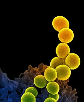
Background imageScanning Electron Microscope Collection: MRSA and dead neutrophil, SEM C018 / 8601

MRSA and dead neutrophil, SEM C018 / 8601
MRSA and dead neutrophil. Coloured scanning electron micrograph (SEM) of methicillin-resistant Staphylococcus aureus bacteria (MRSA, yellow) and a dead neutrophil white blood cell (red)

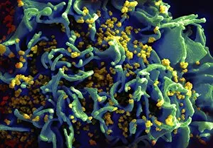
Background imageScanning Electron Microscope Collection: Budding HIV particles, SEM C018 / 8599

Budding HIV particles, SEM C018 / 8599
Budding HIV particles. Coloured scanning electron micrograph (SEM) of human immunodeficiency virus (HIV) particles (yellow) budding from the surface of a T lymphocyte from the H9 cell line

Background imageScanning Electron Microscope Collection: Neutrophil engulfing MRSA, SEM C018 / 8597

Neutrophil engulfing MRSA, SEM C018 / 8597
Neutrophil engulfing MRSA. Coloured scanning electron micrograph (SEM) of a neutrophil white blood cell (bottom) engulfing methicillin-resistant Staphylococcus aureus bacteria (MRSA, yellow)

Background imageScanning Electron Microscope Collection: Silkworm silk fibres (SEM) C014 / 2150

Silkworm silk fibres (SEM) C014 / 2150
Silkworm silk fibres, coloured scanning electron micrograph (SEM). The silkworm is the larva or caterpillar of the silkmoth which has been domesticated from the wild silkmoth Bombyx mandarina

Background imageScanning Electron Microscope Collection: Red Campion Seed (Silene dioica) SEM

Red Campion Seed (Silene dioica) SEM
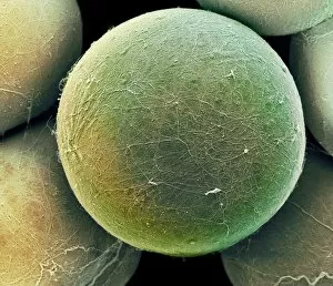
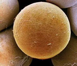
Red campion seed. Coloured scanning electron micrograph (SEM) of a seed from a red campion plant (Silene dioica). Seed shapes

Background imageScanning Electron Microscope Collection: Beaver beetle head, SEM

Beaver beetle head, SEM
Beaver beetle head. Coloured scanning electron micrograph (SEM) showing the underside of the head of a beaver (Platypsyllus sp.) beetle

Background imageScanning Electron Microscope Collection: Basil pollen grain, SEM

Basil pollen grain, SEM
Basil pollen grain. Coloured scanning electron micrograph (SEM) of a pollen grain from the flower of a basil (Ocimum basilicum) plant. Pollen grains are the male gametes (sex cells) of a plant

Background imageScanning Electron Microscope Collection: Spider body hairs, SEM

Spider body hairs, SEM
Spider body hairs. Coloured scanning electron micrograph (SEM) of the hairs on the body of a Nigma walckenaeri spider. This spider grows up to 5 millimetres long

Background imageScanning Electron Microscope Collection: Blood clot, SEM C016 / 9745

Blood clot, SEM C016 / 9745
Blood clot, coloured scanning electron micrograph (SEM). Red blood cells (erythrocytes) are trapped within a fibrin protein mesh (beige)

Background imageScanning Electron Microscope Collection: Hair follicle, SEM C014 / 0382

Hair follicle, SEM C014 / 0382
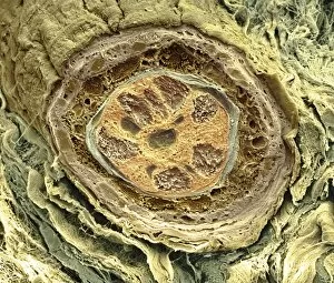
Hair follicle. Coloured scanning electron micrograph (SEM) of a section through a hair shaft and follicle (round structures at centre)

Background imageScanning Electron Microscope Collection: HeLa cell, SEM C014 / 0371

HeLa cell, SEM C014 / 0371
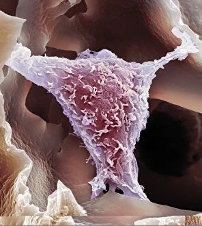
HeLa cell. Coloured scanning electron micrograph (SEM) of a HeLa cell (centre) grown in a 3D matrix (background). HeLa cells are a continuously cultured cell line of immortal human cancer cells

Background imageScanning Electron Microscope Collection: Hibiscus flower pistil, SEM C016 / 9758

Hibiscus flower pistil, SEM C016 / 9758
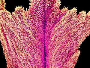
Hibiscus flower pistil. Coloured scanning electron micrograph (SEM) of a Hibiscus sp. flower pistil. Pistils hold a plants female reproductive structures

Background imageScanning Electron Microscope Collection: Pseudomonas aeruginosa bacteria, SEM C017 / 7140

Pseudomonas aeruginosa bacteria, SEM C017 / 7140
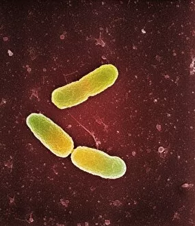
Pseudomonas aeruginosa bacteria, coloured scanning electron micrograph (SEM). These Gram-negative rod-shaped bacteria are found in soil, water and as normal flora in the human intestine

Background imageScanning Electron Microscope Collection: Scenedesmus algae, SEM C014 / 1461

Scenedesmus algae, SEM C014 / 1461
Scenedesmus algae. Coloured scanning electron micrograph (SEM) of Scenedesmus sp. algae. Scenedesmus is a nonmotile colonial green algae that has its cells arranged in a row

Background imageScanning Electron Microscope Collection: Fat cells, SEM C014 / 0364

Fat cells, SEM C014 / 0364
Fat cells. Coloured scanning electron micrograph (SEM) of adipocytes (fat cells). Adipocytes store energy as an insulating layer of fat

Background imageScanning Electron Microscope Collection: Fungal spores on pollen grain, SEM

Fungal spores on pollen grain, SEM
Fungal spores on pollen grain. Coloured scanning electron micrograph (SEM) showing fungal spores (round) on a pollen grain from a goji berry (wolfberry, Lycium sp.) plant

Background imageScanning Electron Microscope Collection: Carnation flower pistil, SEM C016 / 9763

Carnation flower pistil, SEM C016 / 9763
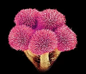
Carnation (Dianthus sp.) pistil, coloured scanning electron micrograph (SEM). Pistils hold a plants female reproductive structures

Background imageScanning Electron Microscope Collection: Fat cells, SEM C014 / 0363

Fat cells, SEM C014 / 0363
Fat cells. Coloured scanning electron micrograph (SEM) of adipocytes (fat cells). Adipocytes store energy as an insulating layer of fat

Background imageScanning Electron Microscope Collection: Hibiscus flower pistil, SEM C016 / 9757

Hibiscus flower pistil, SEM C016 / 9757
Hibiscus flower pistil. Coloured scanning electron micrograph (SEM) of a Hibiscus sp. flower pistil. Pistils hold a plants female reproductive structures

Background imageScanning Electron Microscope Collection: Bread mould, SEM C017 / 7127

Bread mould, SEM C017 / 7127
Bread mould. Coloured scanning electron micrograph (SEM) of a sporangium of a bread mould (Mucor sp.). Sporangia are asexual reproductive structures where the moulds spores develop

Background imageScanning Electron Microscope Collection: Carnation flower pistil, SEM C016 / 9761

Carnation flower pistil, SEM C016 / 9761
Carnation (Dianthus sp.) pistil, coloured scanning electron micrograph (SEM). Pistils hold a plants female reproductive structures

Background imageScanning Electron Microscope Collection: Bread mould, SEM C017 / 7129

Bread mould, SEM C017 / 7129
Bread mould. Coloured scanning electron micrograph (SEM) of a sporangium of a bread mould (Mucor sp.). Sporangia are asexual reproductive structures where the moulds spores develop

Background imageScanning Electron Microscope Collection: Blood clot, SEM C016 / 9748

Blood clot, SEM C016 / 9748
Blood clot, coloured scanning electron micrograph (SEM). Red blood cells (erythrocytes) are trapped within a fibrin protein mesh (beige)


For sale as Licensed Images

Choose your image, Select your licence and Download the media

"Unveiling the Hidden World: Exploring with a Scanning Electron Microscope" The scanning electron microscope (SEM) is an incredible tool that allows us to delve into the microscopic realm, revealing intricate details and unlocking secrets of the unseen. With its high magnification capabilities, it offers a glimpse into fascinating aspects of life and nature. In one captivating image, we witness the beauty of gender determination as X and Y chromosomes are captured under the SEM's lens. The intricate patterns and structures within these chromosomes showcase the complexity of our genetic makeup. Moving on to another astonishing discovery, a Scanning Electron Micrograph presents us with an up-close view of a Praying Mantis at 30 times magnification. Every tiny detail becomes visible, from its delicate limbs to its mesmerizing eyes – reminding us of nature's remarkable diversity. Shifting focus to plant life, lavender pollen grains come alive in another SEM image. These minuscule particles take center stage as their unique shapes and textures become apparent under intense scrutiny. Zooming in even further reveals a Tardigrade or 'Water Bear' at an astounding 1250 times magnification. This resilient creature captures our imagination as we explore its alien-like features through this powerful imaging technique. Delving deeper into biological wonders, T lymphocytes battling cancer cells are showcased in yet another breathtaking SEM capture. This visual representation highlights the ongoing fight within our bodies against diseases like cancer – emphasizing both hope and resilience. Calcareous phytoplankton takes center stage next as we uncover their intricate forms through SEM imagery. These microscopic organisms play crucial roles in marine ecosystems while leaving us awe-inspired by their stunningly detailed structures. Transitioning to neural marvels, nerve cells reveal their intricacy under the watchful eye of an SEM. Their branching extensions create complex networks that enable communication throughout our bodies – showcasing nature's ingenuity at work. Venturing into insect realms once again, a fruit fly is immortalized under the SEM's lens.